Feeds:
Posts
Comments

Archive for February, 2023

LA dimension and volume have become vital parameters in recent times, especially, with the entity of HFpEF is becoming so common. LA not only acts as a live barometer, reflecting all that happens in LV, but it is also a chronic marker of LV diastolic function. (Funnily referred to as HBA1c of diastolic dysfunction) 

What is normal LA dimension & volume ? 

  • Normal left atrial diameter < 4.1 cm in men or < 3.9 cm in women
  • Normal left atrial volume indexed for body surface area (BSA) is 34 ml/m2 for both women and men 

Which part of the cardiac cycle do we measure? 

Ever since Wiggers introduced the overwhelming concept of LV systole and diastole, most of us ignored the fact that atria do have a separate contraction relaxation cycle, independent of what happens in the ventricle. Of course, atria and ventricles act as a single chamber in diastole. In reality, atria lack true boundaries when it acts as a conduit. The LA dimension varies considerably during the atrial cardiac cycle. Look at the  LA pressure-volume loop, which can frighten anyone, with its horizontally lying figure of 8 pattern. During every cardiac cycle, the volume reaches atleast two troughs and one peak.

Don’t get frightened with this graph, spend some time, and you will get it right, Begin at  “3” o clock position with the onset of diastole with a downsloping green loop, that continues as the red line of atrial contraction to end up in systole. The entire black loop, that happens during ventricular systole depicts the true reservoir function. with MV closed. ,


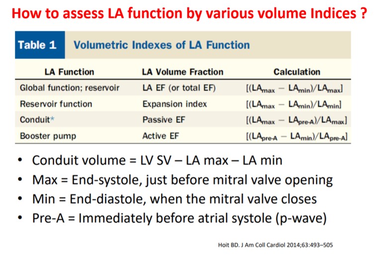
As of now, we have a consensus, LA volume is measured typically in LV end-systolic frame. ( Rather, we measure it at maximum LA volume ) However, we have 4 different LA  volumetric components to assess, as this article excellently depicts. (Hoit BD. Left atrial size and function: role in prognosis. J Am Coll Cardiol. 2014 Feb 18;63(6):493-505.)

What could be the limitations of the traditional end-systolic measurement?

No single measurement will give an overall LA function assessment. But still, Somehow, we have measured the maximum LA  volume as a reference of true diastolic function. This happens in LV end-systolic point where atria reach the maximum size. But, here is a catch, we assess the left atrial function before its main physiological function of emptying takes place.

How about assessing  LA efficiency after it completes its job, ie end diastole? 

In LV function end-systolic dimension has pride of place as it is devoid of influence from loading condition. If applying the same logic, the “end atrial” systolic dimension(Which is the same as LV end-diastolic point/or post A ) should be perfect. It can also help measure the residual LA volume after its systole.

A potential advantage of LV end-diastolic dimension (The Heart & Soul study )

Maybe, this is less affected in the presence of MR systolic jet will spuriously elevate LA volume. In AF also this parameter is less likely to be influenced by LA preload.

Final message

Suddenly, we are debating a fundamental Issue, ie timing of LA measurement. While the end-systolic size/volume is the current standard, the LA dimension in the end diastole also provides useful info. There are at least 4 different LA volumes, at different parts of the LA cycle that need to be studied for a proper understanding of diastology (Unlike LV which has only two).

Now, we may need to ponder, if there is a mean LA volume, measured with the 3D volumetric analysis or MRI, that could be representative of the global LA function. 

Reference

Thadani SR, Shaw RE, Fang Q, Whooley MA, Schiller NB. Left Atrial End-Diastolic Volume Index as a Predictor of Cardiovascular Outcomes: The Heart and Soul Study. Circ Cardiovasc Imaging. 2020 Apr;13(4):e009746. doi: 10.1161/CIRCIMAGING.119.009746. Epub 2020 Apr 20. PMID: 32306763; PMCID: PMC8846436.


Read Full Post »

Myocardial infarction, a gross pathological entity renamed now as STEMI for clinical purposes, continues to be the most famous medical emergency that triggers a series of calls, right from 911 to the ER, that eventually ends up in CCU or a 24/7 cath lab. The heart, can’t wait for all these external responses when it is challenged with a vascular accident. The moment ATO occurs, two things happen. The endogenous fibrinolytic led by native tissue PA (Tpa) tries to get rid of the thrombotic plug by all its means. It succeeds in 15%. We call it spontaneous lysis or aborted MI. Many lives are lost in the remaining before they reach the hospital..

Meanwhile, the immune high-command deputes scores of neutrophils to the ground zero Fig 1) to supervise what is happening and try to heal the injury. It is worth, understanding, that activated WBCs, sort of convert myocardial infarction from a vascular event to inflammatory pathology, as the hour progress.


Fig 1  Infarcted myocardium Invaded by neutrophils


What do these neutrophils do?  

It will be our ignorance, if we think, they simply crowd around the myocytes helplessly, that is hit by ischemia. They are sent with a specific purpose to protect and heal the myocardium. Very often it fails in its mission is a different story. These fighting neutrophils send a subtle signal  of their presence with a mild increase in temperature during ACS  ( Patel  Prognostic usefulness of white blood cell count and temperature in acute myocardial infarction  Am J Cardiol. 2005 Mar 1;95(5):614-8. )

Is neutrophil invasion good or bad for the myocardium?

These are pro-inflammatory cells. meant to promote healing and mitigate Injury. Interleukins and Leukotrienes do have a healing power as well. But, what happens, in reality, is, still a mystery. As of now, it is tempting to think, it does more damage than good. Its role changes over time. 

 Acute reperfusion Injury in Primary PCI 

Neutrophils are quiet obedient cells generally, but once activated, their behavior can’t be predicted. It may start attacking the host cells, We know reperfusion injury is real and poorly understood. Delayed reperfusion, is well known to cause myocyte softening and lysis. What we know for sure is, primary PCI, induced accelerated flushing of these angry neutrophils is definitely related to no flow, microvascular plugging, and cardiogenic shock.  (The fact that no-reflow and reperfusion injury is less of a problem in fibrinolysis  demands introspection)

Diagnostic & prognostic value of neutrophilia 

Neutrophilia is such a nonspecific response, faces ridicule for its clinical utility. But, In reality, it can be a worthy parameter, to time the age of the infarct (or even confirm* the ACS ) in otherwise equivocal clinical presentation.(Ref 2) More importantly, it provides prognostic information. One more potential use is Neutrophil count to guide the timing of surgery post-MI (as in VSR) A neutrophil count could help avoid the active phase of inflammation.

*I recall my surgical professor’s emphasis on leukocytosis to confirm acute appendicitis during the first clinical year. 

Final message 

There is no academic by-law, that forbids full-blown interventional cardiologists from having an affair with basic science. Unless the core science is irreversibly bonded to the bedside, we can never reap the true benefits of translational research. Hematological aspects in STEMI is one such underrated discipline. Also, we must encourage every postgraduate student in pathology/biochemistry/physiology to visit the CCUs or sit in the cath lab gallery more frequently. Watching the cardiology stalwarts plowing through the blocked coronary arteries, racing against time, is sure to kindle young minds, for a potential molecular breakthrough in cell survival and healing following myocardial hypoxia.

Reference

1.Ma Y. Role of Neutrophils in Cardiac Injury and Repair Following Myocardial Infarction. Cells. 2021 Jul 2;10(7):1676. doi: 10.3390/cells10071676. 

2. Thomson SP, Gibbons RJ, Smars PA, Suman VJ, Pierre RV, Santrach PJ, Jiang NS. Incremental value of the leukocyte differential and the rapid creatine kinase-MB isoenzyme for the early diagnosis of myocardial infarction. Annals of internal medicine. 1995;122:335–341. [PubMed] [Google Scholar]

3.. Cannon CP, McCabe CH, Wilcox RG, Bentley JH, Braunwald E. Association of white blood cell count with increased mortality in acute myocardial infarction and unstable angina pectoris. OPUS-TIMI 16 Investigators. Am J Cardiol. 2001;87:636–639. A610. [PubMed] [Google Scholar]

4. Bhatt DL, Chew DP, Lincoff AM, Simoons ML, Harrington RA, Ommen SR, Jia G, Topol EJ. Effect of revascularization on mortality associated with an elevated white blood cell count in acute coronary syndromes. Am J Cardiol. 2003;92:136–140. [PubMed] [Google Scholar]


Read Full Post »