Feeds:
Posts
Comments

Archive for February, 2020

Bernoulli equation is the most critical equation on which the foundation of clinical Doppler echocardiography is built. Bernoulli equation tells about fluid mechanics. Bernoulli’s principle states that the sum of potential and kinetic energy of fluid per unit volume flowing through a tube is constant.

A more detailed explanation regarding Bernoulli equation is linked in this video

Applying Bernoulli in Echocardiography

So, if we can somehow measure the velocity gained across a point inside the heart we can deduce the pressure gradient. Here comes the Doppler principle that helps us calculate the velocity. Doppler is based on the reflection of sound and the Doppler shift. With the Doppler shift, we can arrive difference in velocity across a valve, or conduit. When fluid flows across a narrowed orifice (Valve /Outflow) it accelerates and builds up velocity. This gain in velocity is equal to the pressure lost ie as given by the Bernoulli equation. Since potential energy is related to height and gravity same intracardiac zones it cancels out on either side. Hence, essentially the Bernoulli pressure gradient is equal to the difference between the kinetic energy on either side.

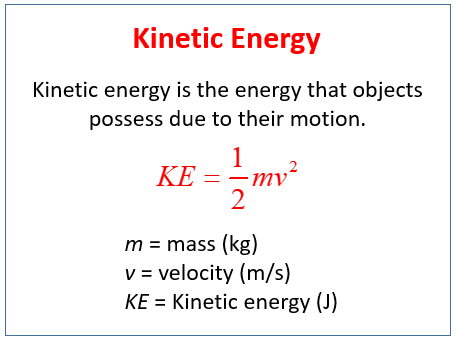
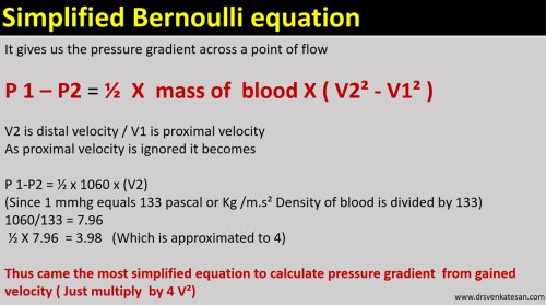
Let us see how this 1/2 of mass becomes 4. We have to convert density of blood which is 1.060 to mass.

mass to density

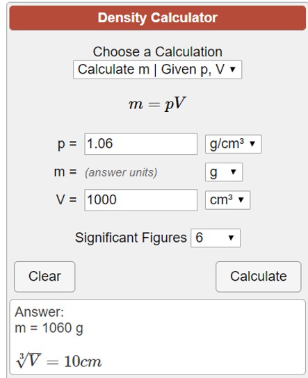
Note : Mass = ρV . Density is mass per unit volume. So the “m” in the equation is some times referred to synonymously with the density of blood.

modified simplified bernouli equation doppler pressure gradient mass density velocity drsvenkatesan madras medical college echocardiography 2 tr jet lvot gradient

Application in clinical echocardiography

There has been pioneering work from Holen, Hatle and Angleson who proved the value of this equation in the clinical situation in the late 1970s. Of course, Gorlin and Gorlin worked on this similar concept in the cath lab derived pressure gradients

Reference

1.Gorlin R, Gorlin SJ. The hydraulic formula for calculation of the area of the stenotic mitral valve, other cardiac valves, and circulatory shunts. I. Am Heart J 1951:41:1-29.

2.Holen J, Aaslfd R, Landmark K, Sknonsen S, Ostrem 1. Determination of effective orifice area in mitral stenosis from noninvasive ultrasound Doppler data and mitral flow rate. Acta Med Stand 1977;201:83-88.

3.Hatle L. Noninvasive assessment and differentiation of left ventricular outflow obstruction with Doppler ultrasound. Circulation 1981;84:381-
4.Hatle L. Brubakk A. Tromsdal A. Angelsen B. Noninvasive assessment of pressure drop p in. mitral stenosis by Doppler ultrasound. Br Heart J 1978:40:131-140.

Read Full Post »

Here is a 3-minute algorithm for the management of acute pulmonary embolism. Just need to ask 3 questions.

Caution: User discretion is advised. Tainted with reasonably acceptable levels of non-scientific content.

Click over the image for a high-resolution slide

Some more critical  questions need to be answered.

What is hemodynamic stability?

It is purely based on clinical signs and judgment.(One need to be doubly sure to rule out hypovolemia and sepsis-related hypotension)

Is RV dysfunction equivalent to hemodynamic stability?

No, it is not. Clinical instability must be associated.( The dogma is,  if the patient is stable even if there is significant RV dysfunction by echo , that RV dysfunction is not attributable to the current episode of PE)

Can we diagnose and proceed with lysis without CT pulmonary angiogram confirmation?

Yes, you can, provided your suspicion is too strong or you have the extraordinary talent to argue/defend even a fatal bleed ( with your boss or in medical audit ) in a patient who was subsequently proven not to suffer from PE .

How to switch over to Lysis from Heparin alone protocol?

Occasionally one may require to do it. There is an added risk of bleeding here. It can’t be avoided in some situations as Initially, it appear as low-risk PE later on becoming more Intense. Generally, high-risk unstable patients should receive lysis straightaway.

Is 60/60 sign is really useful in deciding lysis?

60 /60 sign tell us if Pulmonary artery acceleration time (PAT) and the TR jet both are less than 60 the likely hood of PE is high in a patient with suspected PE.

  • This sign recently got popular not because of its utility, rather because of its simplicity and attractive caption.
  • It may be very specific but least sensitive (<20%) So it can never be used as a screening test.
  • It also fails to differentiate chronic RV dysfunction from acute RV dysfunction.
  • The PAT is strongly influenced by RV dysfunction (It pulls it down below 60 as PAT is dependent on RV Dp/Dt and falsely diagnosing PE
  • 60/60 sign adds up to the value of  Mconllels sign and can confirm PE with almost 100% specificity.

Read Full Post »

Pulmonary atresia with VSD is one of the complex CHD subsets that requires a meticulous understanding of anatomy, physiology of pulmonary circulation. It can be termed as TAPAC -Total anomalous pulmonary arterial connection in extreme cases. Should we attempt to reverse this total chaotic pulmonary blood supply is the question?

It demands a highly focused cath study(hands & brain) and CT Imaging which might actually throw more light. Post-study Interaction with surgeon and team of cardiologist are vital. The decision to take up the challenge of surgery or abandoning poses equal intellectual stress. Continuous and critical decisions need to be taken. Repeat surgeries and cath based Interventions are often a rule.  Very few centers have mastered this surgery.

A single slide presentation


pulmonary atresia 4

In spite of all technological developments in pediatric cardiac surgery, there is considerable variations and expectation of the surgical outcome. The major surprise is the original Melbourne group(Ref 1 )  that advocated the uni-focalization as a  concept has almost abandoned this. Stanford and other groups still continue to use this technique more often as a single-stage procedure to improve the outcome.

Let us hope these children get the best of the right mix of technology and natural survival power and more importantly we must ensure the former do not interfere with the later

Reference 

1

Post ample

Surgery has definitely  revolutionized the outcome in neonates and children in less severe forms of PA with good central pulmonary arteries ( Most of the Barbero Marcial Type A and many type B) The perceived negativity in this post regarding the outcome of surgery is primarily belong to some of the  Barbero Type B and many of  C.

gr1

Barbero-Marcial M , Jatene A Semin Thorac Cardiovasc Surg. Surgical management of the anomalies of the pulmonary arteries in the tetralogy of Fallot with pulmonary atresia.1990 Jan;2(1):93-107. 


Read Full Post »