Feeds:
Posts
Comments

Archive for March, 2015

We generally believe ischemia and it’s clinical counterpart  angina would  go together .It is not true .Most patients with ischemic cardiomyopathy do not have any significant  angina in spite of  having one or more critically  narrowed coronary arteries.

The reasons could be many ,

  1. Little viable tissue to generate Ischemia.
  2. Less contractile elements and less MVO2 consumption.
  3. Severe LV dysfunction makes these patients adopt a very restrictive lifestyle.
  4. Loss of nerve fibers  along with myocyte necrosis and apoptosis.
  5. Post CABG patients often have no angina due to denervation..

The benefits of revascularisation in ischemic DCM is not clear. As the cardiomyopathy  progresses , intensity of angina regresses and dyspnea dominates .Presence of angina makes the decision to  revascularise easy .To consider dyspnea as an anginal equivalent in ischemic DCM and advising revascularisation can not be  justified .


Read Full Post »

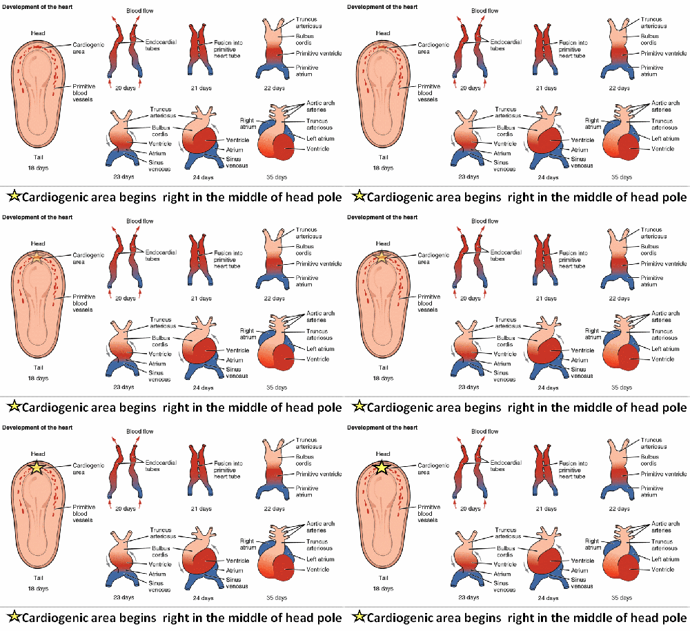
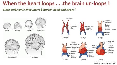
Human facial configuration  is formed by fusion and absorption of  different  tissue structures in a preplanned genetically programmed planes . The developing cardiogenic area  lies right in the cranial end of the  neural tube which enlarges to grow as face .The timing as well the location of facial plates are closely related to cardiac development. As the head end unfolds  to take it’s shape , the heart folds to form the chambers and the great vessels , septum and valves are subsequently  cleaved . Even minor genetic errors in the codes that  initiate and coordinate  the bio-genetic forces in the cardiogenic  area ,  results in simultaneous defects in  facial as well as cardiac contour .

There is a proposed embryonal classification for congenital heart disease (Clarkes ) The genetic  defect responsible for cono-truncal anomalies are located in  chromosome Q22.


cono truncal facies heart face abnormality catch 22 tof cardiac looping


cardiac development embryology of heart

Reference

1.Conotruncal anomaly face syndrome is associated with a deletion within chromosome 22q11.J Med Genet. 1993 Oct; 30(10): 822–824

Parallel reading
geentic basis of face development cono truncal facies

It is stunning to note some of the plant  species (Chinese Ginseng)  express unusual human phenotypes  , indicating the genetic codes responsible for  such things are beyond human comprehension . If one can  demystify  the genetic basis of this  perfectly smiling fruit plant with a human face ,  we may also know  what makes  it to go awry in human CHD  !

ginseng fruit cono truncal facies geentics of facial development

Read Full Post »

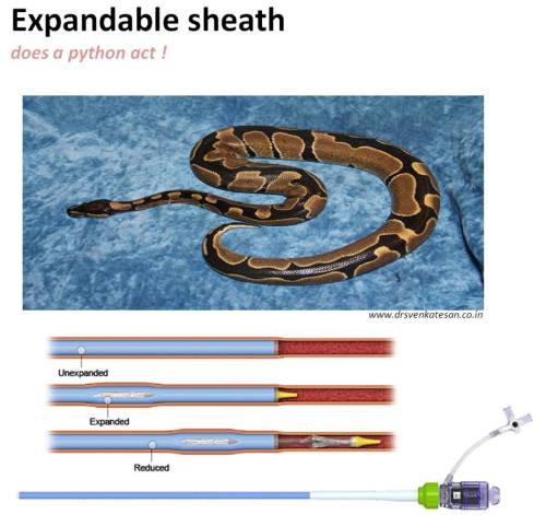
Interventional cardiologists in one way be labelled as intra-cardiac and intra-vascular civil engineers.Their primary  job is to create ,or close vascular tunnels and holes in various locations within the heart.How to deliver the  working hardware  to the  site of action ?. Temporary bridges ? .The vascular access is through long sheaths though which , wires, catheters, and devices , valves  are transported. It’s the key supply line to the ultimate battle field of life , right inside the beating heart.  .

So far,the sheaths  and catheters were rigid tubes with a fixed diameter.Innovative sparks come from  strange thoughts.As we struggled to take the per-cutaneous valve for  TAVR  through small caliber sheaths , some one thought why should the sheath be fixed and static .Why can’t it accommodate  liberal sized devices just by expanding its shaft like a python ,come back to its original state once the device passes by ?

Expandable sheath 164 solo path tavi tavr cathlab hardware

Thus came the expandable sheaths. Soon this concept is going to come in a big way and most complex and large device interventions will be benefited by this.

Read Full Post »

We have been taught right from first year cardiology residency  how to trouble shoot a pacemaker .It has been a real complex thing for us. Now looking  back ,all the troubles we took to understand seems to be redundant.Here is a summary of my thought process on the issue. It can be approached  with reference  to time, symptoms and ECG features.  With due respects to all those brainy hardworking   EP experts  , I have taken few academic liberties!

pacemaker trouble shooting

Timing

  • Within 24 hours -100% technical or procedural Issues , like lead dislodgement/Screws and nuts.
  • Within 1-2 week – Again technical , Pocket issues , Infections.
  • Within 6 months – Benign pacemaker syndrome ,Threshold settings, Scars
  • After  first year – Generally Issues are rare , Lead issues , Associate disease progression.
  • Beyond 8-10  years /Near end of life – 95% Energy depletion leads issues .( Please note , pacemakers do not stop all of a sudden it has a intrinsic end of life indicators .We have to look for it. May be ,we can expect a  warning siren in the future ? )

Symptoms

  • Vague dizziness – Pacemaker syndrome ? Anxiety ?
  • Near syncope – Show some concern (For many , Impending true syncope is a non existent entity )
  • True syncope  – Real emergency*

* Syncope can be unrelated to pacemaker but always consider  them electrical  unless proved otherwise . Few patients  may continue to have significant symptoms  in-spite of   normal pacemaker parameters. This would  mean , the original symptom for which  pacemaker was put is not related to the Brady-arrhythmia .It could   suggest alternative hemodynamic explanation  like vaso-depressive component of vagal syncope ,autonomic dysfunction , orthostatic intolerance or  a coexisting neurological /systemic condition.

**Never forget syncope is not an exclusive symptom of bradycardia .A new onset  tachycardia  , which is either a part of  brady- tachy syndrome or separate arrhythmia can continue to provoke the symptom.

Gross ECG findings

Bradycardia /Often implies back to original rhythm –  Indicates real trouble . Since ,in a paced patient HR cannot be less than programmed rate of 70.

Tachycardia -No spike.( Not to worry ?) A common  situation if the original indication was  sinus node dysfunction . Many of them are  in own sinus rhythm or AF . Just ensure spikes reappear when the rate falls below 70 . If the rate never goes down , what to do ? Try a carotid massage or observe a nocturnal ECG  or call analyst and increase the rate to document pacing . (In DDD mode we have a rare PM mediated re-entrant tachycardia , which is mainly used to grill cardiology fellows in their board exams with all those PVARP stuff !)

Simple pauses – Any pause more than the pacing interval is a definite concern .

Spikes more than QRS  – Indicate capture failure.

No spikes (Can be so benign  to ultimate danger )

  • No spikes , but  excellent own rhythm – Good  functional  SA node
  • Regular  spikes ,but intermittent own rhythm or only random spikes with good own rhythm – Needs bedside hairsplitting and  EP assistance !
  • “No spikes -No Own rhythm” -Most dangerous .Sudden lead issues or hyper sensing .(Emergency  switch off  by magnet  application before inserting temporary pacing advised )

* Anatomical issues like lead dislodgement , fracture , compression ,  perforation are to be ruled out in every pateitn with intermittent capture or failure .This is done by combinations of imaging as well physiological assessment.Dislodgement must be visualized .The term micro dislodgement may not exist.

Other Investigations

  • X ray
  • Echo for any new structural lesion (RA,RV dilatation , TR RV clots or vegetation )
  • Holter
  • Event monitors ,Loop recorders.

Pacemaker analysis

  • Battery life ( Very important parameter .Usually around 10 -12 years.Unexpected early drain can occur.)
  • Threshold (Most failure to capture associated with high threshold Note :Threshold will be normal in battery depletion Acute threshold can increase marginally .Should be reasonable other wise battery will drain.New protocols like auto capture and managed pacing will help optimal threshold
  • Impedance – Normal in battery depletion , dislodgement and exit block,  Increased in lead fracture and loose screws.Decreased or lost in insulation failure.

Management

The principle of management are simple. Few logical questions ,

  • Is the pacemaker generator is alive and has has enough energy ?
  • Are the  leads okay ?
  • The problem is in the settings ? can it be rectifies by the programmer
  • Or should we replace the pacemaker ?

Technical jargon like  under sensing , over sensing or no sensing  , fusion beats , micro dis-lodgement  etc are important for  academic reasons . We may talk any thing , realistically , what  the ventricle want  is a non stop heart beat  every second or so !

Emergency

Bradycardia – Insert a temporary pacemaker /Call the analyst  /Inform the  electrophysiologist /Senior cardiologists /(Please realise ,  some fellows  can be better than the personnel mentioned above in tackling emergencies !)

Tachycardia : Native or machine induced ?

Native – Mostly safe ,  Ignore  or treat with drugs.

Machine induced :(very rare) Switch of the pacemaker . No off switch available as in a mobile phone ? *What to do ? if unclear about the  whereabouts  tachycardia origin . If hemodynamically unstable no harm in shocking .Nothing will happen .Call the EP  guys on hot line and decide.

Elective symptom guided.

  • Asymptomatic -Normal ECG : Reassure and send home.
  • Vague symptoms   -Do Holter and Observe
  • Syncope -Normal ECG needs extensive all system investigation.
  • Syncope -With pauses /Bradycardia /Asystole  – Ironically ,decision  making is easier. Temporary pacing is the  ultimate savior. Later , check the lead,  generator .One may need to change  either one or both of of them.

** While the above principles apply  for both single  and dual chamber pacemakers , the later doubles our thinking burden . While atrial tracking is a great technological advancement , what to  do with those sensed event can be really  tricky .The response of  ventricles and the AV intervals  can be tentative at times. Cross talks from unexpected atrial and ventricular arrhythmia can occur.  Further , mechanical atrial lead  issues are far  more common . When confronted with recurrent atrial lead related  problems , one  simple solution is  silently convert the mode to  single chamber VVI mode.

Final message

Pacemaker trouble shooting appears complex at the first look .It’s all common sense.Thinking with simple state of mind and  being clear about the intended  goal is vital. Electrical intricacies are tough to understand  but most situations do not require them. However ,If the initial indication was for complete heart block one has  to be very alert.

Principles of medicine argue us to make an exact diagnosis before treating . But,realise  this is rarely  possible or even desirable in emergency .Curiously , most pacemaker troubles can be solved successfully without making a proper trouble shoot !

If  we can summarize in one line  , a prompt emergency  back up temporary pacemaker insertion  is key to  management most of the serious  pacemaker related problems.It ,not only tackles the emergency ,  buys time till we decode the real problem . . .  if we wish to !

Related article.

Role of magnet application in pacemaker trouble shoot


Read Full Post »
We know , any wide QRS tachycardia  would argue us to make a default diagnosis of VT.But,  one has to be extremely cautious to apply this rule if  wide QRS  tachycardia shows  significant irregularity in RR interval .

All classical VTs are fairly regular tachycardia (Note the  key words , fair and regular) . Small cycle length variations are observed in VT,  but they are usually not discernible in surface ECG.

There are no practical rules .A well  appreciable  irregularity in RR interval will seriously  question the diagnosis of classical VT. To make an another statement, most of the  irregular wide QRS tachycardias infact turns out to be  atrial fibrillation with some form distal widening mechanism .(Preexisting blocks, or rate dependent  or antidromic conduction through accessory  pathways)

However , irregularity  is still possible during VT .(May be less than 10% of times)

When can VT can be irregular ?

  1. Irregularity is observed  immediately at the onset of VT as the re- entrant circuit warms up and tries tosettle down
  2. AV dissociation  can make the VT irregular but it is subtle .(This AV dissociation is absent if retrograde VA conduction is intact)
  3. Multiple reentry circuits with two morphological VTs dissociating themselves
  4. VT with multiple exit points and epicardial breakthroughs
  5. A drugged VT.Amiodarone modified VT can be irregular as it can variably lengthens  the re-entrant  circuits and inducing VA block and precipitating AV dissociation.
  6. Multi- focal VT  (We have MAT in atria do we really have MVT ? (Why not , are we missing it ?)

Final message

Statistically , as well as realistically  , the commonest cause for  any highly irregular tachycardia turns out to be AF , whether  QRS is wide or narrow !

Read Full Post »

ICDs are primarily life saving devices.Whether single or dual chamber  it does this function  effectively.They will also  take care of  bradycardia by  default  back up pacing .For most indications single chamber ICDs are good enough.

My professor used to tell us , dual lead  means ,  dual expertise , dual cost , dual caution and  dual set of complication . One should avoid it whenever possible. Make things as simple as  it could be , without compromising the main goal (Here prevention of SCD) . The incidence of inappropriate shocks being lesser with dual chamber ICD  has not been truly  realised in real world scenario.

Recent studies  tend to give  credence to this  perception .(Peterson JAMA 2013)

Dual  Chamber ICDs  may have an edge only in few situations .

  • When there are both indication for pacing as well as ICD like heart block and LV dysfunction.
  • In extreme LV dysfunction were benefits of dual chamber pacing may have advantage.(If CRT is not an option )

Reference.

1 .Peterson PN, Varosy PD, Heidenreich PA, et al. Association of single- vs dual-chamber ICDs with mortality, readmissions, and complications among patients receiving an ICD for primary prevention. JAMA 2013; 309:2025-2034.

2.Medscape review


single vs dual chamber pacing indication


Read Full Post »

We are aware  , modern day cardiologists literally live within the patients coronary artery and vascular system .  It ‘s not at all surprising then , man made cardio vascular accidents  are becoming more common  , where pieces of hard ware like guide wires catheters and stents  get trapped .

Knowing about the hardware and techniques of retrieval of foreign bodies within vascular system is so important .It would appear  indulging in cath lab work with out proper salvage hardware and expertise is a  near  serious offense.Apart from this , many complex procedures require intentional snaring of wires and gadgets .

How to retrieve a foreign body from  coronary artery ?

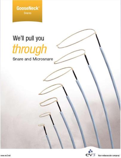
There are few snares availablew  with  single or multiple loops  and comes in various sizes .

1.Goose neck EV3 snare (Covidien /Medtronic)

2.En snare -Multiple loops (Merit Medica)

3.Micro elite snare (Vascular solutions)


ev3 microsnare covidien

The snare is constructed of Nitinol cable and a gold plated tungsten loop. The pre-formed snare loop can be introduced through catheters without risk of snare deformation because of the snare’s super-elastic construction. The snare catheter contains a platinum-iridium radio opaque marker band.

  • Nitinol Shaft for durability and kink resistance
  • Super-elastic and shape memory properties of nitinol provide kink resistance.
  • Ideal for challenging or unplanned foreign body retrieval and manipulation cases.

goose neck snare amplatz ev3
True 90° snare loop remains coaxial to the lumen

  • Snare loop forms a true 90° angle.
  • Device remains coaxial to the lumen for proper insertion and successful retrieval or manipulation of atraumatic foreign bodies.


Hardware specification

ev3 goose neck

2.Ensnare


coronary snare ensnare merit medica


Micro elite snare

micro elite coronary snare vascular solutions


micro elite snare

Other retrieval devices

  1. Bioptome* (Cook medical)
  2. Needle and eye snare
  3. Multi snare
  4. Welter loop catheter
  5. Expo retrieval catheter
  6. Curry snare
  7. Simple  alternate option : 2 or three wire guide wire trapping  technique.
  8. The cheapest option :To make a custom made snare with  .014 PTCA   guidewire  with a flexible loop .
biopsy forceps

Intra cardiac biopsy forceps may help to retrieve some of the foreign bodies


Final message

At least few of  these retrieval devices  should be  available in  every cath lab .  Attempting to do sophisticated procedures  in your cath lab without essential hardware is akin to driving a car with defective breaks  or like flying airplane with a single engine .

Acknowledgement

Image source , content and courtesy respective manufacture web site

Read Full Post »