Feeds:
Posts
Comments

Archive for October, 2022

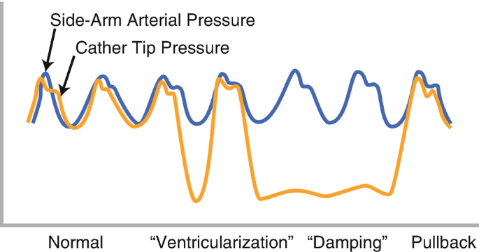
“Never take your eyes off the monitor and the pressure curve” It is one of the basic instructions given to the fellows & technicians as they start engaging the coronary artery and Intubating the coronary ostium in their early cath lab postings. There are two commonly heard noise bites in the cath lab for the beginner. 1. Damping 2.Ventricularization.

Damping

It is the deformation of the normal arterial pressure curve, with a blunting of both systolic and diastolic pressure that drops compared to aortic pressure. Extreme damping can mimic a straight line with few wavy undulations. It means the forward flow is impeded as the catheter might be blocking the coronary flow at least partially, if not completely. Immediate pull back and adjustment of catheter is required to prevent adverse events that includes cardiac arrest. (Of course, catheter kinks, blocks, and air in a circuit are to be ruled out first)

Mechanism of damping

  1. The catheter tip is too large for the ostium.(Technical)
  2. A left main or RCA ostial lesion*(Most significant clinically)
  3. The coronary artery wall is thin and goes for spasm
  4. Catheter diameter is too small and glides into the coronary artery (The catheter tip hitches against the lateral wall of the coronary artery often over a plaque, a silently staged perfect setting for a dissection )

Unique features in RCA damping

In RCA, damping could simply mean it has engaged conus branch. Damping is more common in RCA and it tends to get sucked in deeper for two reasons. We know , the RCA catheter is not preformed, the tip seeks the ostium, looking for a negative pulling pressure from within the RCA. Further, unlike the LCA, the flow in RCA is continuous in both systole and diastole. Another possible factor is the inspiratory swings of RV transmural pressure is more than LV.

Transient bradycardia due to compromise of SA /AV nodal flow is common. It is well known that RVOT is a thin and VPD-prone zone , compared toLVOT. Hence it is more vulnerable to ischemia and triggers a VT/VF if damping is prolonged in RCA.

Ventricularization

This is tracing during the engaging of a left main ostial stenosis. The diastolic pressure drops on the third complex, and the pulse pressure becomes large . Note the small “a ” wave slur as well near the diastolic nadir. The aortic pressure curve is restored once tip adjustment is done. Image courtesy Thoraci key.https://thoracickey.com/coronary-angiography-3/

It simply means the pressure curve starts looking like a ventricular pressure curve. The issue is not that simple. It is still a mystery, how could the coronary artery pick up ventricular pressure. In fact, it doesn’t. It was initially thought to be either deformed aortic pressure or a wedged coronary artery pressure .or a combination of the two.

It s now accepted it’s due to total occlusion of the coronary artery the ventricle muscle sucks the catheter and pulls the diastolic pressure markedly down even lower than LVEDP. The systolic pressure also falls but not much of it is noticed.The fact that ventriculaization happens less commonly with RCA gave us a clue to its mechanism. (Most of the RCA travels over RV and since RV muscle mass wields less negative suction force )

How to differentiate true LV location from ventricularization?

If the catheter enters the LV cavity, the systolic pressure will match that of aortic systolic pressure and diastole will reach the LVEDP. In ventricularization there can be a slight drop of the systolic pressure, but the diastolic is the one goes far down. Further, the ascending limb is shallower, the descending limb is steeper in coronary ventricularization. Apart from this if we carefully look “a” waves will be visible in ventricularisation in the late diastolic phase.(Could it be the modified Incisura ?)

Relationship between damping and ventricularization ?

Discussing damping and ventricularization together is more of an academic tradition. Please realize, both may not be two exclusive entities. In fact, they can occur at the same time, or sequentially or interchangeably if the catheter tip swings back to partial to total occlusion and vice versa. if complete wedging occurs it becomes ventriculariszation.

Which is more dangerous damping or ventricularization?

How can you ask such a question? Both can be harbingers of serious hemodynamic issues if ignored and accidentally injected, The dreaded one is the dissection. Apart from mechanical injury, Injecting dye during damping or ventricularization causes dye stasis, and may trigger VT or VF if it is prolonged. There is a biochemical component too. As the contrast swirls around for a few seconds it can cause transient hypocalcemia by chelating this cation.

I don’t have a clear answer to the above query. In my understanding, forceful injections during ventricularization could be more riskier as injection happens over a total occlusion in a wedged position and dye has direct access to the microcirculatory bed. It is true, expert cardiologists do shoot in damped positions occasionally to see the anatomy quickly. They may be your mentors, still don’t learn this trick, till you become a true expert (Famously referred to as hit and run technique)

How to overcome these pressure curve malformations?

Most times, it is a simple technical artifact issue. Deep Intubation with an oversized catheter could be the commonest cause in the otherwise normal coronary artery. Proper catheter sizing, angle of alignment, and adjustment is the key. If it is a spasm nitroglycerine might help. Using catheters with side-hole perfusion catheters is an option in difficult anatomies. Mind you, side holed guide is a two-edged weapon, it can mask true hemodynamic adversaries by falsely showing a good aortic pressure tracing.

(One of the good cath lab habits is to have a look around the patient face at least once in a while when performing complex procedures. We have many times recognized serious hemodynamic issues only after the patient starts behaving bizarrely due to hypotension. Pressure curves can fool you, but patience will never.)

Final message

Recognizing abnormal waveforms during engagement is a crucial step for the beginner (Experts can’t ignore though) Please remember LMCA stenosis should be the default alarm for any damped pressure curve as you engage the LCA. If you take things casually consequences can be lethal for the patient as well as the consultant in charge.(Fellows can’t take it lighter though). It is an undisclosed fact, that many of the serious complications in the cath lab happen not out of ignorance or lack of expertise, but because of ignoring some basic principles and lack of cath lab discipline.

Reference

1.Baim DS, ed. Grossman’s Cardiac Catheterization, Angiography, and Intervention. 7th ed. Philadelphia, PA: Lippincott Williams & Wilkins, 2006. 2. Judkins MP. Selective coronary arteriography, a percutaneous transfemoral technique. Radiology 1967;89:815.

2.T Pacold I, Hwang MH, Piao ZE, . The mechanism and significance of ventricularization of intracoronary pressure during coronary angiography.Am Heart J. 1989 Dec;118(6):1160-

3.A good review from Journal of Invasive cardiology .

Read Full Post »

Read Full Post »

A forbidden quote in medical science

Read Full Post »

Though all of us are aware, the incidence of heart failure is increasing exponentially and is the leading cause of global disease mortality, what we fail to understand is, we still lack a good definition cardiac failure. 

Defining HF based on EF% is convenient but adds more complexity, and is less scientific too. Still, as of now, we have adopted this. I think, one of the important factors that apparently increased the incidence of HF is the creation of an entity called HFpEF. (Formerly diastolic heart failure)

Thanks to ESC, we have a consensus document, which has defined HFpEF based on functional, morphological, and biochemical features. This is a more refined model from the original Mayo clinic H2FpEF score.

Both are given below.


The problem with both these criteria is the disproportionate importance given to AF. The knowledge gap here is, AF can be initiated at any degree of increased atrial strain which can independently raise LA mean pressure without persistent elevation of LVEDP. We recognize now, left atrial obesity (fatty atrium)  is a powerful trigger of AF, still, in this situation, an Innocent LV may get blamed with a tag of HFpEF. Likewise, many HFpEF may turn out to be primary  LA dysfunction than LV failure. To make things more confusing(scientific) for diagnosing true HFpPEF, we may soon need to look into LA-EF as well. (LA-HFpEF)

Can we diagnose clinically significant HFpEF, without pulmonary hypertension?


In my understanding, the answer is No.

Looking at the two schemes (Mayo & ESC) one thing is clear. Pulmonary hypertension is the key hemodynamic expression of HFpEF. It could be either resting and persistent or exertional and transient.t is obvious the PH in HFpEF is post-capillary. (The modern term for pulmonary venous HT). Mind you, while PVH is mandatory to diagnose HFpEF, PAH (precapillary ) is also observed in most patients with significant HFpEF. This is the reason TR jet velocity is included as one of the criteria.  (To make things simple, we may need to create a new classification of HFpEF, ie resting vs exertional HFpEF.This is what the diastolic stress testing is all about.)

Final message

It is back to basics & time to dig into the fundamentals, of what exactly we mean by heart failure. Is the elevation of LV filling pressure alone sufficient? Should it happen at rest or at exertion, and whether neuroendocrine activation is necessary? Is RASS activation similar in both HFrEF and HFpEF? Try to find the answer to this. How often does HFpEF fulfill Framingham’s criteria of HF.? ( Löfström et al  ESC Heart Fail. 2019) 

Trying to understand the nuances of HFpEF, I think, we can make a statement,- HFpEF can not be diagnosed without pulmonary hypertension. It makes a lot of sense the P in the H2FpEF  scoring system denotes PH, however, It is assigned only a single point, which needs revision. In fact, there is a strong case to argue and make it an essential criterion.

Paradoxically & curiously HF with reduced ejection fraction (which is the most common form of HF) doesn’t require the presence of PAH to diagnose it. This issue may also be examined.

Reference 

1.How to Diagnose Heart Failure With Preserved Ejection Fraction: The HFA–PEFF Diagnostic Algorithm: A Consensus Recommendation From the Heart Failure Association (HFA) of the European Society of Cardiology (ESC). Eur Heart J 2019;40:3297-3317.

2.Löfström U, Hage C, Savarese G, Donal E, Daubert JC, Lund LH, Linde C. Prognostic impact of Framingham heart failure criteria in heart failure with preserved ejection fraction. ESC Heart Fail. 2019 Aug;6(4):830-839. doi: 10.1002/ehf2.12458. Epub 2019 Jun 17. PMID: 31207140; PMCID: PMC6676283.

Read Full Post »

Does the Aortic root contract or relax during ventricular systole? Some time back, I asked this question in one of my classes for the fellows and found no takers. Not even a guess? I realized later, it was indeed a tough question. The heart is not the only dynamic organ, as we generally believe. The entire aorta which is an extension of the left ventricle has to be dynamic according to the physics of ventricular-arterial coupling and the momentum of blood flow.

What happens to the aortic dimension with systole?

Even prior to systole, there is evidence, the aorta gets ready to receive the blood from the LV. So, the Aortic root must be larger at the onset of systole. (Ref 2 ) It is been generally agreed now, that the systolic dimension is slightly more by a few mm. That is why aortic dimensions are measured in peak systole as per the American society of echocardiography.

There has been an opposite argument as well. The diastolic aortic dimension could be larger, as the aortic valve is in the closed position and the aortic root distended, & becomes a reservoir of blood that’s meant to be distributed during diastole. What determines the aortic dimension on a moment-to-moment basis?  Is it the, LV contractility, pressure, or volume, or the compliance of the aortic wall that determines the aortic dimension and pulsatility? How does a prosthetic valve alter this?

So, what exactly happens to the aortic dimension during the cardiac cycle ? 

A wonderfully done study from University Medical Center Utrecht, The Netherlands throws some important facts with their analysis of ECG-gated CT scans in 108 Aortas.(Ref 1)

It is surprising, to note the aortic root behaves independently. It either contracts or relaxes with a range of 4 mm swing on either side of the systole and diastole. Another stunning fact is, it remains static in a significant number. (One possible explanation is the true aortic annulus is less dynamic because it is bordered by the fibrous skeleton, while the rest of the aortic root only can distend or shrink )

Clinical implication of aortic pulsatility

The implication of knowing (or not knowing )the dynamism of the aorta can be huge.

  • Age-related stiffening and onset of systolic hypertension
  • Aortic diameter, pulsatility, and shear stress are the key parameters in initiating dissection and its propagation
  • Choosing the right sized valve for AVR
  • Current interventional heart-throb TAVI involves just a passive placement of the valve in the aortic root. Imagine what will happen, if the foundations are excessively dynamic and shaky   

It is surprising, even after decades of vascular research, we lack clarity on what exactly happens to aortic root during various phases of the cardiac cycle. (Currently, paravalvular leak, & migration of TAVI remains a major worry, which has a direct relationship with pulsatility of the aortic root ) One thing is obvious,.Young cardiologists have a lot of work to do in this arena.

Final message

 Though the aorta is a direct extension of LVOT, its vaso-motion doesn’t seem to be in complete sync with the cardiac cycle. It tends to have an independent behavior, out of phase with the heart. 

As per available evidence, the aortic root dimension can either increase, decrease, or be static in response to LV contractility.

Reference

1.de Heer LM, Budde RP, Mali WP, de Vos AM, van Herwerden LA, Kluin J. Aortic root dimension changes during systole and diastole: evaluation with ECG-gated multidetector row computed tomography. Int J Cardiovasc Imaging. 2011 Dec;27(8):1195-204.

2. Pang DC, Choo SJ, Luo HH, et al. Significant increase of aortic root volume and commissural occurs prior to aortic valve opening. J Heart Valve Dis. 2000;9:9–15. [PubMed] [Google Scholar]

3. Vesely I. Aortic root dilation prior to valve opening is explained by passive hemodynamics. J Heart Valve Dis. 2000;9:16–20. [PubMed] [Google Scholar]


Read Full Post »