Feeds:
Posts
Comments

Archive for December, 2010

LVH is supposed to produce tall R waves . But , we know  often LVH is misdiagnosed as   myocardial infarction especially  anterior MI.  (With deep q waves*  in v1 to v3 and sometimes q in inferior leads as well)

Infarct tissue  is a  cluster of dead cells  , while  LVH is a bundle of live cells . How can the ECG produce similar changes  in both ?

One need to realise ,  ECG does not function  as  a tissue identifying  machine.  It’s job is to simply  tell which direction the current  is traveling with reference to the  recording electrode .

If it comes towards  the electrode ,  R  wave is recorded and  if it goes away Q is recorded.

In infarction it is obvious the dead cells  form a distinct electrically inert  window so that the  muscle  mass located in the opposite pole  (If viable ) will record  q waves.

In LVH  how the  direction of  current get reversed ?

We know,  cardiac muscle  is made  up of not only myocytes , it is enriched with, fibroblasts, interstitial cells, collagen and other extracellular matrix .These non contractile cells have little electrical energy to show off.  In physiological LVH there is  not much proliferation of interstitium . It simply  reflects hypertrophy of  individual contractile units. It robustly produce good quality electricity and the ECG inscribes a tall r waves

Causes of  physiological LVH include

  • Athletic heart
  • Many of the hypertensive patients
  • Early stages of Aortic stenosis
  • Any LVH due to increased loading conditions( In the initial stages )

Pathological LVH

Here  LVH  is predominately  due to  proliferation of fibroblasts  and interstitial cells  .This interferes with the alignment of sarcomeres of myocytes. When the  architecture of contractile units  are  altered ,  it finds difficult to generate good quality action potentials  . Since the ECG is the summation of action potentials  ,  it gets distorted  with local delay,   notch ,slur etc . Ultimately it many  cases q waves are inscribed .

Th  q waves ,  gets amplified by the fibrotic process which is  technically dead cells for the ECG machine at least !.

Note: Pathological LVH grows well with excellent nourishment from ACE gene dependent growth factors. In fact , who will develop pathological LVH  (and who will not  )  is  predetermined by our ancestral genes.  (Other wise called fate or destiny  !)

Conditions  causing pathological q waves

  • About 10% of  LVH due HT can manifest q waves
  • HOCM
  • Late stages of Aortic stenosis
  • Some cases of Diabetic HT combination
  • HT with CKD

* There is one more cause for q in LVH .This is technical .   As  the  heart rotates counterclockwise ,  septal activity instead of  recording a r wave  ,  merges  with the s wave mimicking q waves. In fact this could be very common cause for labeling LVH as MI.

Final message

Q waves are not sacred to diagnose MI.It can be generated  even by live myocytes  when it behaves like an  electrically dead ones.

Read Full Post »

Calculating the pulmonary vascular  resistance (PVR)  has been a  big head ache for all those involved in pediatric and (for many )in adult cardiology as well  . The complex formulas , the delicate  oximeter samples, the catheters, a sick child , an arrhythmia prone right ventricle , restless staff  nurses , and  finally the mathematics  !  all make it a dreaded exercise .

Echo is a great physiological tool  . . . It is now been used  over 50 years . It is our earnest belief ,   Doppler can measure the flow and pressure any where within the heart   however dynamic  the chambers may be !

Then ,why can’t  we have a simple formula  by  this  non invasive method  to calculate PVR  ?

Yes ,  Dr Abbas et all  from the desert hospital  of Arizona  raised  this question and  reported a new equation to calculate PVR.

Their  hypothesis is as simple as this . . .

  • Pressure     =    Resistance X Flow
  • Resistance = Pressure /Flow

Pulmonary vascular resistance = PA Pressure/PA FLOW

Substitute PA pressure with TR jet

Substitute PA flow by   RVOT VTI (  Velocity time integral )

And  we  get

PVR = TR Jet velocity/ RVOT VTI x  10

This  is the simplest way to arrive at PVR at the bedside .

An example

Is it validated ?

Yes .

Then,why it is not being followed widely ?

It is  a  too  simple  method to  use   !  That  is  the biggest excuse ! We are tuned  to  think  ,  a  complex parameter can not be measured in a  simple manner  .  Any thing simple must be  wrong !

But the reality is  . . .

Cath calculations are   much more  complex with so many variables   which  can  get terribly wrong .

The irony  about this  hypothetical  science of PVR is ,  we do not know  which is  gold the standard ?   In fact , none can be  a standard .  So ,  to label PVR  derived by echo ,  as an   inferior modality  can not be accepted  .It is all the more funny ,  as  we are  trying to  define a new  formula    with  the help of   flawed and battered   parameter  namely  the cath derived PVR .

Final message

Abbas’s  formula  is  indeed a  realistic way of arriving at PVR by echocardiogram. If only we measure it routinely  /serially in as many patients as we can , a new data base  will  be created .Which can later be  proven as a fact.It is suggested every cath lab should try to validate this formula.

Link to full text article : Courtesy of JACC

Abbas AE, Fortuin FD, Schiller NB, Appleton CP, Moreno CA, Lester SJ. A simple method for noninvasive estimation of pulmonary vascular resistance. J Am Coll Cardiol 2003;41:1021-7.

Read Full Post »

Doctors are obsessed  with science .  Science is man-made , often , the quality of which is far from perfect (Apart from scientific inaccuracies , personal and commercial conflicts creep in )  .Hence  ,  patients   may not get  the true benefits of genuine science  today . This has a huge moral and economic implications .

The entire life time savings  of  our population , is threatened to be consumed by the vagaries of modern medicine. A recent WHO  report  reveals  , the  major cause for  poverty  is attributable to  the frivolous and  greedy   modern health care delivery system. Many times,  bulk of the nation’s wealth is  being  spent on  prolonging the final   few  months  of     lives(Often unproductive ! )  of their fellow citizens.

The irony is ,  many of these expenditures have questionable benefits.

  • A simple car is prone for fewer errors but it still serves it’s purpose .A hybrid car which switches between hydrogen, petrol and electricity  is obviously vested with numerous unexpected issues.
  • An ordinary  cell phone is easy to operate,  while an Andorid 2.2 phone  is loaded with great  applications  ,  but the  original purpose of a phone  ,  namely communicating with others is often   compromised.
  • Modern medicine  is a monster machine  with  thousands of  visible and invisible switches . The funny thing is most of us do not even know jobs assigned to these switches .Worst of  all , these controls  can self ignite  or put off  on its own . One can imagine . . .  the potential errors  from this monster controlled by a  minuscule master of medicine .

Does your patient aware of all those uncertainties ! Why is it so difficult for us to communicate  the above facts to our patients ? Mankind can benefit ,    if we put across the  following  doctrine to our public domain.

A medical  non  intervention can be as safe  as  an  intervention ,  but one has to accept the occasional complications  arising out of a  non intervention . In this context  it should be  realised  ,  we never hesitate to  accept the   consequence of  a  modern  intervention.

Why and how our  mind is readily accepting even deaths  during an  inappropriate procedure , while we struggle to accept  even a temporary set back  for not doing a needy intervention.

What is the solution ? We need to uncomplicate  medicine . . . simplify them .When doctors intervene with common sense as a weapon  to tackle the  scientific excesses patient is bound to  benefit.

Don’t ask don’t tell  dogma   should be replaced by ‘”Tell  without asking” .Be transparent about the limitation  of science.

Documenting and adhering to protocols is satisfying for upholders of science , but one should realize being unscientific also   can help our patients many times.

When your hospital protocol says check for hypoxia in every patient  with dyspnea ,  mind you it may land your patient to a totally   unwarranted  ventilator assistance for a very transient hypoxia reported your fellow over phone.

Here is an article that reveals ,   how a  few oral words of   advice could help  both financially and academically in critical care.

http://chestjournal.chestpubs.org/content/138/6/1475.abstract


Final message

William Osler said   ” Lesser is better” in medical  communication  . It may not apply today.

Did Osler  was  referring to  falsehoods in medicine ? , Then ,  probably he  is 100 %  is right . . . for the current times !

Read Full Post »

STEMI is an cardiovascular emergency . We alert  instantly the services  of  911/108/1066 . What we fail to realise is , the  physician’s thought process  should also  be equally fast  . Before dispatching  a STEMI patient by ‘air mail” to the nearest cath lab ,   spend few thought full moments ! We have  a simple and   equally effective option  ,(If not superior )  of revascularisation  , right in your clinic or hospital ! Use it liberally , it is not a inferior treatment !

This article  is from the prestigious  journal  “Annals of emergency medicine” .It confirms a longstanding doubt regarding the efficacy of  air lifting of STEMI patients.

A shocking observation . . . is . . .  many of those patients who are air lifted  for primary PCI   receive none of revascularisation  modalities !  Learn  how  many  of our patients have a futile helicopter ride in the golden hour of STEMI  !

Final message

Even airdropping of STEMI patients to cath lab for primary PCI is not  enough in this unique   race against time  . So , in the management of STEMI ,  we need lots of non -medical  sense* !  .Please judge    “the delay”   due to transfer carefully   and  always consider administering  the thrombolytic agent  when a patient  with STEMI  arrives   within 30 minutes.

A definite primary thromolysis (Within 30 minutes ) is  often  better than a PPCI  (Potential primary  PCI), which  may  or may not materialize in time  !)

*Please  realise  even  inclement weather  and traffic jams can have a impact on coronary patency !

See  comment about this article in Heart wire

What next ?   On site  mobile cathlab ?

Read Full Post »

Sharing and caring for  others  is the  unique human nature . Some believe this is  now gradually   becoming  rare in human domain ,   but still  found in plenty among  animal species. While modern human  likes to live independently  wants to stand on his own legs  our  biological system still  think differently .

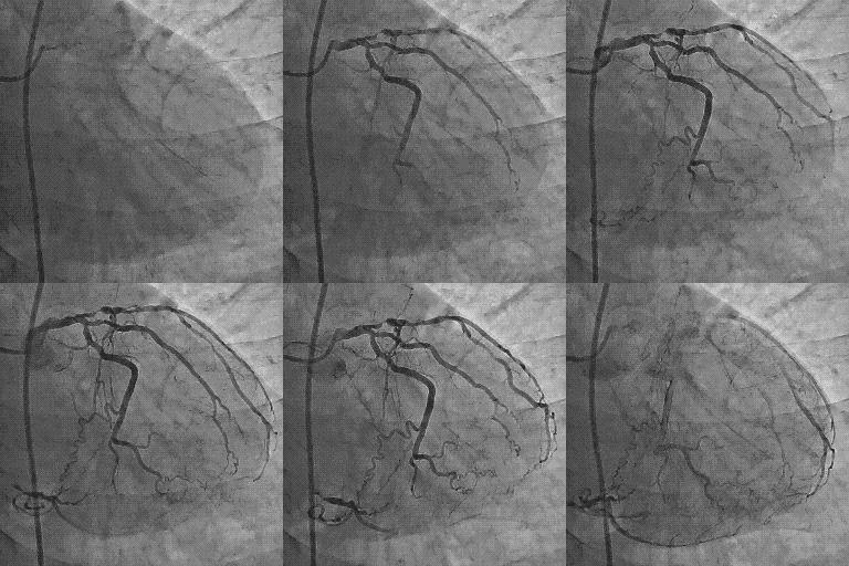
A 40-year-old man with diabetes and hypertension with class 3 angina had this angiogram

RCA to LAD collaterals

A different view

RCA to LAD in RAO caudal view

See , how a  pair of  human coronary arteries  mutually  help   their  colleague  at times of distress !

The astonishing observation is ,  the  RCA even as its suffering  with  a severe,   long segment disease it  helps out-of-the-way ,  with a long arm  of  support to  the entire LAD . While , the LCX reciprocates  the RCA by sending  thank you twigs to distal RCA

LCX sending reciprocating twigs to RCA

By the way , this patient was referred  for CABG after an   intense  debate in the cath meeting  .The argument ranged from medical management /PCI/CABG.

The key question were

  • How good is the collateral’s and what  are the chances of  graft flow  exceeding the collateral  blood flow ?
  • What is the effect of CABG on the existing collateral’s ?

Final message

Coronary arteries  has unique sense of sharing and friendship at times of vascular crises.

This is the fundamental basis for   coronary collateral circulation .

Read Full Post »

Cleveland clinic is a leading centre for cardiac care .Major technological breakthrough occurs from this institute than any other place. Thousands of articles come out every year. Some articles , get global attention and make  a huge impact. These are usually related to a new hi- tech modality like CRT devices or percutaneous aortic valve deployment etc ,etc.

                                                Some articles , which are very important  may not get the due  attention . Journal editorial boards often  have a scorecard called impact factor .That is ,   how  a  journal  is  impacting the practice habits of  medical professionals . Ideally we need to have to grade individual   articles with impact factor .Many articles may not have any significant  impact  however good the impact factor of the journal.

Here is an article,  which excellently depicts the principles of management of ACS.  It was published in 2003 JACC,  by Steven Nissen  from Cleveland,  Ohio .It deserves more attention . Every cardiologist , involved in ACS management should read this, especially the interventionist.

Link to article placed her with courtesey of JACC

Read Full Post »

Acute coronary syndrome  is primarily a disease of blood vessel , which perfuses  the heart.  It can even be a disorder of blood, often called vulnerable blood which predispose  for intra- coronary thrombus .

Mind you  , heart is an innocent bystander ! to the onslaught of  coronary atherosclerosis !

Hence , we  often use two terminologies .

CAD : Pure vascular (Coronary )  disease without  any structural and functional impairment of heart  ( No Angina, No myocardial damage ) Most of the asymptomatic plaques  , non flow limiting  lesions, incidentally detected by the modern coronary imaging gadgets  fall in this category.

When does  CAD becomes CAHD ?

CAHD : Coronary artery heart disease .Here not only the coronary artery is diseased , but it has it’s mission fulfilled   ie target organ either damaged structurally (STEMI, NSTEMI ) or functionally (EST positive , Chronic stable angina CSA )

Does the heart does any wrong to suffer from Acute coronary  syndrome  ?

No, it is simply not .The fault lies in one or more  of the following   .Generally at-least two these factors are enough to impede blood flow )  . They  combine to produce an ACS.

  • Blood defect
  • Vessel wall defect
  • Slowing of flow (Stasis)

This is called as Virchow’s triad   suggested over 100 years ago . Still valid in the era of per cutaneous  aortic valve implantation.

* The concept of de-linking  disorders of  coronary  vascular disease  from myocardial disease  is vital  in understanding the implications of current modalities of treatment. 

Even though we PCIs target the culprit ie blood vessel , it need to  realised , we  always fall short of real target . . .namely the heart . In coronary interventions  the catheters and wires roam around superficially over the heart  and they never even touch the heart .This is the reason PCIs are struggling to prove it’s  worthiness over medical therapy in many CAHD patients , which can reach deep  into the vessel, heart  and even every individual cells of heart.

Many (or . . . is it most ?)  Interventional  cardiologists have a bad  reputation for ” failing to look  look beyond the lesion” .  It is estimated  a vast  number  of cathlabs  and CABG theaters worldwide  are engaged in futile  attempt to restore coronary artery patency after a target organ damage is done .This is akin to building flyovers  to dead and closed highways .

Salvaging a coronary  artery and reliving a coronary obstruction is an entirely unrelated and futile  exercise to  a patient who has a problem  primarily in  musculature .

The much debated concept of  documenting  myocardial viability  , before revascularisation  died a premature death as the concept  by itself , was not viable commercially . (Viability studies   , tend to tie down the hands of device industry further , some  interventional   cardiologists began to see this concept  as an  interference to their freedom to adventure  )

Of-course , now  we have  other parameters  phenomenon  like  FFR estimation by Doppler , epicardial  -myocardial dissociation, slow  flow , no re-flow are  gaining importance.

Final message

ACS is primarily a disease of blood vessel but it’s impact is huge on heart. We need to look beyond the lesion .Restoring  a blood vessel  patency  to an ailing organ (Heart ) is not synonymous with total  cardiac intervention  and protection . There is lot more to cardiac physiology other than it’s blood flow. Heart muscle is a too complex organ to be controlled by few balloons and wires  which beat around the bush.

Read Full Post »

Heart is the first organ to differentiate in the fetus.The first heart beat begins on the 22nd day of fetal life ! . From that day , it’s function is linearly determine the   fetal growth  .It continues in the new-born,  infancy and  up to late child hood.

It is no surprise  then, to detect growth retardation in congenital heart disease. A proper evaluation begins right from fetal mass / birth weight estimation  .Motor and cognitive milestones should be assessed meticulously .

The pattern of growth affliction is  complex  and poorly understood.Few  working rules are  often taught in cardiology classrooms.

Caution : This is a too simplified version. Discerned readers should consult all sources cited here.

Acyanotic heart disease

ASD, VSD, PDA  tend to affect weight gain  more but generally do not affect height  much  . But, the onset of pulmonary  hypertension early in the course will severely  affect height as well.

Co arctation of  Aorta and other Aortic interruptive   diseases  can have a differential  affection of growth . (Upper part of body > Lower part)

Cyanotic heart disease

CHD  affects both height and weight proportionately. Cyanotic  heart disease with increased pulmonary blood flow  the overall survival is less ,  recurrent failure is common  and hence growth and development is more affected.

The mechanism of stunted  growth.

The often used terminology  ‘failure to thrive” , may not be attributed to heart disease per-se. It has to be multi factorial and  is related to  social well-being  ,  feeding habits , and mother’s effort  , interruptions due to co existing illness , effects of surgery  etc. Obviously these factors operate  more  in infants with increased pulmonary blood flow.

Effect  on cognitive function

Contrary  to the expectations  even chronic hypoxia and cyanosis has no compelling effects  on the child’s intelligence . Unless there is co existing  neurological defects severe compromise of cognition  is  uncommon.

However now we realise , brain development do  suffer   in hypoxic environment.In fact, the damage  to cognition could start right from the fetus .

New evidence is coming  up.

*Recurrent hypoxia spells and convulsions in TOF  can lead to reduced cognition

Is the normal  growth  and development   restored after complete correction of the disease by  cardiac surgery ?

The expected benefit is usually achieved  . The catch up occurs . But it is not guaranteed,   especially in  cyanotic heart disease. As , many times the  destined growth of a child   is  reprogrammed and  predetermined  in the fetus itself.


Link to a rare review article on the topic

References  on Growth impairment in congenital heart disease


R. L. Naeye, “Anatomic features of growth failure in congenital heart disease,” Pediatrics, vol. 39, no. 3, pp. 433–440, 1967.

A. Mehrizi and A. Drash, “Birth weight of infants with cyanotic and acyanotic congenital malformations of the heart,” Journal of Pediatrics, vol. 59, no. 5, pp. 715–718, 1961.

R. J. Levy, A. Rosenthal, D. C. Fyler, and A. S. Nadas, “Birthweight of infants with congenital heart disease,” American Journal of Diseases of Children, vol. 132, no. 3, pp. 249–254, 1978.

H. H. Kramer, H. J. Trampisch, S. Rammos, and A. Giese, “Birth weight of children with congenital heart disease,” European Journal of Pediatrics, vol. 149, no. 11, pp. 752–757, 1990.

Read Full Post »

y

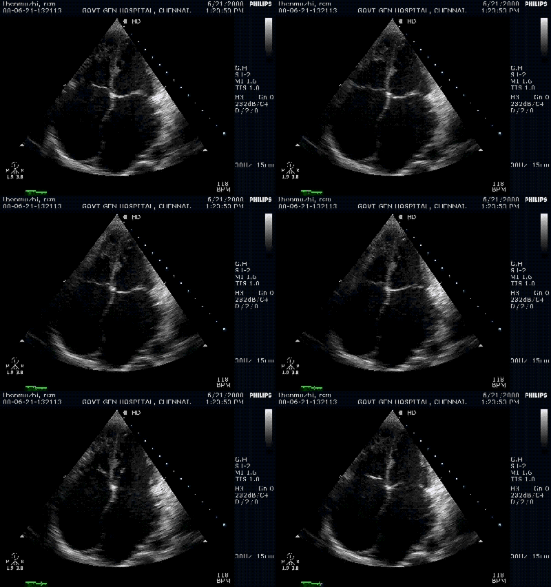
A classical restrictive cardiomyopathy . Both atria balloon out as they face stiff resistance from the ventricles

Restrictive cardiomyopathy(RCM) is a common form of myocardial disease which was difficult to recognise  in the past.It was also commonly confused with constrictive pericarditis. Today , one can easily recognise this entity.A simple clue is bi- atrial enlargement  with relatively normal ventricle size.The above case is a classical form of RCM.

In late stages of RCM ,both LV, RV begins to dilate and can mimic a dilated cardiomyopathy. Doppler filling pattern  , and tissue Doppler motion of mitral annulus are recently validated methods to identify RCM. Still ,  2D features  are  very useful .This implies , the anatomical changes in the chamber size  are as important as physiological AV filling profiles. It is generally believed , physiological impairment precedes anatomical defects.( But not proven concept yet ! )

Note: The ventricles are not dilated and retain good systolic function

In the above patient  , etiology could not be confirmed and was labeled as idiopathic RCM  as tests for amyloid and eosinophilic infiltrations were negative.

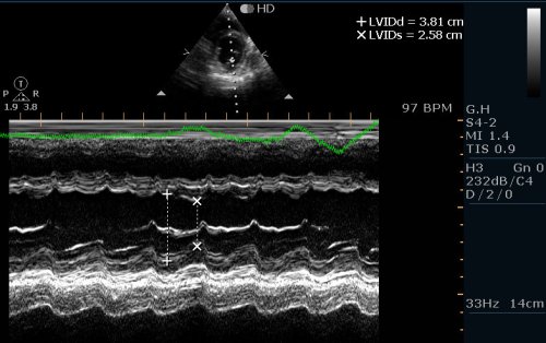
M -Mode of the same patient confirms good systolic function

One would  call  for doppler mitral filling profile here ,  to confirm restrictive physiology ( A short DT , Short IVRT  . A reversal in  pulmonary veins , E/E’ ratio etc etc ) But all these are redundant here.

How is RCM different from non dilated cardiomyopathy ?

A new entity is being recognised in the cardiac  muscle disease.This is often referred to as NDCM (Non dilated cardiomyopathy)  .Global  systolic LV dysfunction  with normal  LV dimension.This a similar to the terminology MDCM (Minimally dilated  cardiomyopathy  where LV dimension increases  not more than  15 % of basal size ) .

This is seen in CKD and diabetic individuals.Atria may be enlarged .Diastolic dysfunction may co exist.  It is no surprise,  this entity closely mimics RCM. But in RCM LV systolic function  is not greatly compromised till late stages , while NDCM it begins with systolic dysfunction. This is the only difference .There can be overlaps .

Many strongly believe ,  both  RCM  and NDCM are one and the same entities ,  that present in different time frames in their natural history.

Final message

Diagnosing RCM is no longer difficult in established cases* . The message from  this article is , 2 D echo can  strongly suggest  the possibility of RCM (or even clinch it)  .  Never ever  diagnose RCM with normal 2 D echo. Doppler filling profiles are useful  additional tools . We  can not diagnose RCM with doppler features alone , but we can  be fairly certain about RCM when we encounter  typical bi-atrial enlargement and a normal LV by  2 D echo.

Caution : Patients  with longstanding atrial fibrillation of any cause , can dilate their atria and could mimic RCM .They can be some compromise in LV function due to chronic tachycardia .

* Recognizing RCM ,  very early in the course is still a problem . Here the  newer modalities like Phase  MRI, tissue doppler, speckle tracking, and velocity vector imaging may be useful.

Read Full Post »

Dilated cardiomyopathy (DCM )  is the commonest  cardiac muscle disease .Diagnosis is simple and straight forward. All that we  require is,   documentation of LV dilatation and contractile dysfunction.The nomenclature of cardiomyopathy has remained a difficult exercise .This   is primarily  due to   iatrogenic  & intellectual confusion  among  cardiologists . They mixed up etiological and morphological entities together ,   later on  they  wanted to de-link  etiology from morphology  ,  ultimately they realised  when illness strikes the heart ,   it   can not differentiate the  morphology, etiology and pathology as we would want to   . So , whenever possible we have to label  cardiomyopathy with all components (Dilated cardiomyopathy due to alcohol with some restrictive features.)

This article  tries to evoke  some thoughts    about  why  LV dilatation  is central to the understanding of cardiomyopathy.

DCM is the prototype where LV dilates with global hypokinesia.The upper limit of   LV diameter is generally considered to be 56mm in diastole.  (Range 35 -56mm) .This cut off  point is too empirical  for the simple reason, the  left ventricle can dilate   up to  50 %  from it’s basal diameter and still technically  be within normal limits.( A 3.5 cm LV ( end diastolic diameter ) can dilate to 5.6 cm ,i e  a 50 % dilatation , still LV  has not reached the upper limit of normal  )

Even as we do not have a clear  answer to the above issue , we  recognise  left ventricle muscle can hypertrophy, progressively  dilate , transiently dilate, fail to  dilate ,  regressively  dilate  or  hypotrophy .These changes can be dynamic and heavily influenced by hemodynamic and local pathologic factors like fibrois, interstitial proliferation etc. Meanwhile , the pharmacological ,   surgical /catheter injuries we  inflict    , modifies  the muscle behavior in a positive or negative manner.

In this back ground ,  we have found a new entity called NDCM .

Apart from  DCM, a newer form of  cardiomyopathy  is being recognized  .This is often referred to as NDCM (Non dilated cardiomyopathy)  .Global  systolic LV dysfunction  with normal  LV dimension.This a similar to the terminology MDCM (Minimally/Mildly  dilated  cardiomyopathy  where LV dimension  do not  increase  beyond   15 %  upper limit of normal  ) .

This is seen in CKD and diabetic individuals.Atria may be enlarged .Diastolic dysfunction may co exist.  It is no surprise,  this entity closely mimics RCM. But in RCM LV systolic function  is not greatly compromised till the  late stages , while NDCM it begins with systolic dysfunction. This is the only difference .There can be overlaps .

MDCM was reported in 1990  . Keren gave a excellent insight about the condition  ,  It is unfortunate it failed to take off as a popular  clinical entity .  Cardiologists are argued to use this term liberally in their clinical practice .

Final message

Cardiology is not  that  simple as one would like to ! The two components of cardiomyopathy ie   LV dilatation and LV dysfunction can be temporally dissociated  one may precede the other. To  complicate the matter further, one of them may not manifest at all !

Few ,  still consider many of the RCMs and NDCM are one and the same entities that present in different time frames in their natural history.

So the simplified  concept  to decode the cardiomyopathy conundrum  could be

  • When both  dilatation and dysfunction occur it is classical DCM
  • When dysfunction  alone occur without dilatation it is NDCM
  • When both dysfunction  and dilatation are less it is RCM*(Relaxyl dysfunction must)
  • When dilatation is mild and dysfunction is severe it  is MDCM

Read Full Post »

Older Posts »