Feeds:
Posts
Comments

Archive for October, 2021

With deep regret, reporting the demise of Dr. KA Abraham, cardiologist, par excellence in our part of the world (Chennai, Tamil Nadu, India.) He was a man of great knowledge,  wisdom, and integrity and was an inspiration to generations of cardiologists like us.

Dr.K.A Abraham 1942-2021

A life, that was fully dedicated to all those heart patients, many of them sick children in his den, the Railway hospital, Chennai for three decades. Though, never had the privilege to be associated with him either directly, nor does he knew me, one could feel instantly the greatness and simplicity in him. It’s 1994-95, vividly recall, the early morning classes, he took in the small auditorium in the Railway hospital,(For which, we used to rush to the far away Perambur from MMC in the peak Chennai traffic). His passion for teaching the basics of cardiac catheterization was phenomenal. Somehow, I used to think he was in the league of the Nobel trio of “Forssman, Richards & Cournand” who invented cardiac catheterization.

Those were ordinary days when the quest for knowledge and teaching was pure and Dr. Abraham was one of the great souls in pursuit of genuine and quality cardiac care. No surprise, his services were recognized and conferred a top award of India,  Padmashree.

Yes, India truly lost a pioneering cardiologist. 

Reference

https://en.wikipedia.org/wiki/K._A._Abraham


Read Full Post »

This piece of article by Mr. Arun Maira,(The Pakistan-born British Indian ex-planning commission member) is a real eye-opener in the manner we have understood science. All socially conscious scientists must-read. (If properly appreciated, the 15 minutes  you are going to spend on this is worth the time of one full semester in economics at a top-notch university )

Was the past perfect?  & Will the future be tense?

No is the answer to both questions. Noble prizes are increasingly given for some soul-searching simple researches. Complex research methodology is looked down on, especially in economics. Contributors of simple observational studies bordering on common sense shall be rewarded. Incidentally, this year’s physics prize was also different from other years (Given for finding faultlines in working models of climate change). It is heartening to note the shift in thinking and points to good times for true science. We have finally started to question the genuineness in the foundations of existing research models and epistemological purity of knowledge.Very soon, major global awards are waiting for the Innocuous looking amateurish research that is willing to expose trivia and the flawed understanding of science itself.

High stakes in the noble profession

Now, this has major Implications in the terrain of medical practice, a fragile scientific art that is dangling between facts and fakes, uncertainties of nature & certainty of greedy monetization, social inequalities, and finally the stupidity of half-baked knowledge.

I strongly believe the following two concepts if proven properly deserve the Nobel prize in medicine or economics with a huge Implication for humankind. 

1. In the global health care delivery, nurses and para-medical health workers have a multi-fold positive impact on universal health goals than the highly specialized doctors, who are at best have a minuscule role. There should be intensive restudy of their actual requirements and redefining  doctor vs nurse vs population ratio (What a big revelation,  even a novice can say this, but that is exactly  is the reason which makes it eligible for the Nobel award)

2. Specific treatment modalities are either lacking or trail behind the hyped-up diagnostic methods for a good number of illnesses. They are not only redundant but also malignantly consume the global economic resources without a real purpose. What is the big deal of accurately diagnosing and labeling a disease if there is no treatment? (Typical example in recent times,100s of millions of costly RTPCR tests are Indiscriminately used for an incurable self behaving pandemic).

Who is willing to do the above studies? I wish WHO can sponsor this. Research questions, methods, statistics, and even conclusions are ready with 100% accuracy, I am sure, they will withstand any rigorous scientific scrutiny. Though every Tom, Dick  & Harry can do this research from any academic garage, the chances of it getting noticed by Karolinska institute is low, unless It comes from an Ivy League or an elite European university. When someone receives this coveted award down the lane of time, hope this cranky post gets some credit.

Reference

https://www.thehindu.com/opinion/lead/over-simplified-models-complex-social-systems/article37061493.ece

Nobel Prize economics list

Noble prize in Medicine list 


Read Full Post »

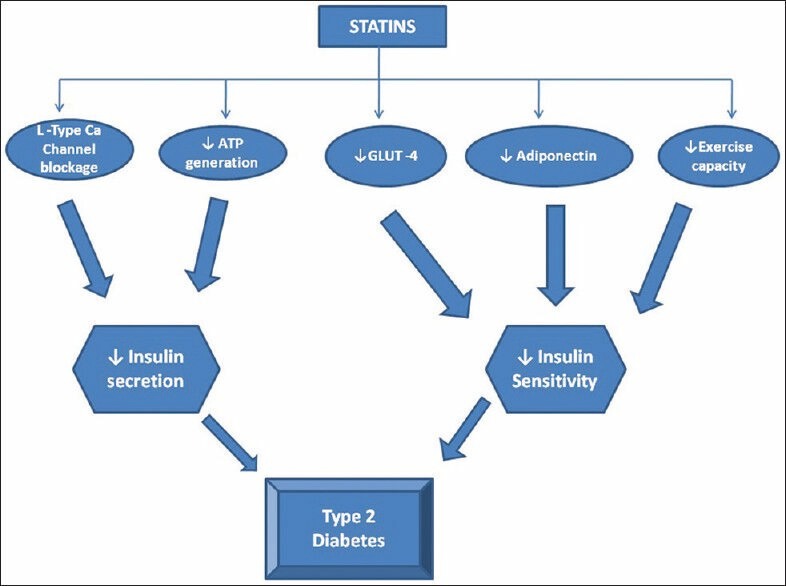
Aortic stenosis evaluation was simple in our days. Gradients across the valve were the key. Now, we have more parameters to bother about. Dynamic AVOs, flow state, resting LV function, contractile reserves, GLS, dobutamine response, etc. MRI assessment will soon overtake echocardiography. 

Hemodynamics of flow across LVOT. MRI 4D volumetric model of normal Aortic stenotic flow in the bicuspid valve (On the left). The more we know, the more we tend to miss! Image courtesy: Northwestern Medicine

The current AS algorithms, though scientific, I am afraid, appear much complicated with some frightening terminologies at least for the beginner. One such category is PLF-LG-AG.

Let us first answer this. What is LG-AS?

We diagnose a case of significant aortic stenosis but desperately miss to pick an adequate gradient across the valve. It is indeed a low gradient AS, still, you are not convinced, since the 2D look of the valve looks severe. Then you find the culprit. It is the dysfunctional ventricle pulling down the gradient. This is called  LG-AS.(one type )

*What is paradoxical -Low flow-Low gradient Aortic stenosis? (PLF-LG AS)

Now, we have another patient. Again it is  LG-AS, but the LV function and EF are normal. Now, you get confused and label it as PLF-LG-AS. 

PLF-LG AS is known to account for approximately one-third of patients with severe AS and preserved LV EF.PLF-LG severe AS is defined by AVA <1.0 cm2, indexed AVA <0.6 cm2/m2, mean gradient <40 mmHg, LV EF ≥50%, and low transvalvular flow (indexed stroke volume <35 mL/m2).

What is the paradox?

AS is severe, but the gradient is not showing at the valve. Wait, don’t think that is the paradox, since this can happen in any category of LG-AS.  So, the real paradox in PLF-LG-AS denotes to the fact that the gradient is low, in spite of normal LV  function (Rather, normal EF %)

Why the paradox? Why gradient is not showing? (In spite of good Ejection fraction ?) 

  • Reduced stroke volume increased afterload due to associated HT are the major hemodynamic mechanisms of PLF-LG AS.
  • The low-flow state can be related to small LV cavity 
  • Significant diastolic dysfunction (Associated Amyloidosis is a new age problem kid on the block. EF is not the only parameter that can compromise the flow you know !)
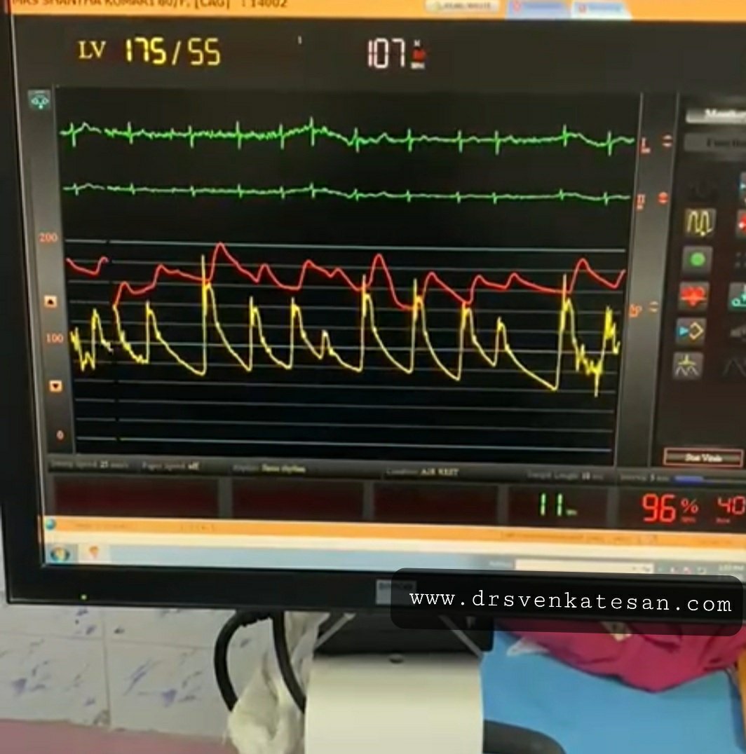
  • Atrial fibrillation (Ofcourse some cycles will pick up the high gradients, but are they spuriously low or high is the question  ) 
  • Associated mitral valve disease leaks, as well as blocks, will ration the flow to LVOT.

*Finally, every cardiologist should be aware of the following two subsets that could wrongly enter this conundrum of PLF-LG-AS  without much fanfare but with lots of implications.

1. Most importantly, technical issues are the key confounders. Malaligned  LV / LVOT with that of distorted out-of-plane AVO is the commonest cause of failure to pick up the gradient. (Never diagnose PLF-LG-AS  without confirming it is not due to technical ). A tip: Don’t complete AS doppler study without a meticulous search for a good doppler signal from the suprasternal/right parasternal window. 

2. Next one can be named with a provocative term “Paranoid aortic stenosis*”. Once, a  fellow was reporting a shabby-looking calcific valve as low gradient, severe AS with normal LV function. He made this diagnosis solely based on AVO. Later, when it was assessed more scrupulously it turned out to be a true mild aortic stenosis, and none of the decorative echo features he was showing were really pathological. It was an error in the aortic valve area calculation due to LVOT area/VTI measurement errors that got mathematically amplified. An important teaching point emerged from this echo lab fiasco. Mind you, any true mild aortic stenosis (If the area calculation is wrong for some reason) will readily fulfill the criteria of PLF-LG-AS. One simple tip: Never diagnose severe Aortic stenosis without significant LVH.

What is the role of Doubtamine stress echo in PLF-LG-AS ?

It does help in both forms of LG-AS. You need to read about the contractile reserve, the response of dobutamine to flow (Cardiac index), gradient, and the valve area.

How to manage PLF-LG-AS ?

Many of them might be argued to end up in TAVR. So, follow the guidelines carefully but don’t apply them blindly

Final message 

In the evaluation of Aortic stenosis let’s make things simple. Gradients are indeed important. But, realize Doppler gives only pressure data. Converting them to flow and volume data is always error-prone. Instead, let us believe our eyes too.(Need not always depend on o MDCT vision)  Concentrate more on LV morphology, valve pathology, and careful assessment of LV function, finally take a decision to intervene based on true symptomatology and comorbidity.

Reference 

1. For the most authentic knowledge base

2.An excellent review on the topic 

Guzzetti Ezequiel Frontiers in Cardiovascular Medicine 2020

Further thoughts

Is there a “high flow-high gradient”- True mild AS?

High gradients across the AV  generally do not cause much confusion. If the mean gradient is > 40mmhg Aortic stenosis is always severe. Is that right ?. I think so. Exercise-induced paradoxical high flow high gradient AS as an entity is not reported as such. But, what really can happen in high output states, hyper contractile ventricles with high EF? We have observed doppler gradients overestimate the severity of Aortic stenosis. I think there is some dynamic component even in the so-called fixed valvular AS that alters the gradient in response to flow. Do we have proof for this?  OMG, it’s right there in echo lab every day, we are failing to notice it. Look at the AS gradients during AF. It is changing every beat, right.


Read Full Post »

Background

“Your husband is really lucky, his heart attack got spontaneously aborted. His ECG is near normal now. The angiogram is normal.No lysis, no stent is required. He secreted his own TPA and got rid of the clot. We will discharge him to tomorrow.”

Thank you very much, Doctor. How did this happen, doctor?

Don’t thank me. Definitely, I don’t have an answer. Spontaneous successful thrombolysis (Ref 4) happens up to 15 % of ACS. All I can say is he has a very disciplined  fibrinolytic system backed up with an agile mast cell and basophil function.

Vascular events: Pathobiology

Vascular highway accidents that happen due to the sudden freezing of blood in any vital organs consume more human lives than any other disease. This can happen with or without a trigger from the vessel wall, the phenomenon which Virchow’s taught us a century ago.

By the way, who is keeping the 5 liters of blood in our body in a fluid status life long?

Blood coagulation and lysis is a fascinating balance of two pairs opposing hematological forces.

  1. Coagulant system (The 13-factor Intrinsic /extrinsic clotting cascade )
  2. Anticoagulation system  (Antithrombin, protein  C and S, and tissue factor pathway inhibitor (TFPI) )
  3. The fibrinolytic system (Commander in chief is Plasmin, It has the ability to feed and digest clots just like that It comes from plasminogen the activation of this the core concept of thrombolytic or fibrinolytic agents)
  4. Anti-fibrinolytic system  Haemostasis 1986;16:16–20

* Among the blood component cells platelet is the only cell that plays a critical role in the clotting process. Paradoxically the fiery red clot, that is loaded with RBCs has a little direct role in the clotting process while biochemical molecules like fibrinogen, thrombin plays a major role. The fact that blood is liquid forever, tells us a compelling fact, that innate anticoagulation and the fibrinolytic system are more critical than the extensively understood clotting mechanisms. 

Are you aware we all have some amount of naturally secreted heparin in blood?

Circulation, Volume XXIII, April 1961

Which cells secrete heparin? 

Basophils of blood and mast cells continuously secrete heparin that keep the blood fluid. Heparin is normally present in human plasma in values ranging from 10 to 24 units percent (1 to 2.4 mg. per liter). The range of average values is from 1.53 to 1.77 mg. The native heparin secretion by mast cell population is directly related to the anticoagulation activity (Bill Eksp Biol Med. 1984 Feb;97(2):131-4. Russian. PMID: 6230117.)

Where does TPA come from?

TPA normal levels not only confers the capacity to spontaneously abort  a vascular event, but it is also a critical determinant of how streptokinase is going to act as this drug can’t act alone it simple accelerates the action of Tpa complex

Mind you, the basal levels of TPA are injected instantly into the coronary thrombus with a zero-second time window. Now guess what is the potency of this enzyme cascade and who directs it?

Yamada R, Yamada S, Ishii A, Sasamata M, Watanabe S. Association between tissue plasminogen activator and serum lipids in healthy volunteers. Ann Med. 1990;22(5):313-8. doi: 10.3109/07853899009147913. PMID: 2127223.

Final message

If we think with a hematologic acumen, all vascular accidents are triggered by temporary aberrant behavior of blood or the interaction of its components with the immediate tissue interface. It is the inability to either prevent or fight the sudden occurrence of thrombus that is a primary problem than the tendency of the blood to clot spontaneously. The basal levels of natural Heparin, TPA, or anti thrombins and the way in which they get activated are much more important than the procoagulant forces, which we tend to blame often. (the famous vulnerable plaque vs vulnerable patient need to be reignited) 

I think it’s time we dwell deeper into the native lytic mechanisms. This will throw an important vision on how we can replicate it in a pharmacological way. If only we have a drug that melts intravascular clots locally, vascular deaths of many vital organs can be prevented. The research in newer lytic agents has almost ceased & needs fresh Impetus.(TPA is a 25-year-old Invention) 

Reference

1.Longstaff, C. & Kolev, K. Basic mechanisms and regulation of fibrinolysis. J. Thromb. Haemost. 13(Suppl 1), S98-105.  

2. Wiman B. Predictive value of fibrinolytic factors in coronary heart disease. Scand J Clin Lab Invest Suppl. 1999;230:23-31. PMID: 10389198.

3.Wiman B, Andersson T, Hallqvist J, Reuterwall C, Ahlbom A, deFaire U. Plasma levels of tissue plasminogen activator/plasminogen activator inhibitor-1 complex and von Willebrand factor are significant risk markers for recurrent myocardial infarction in the Stockholm Heart Epidemiology Program (SHEEP) study. Arterioscler Thromb Vasc Biol. 2000 Aug;20(8):2019-23. doi: 10.1161/01.atv.20.8.2019. PMID: 10938026.

4.Kovacs IB, Yamamoto J. Spontaneous thrombolysis: a forgotten determinant of life or death. Clin Appl Thromb Hemost. 2006 Jul;12(3):358-63. doi: 10.1177/1076029606291410. PMID: 16959691

Postamble

I think we have a problem with our basic science teaching methods. Tried a fact check. When I asked a few passing-out medical graduates, they could rattle everything about clotting factors but struggled to recall any anti-clotting mechanisms. We are still a long way to go, understanding the mysteries of how the blood keeps itself fluid, but always on the alert mode to clot whenever necessary.

DIC: The sine-qua-non of lytic dysfunction

DIC is one of the famous diagnoses in critical care medicine. It is a perfect example of defective fibrinolysis. (Also referred to as acute fibrinolytic shutdown*) classically occurs in septic shock and related conditions. We still lack good criteria to diagnose non-overt DIC-prone patients. A good review was written in 1999, still helps us understand the core concept. 

*Both shutdown as well as Inappropriate activation 

Levi M, Ten Cate H. Disseminated intravascular coagulation. N Engl J Med. 1999;341(8):586–92.

Return to ref 3 in article


Read Full Post »

Doppler E/A ratio reversal is probably the most reported abnormality in clinical echocardiography. We are also pleased to label it as a grade 1 diastolic dysfunction. Making a significant population who come for regular health checks anxious and worried.

Sharing a presentation from the Annual conference ECHO INDIA 2019, I participated in a symposium on Diastolic dysfunction.

Topic : Issues in diagnosing grade 1 diastolic dysfunction: Pearls and Perils

How did we get into this academic trap? Should we continue this practice?

The current ASE guidelines 2016 have a clear message. It has taken off the E/A ratio from the Initial screening for diastolic dysfunction.

Summary & Final message 

Are we ready for the change? By understanding a simple concept, one can reduce the incidence of indiscriminate diagnosis of grade 1 diastolic dysfunction.

  • E/A ratio apparently has a no role in diagnosing diastolic dysfunction in the normal population who have normal EF %.
  • Hence, never report E/A ratio in Isolation as grade 1 diastolic dysfunction.
  • However, in patients with HFrEF it does help in triaging diastolic dysfunction.
  • Always look for symptoms and 2D features  (Unexplained dyspnea, LA enlargement, LVH ) before considering diastolic dysfunction.

*For advanced readers and researchers grade 1 diastolic dysfunction does have a deep meaning and always continues to puzzle.

Patient corner

For all those anxious patients who ramble around with a report of grade 1 diastolic dysfunction, I can assure you this. Please realize, 9/10 times, this is just a decorative echocardiography abnormality meant to add some spice to the report  does not have any significance.

*Will post a PPT presentation shortly.

Read Full Post »

This is not a breaking news story. It’s the same old secret that was exposed in JUPITER  trial with Rosuvostatin 14 years ago. Yes, I am talking about the relationship between the usage of statin and the occurrence of diabetes. Now, we have this huge study on possible diabetes progression with statins. It’s not from a small journal to ignore. 

83 thousand patients data, the world’s largest series on link between statin therapy and diabetes.


This study has this to conclude

Diabetogenic statins

Something* happens as the statins antagonize the HMG COA  enzyme that resides within the delicate membranes of the endoplasmic reticulum inside the most specialized cells in our human body, called hepatocytes.

*What is that something?

Image source Umme Aiman et al Journal of Pharmacology and Pharmacotherapeutics 5(3):181-5DOI: 10.4103/0976-500X.136097

How to go about this issue?

With-holding statin in as many as possible is the best thing for such diabetic  (non-diabetic?) patients. But, the more pragmatic option is to ignore these negative studies, and instead intensify diabetes management if it worsens. After all, we can’t afford to lose the prodigious evidence-based cardio-vascular protective effects of statins and earn the wrath of our patients and peers you know!

Further Interest

1.Mansi IA, Chansard M, Lingvay I, Zhang S, Halm EA, Alvarez CA. Association of Statin Therapy Initiation With Diabetes ProgressionA Retrospective Matched-Cohort StudyJAMA Intern Med. Published online October 04, 2021. doi:10.1001/jamainternmed.2021.5714

2.Aiman U, Najmi A, Khan RA. Statin induced diabetes and its clinical implications. J Pharmacol Pharmacother. 2014 Jul;5(3):181-5. doi: 10.4103/0976-500X.136097. PMID: 25210397; PMCID: PMC4156828.


Read Full Post »

Atrial fibrillation has a direct effect on systemic blood pressure as stroke volume swings from beat to beat because of changing  RR Interval ( preload ). The variation in systolic pressure actually reflects not only the changing stroke volume but also the enhanced contractility of the ventricle to the preload( Frank-Starling principle ). The net effect is reflected in the pulse as an irregularly irregular pulse (Both rate and volume /Amplitude).* However, In dysfunctional ventricles or in acute AF* this variation in systolic  BP can be significant. Also realize, If the preload is changing every beat, there is a considerable dynamism in the afterload as well because of ventricular arterial coupling.

(*Acute effects on BP with the onset of AF : There can be transient hypotension with loss of atrial booster pump.This is not significant in otherwise healthy hearts. Inpatient with baseline LV or RV dysfunction, the onset of AF can be detrimental. The ventricular rate is also a determinant of blood pressure. At fast rates, there can be a fall in BP)

How to record BP during AF ? 

As you record the BP by cuff, the Phase 1 Korotkoff sound floats up and down with each beat. If the variation in RR interval is huge one may get a beat-to-beat variation even up to 40 mmHg.We also know, AF can cause pulse drop /deficit intermittently.

What happens to Korotkoff sound during pulse deficit?

Obviously, there will be a loss of these phase 1 sounds, though the other phases of sound may be heard, which are generated by the previous cardiac cycle. So, measuring blood pressure in AF is not a clinically pleasant task. That’s why we are asked to record 3 times and take an average.

Now, coming to diastolic BP in AF. It’s a real hemodynamic riddle. Traditional teaching is, systolic BP is determined by cardiac output and diastolic BP by peripheral vascular resistance. This is at best a gross understanding of circulatory physiology. Both systole and diastole are coupled together as blood flow across the system of varying resistance. In fact. The preceding systolic pressure head stores the elastic energy in large vessels that are thrown back as diastolic BP.

So what happens to BP during AF? What does the literature say? It doesn’t say much. So we decided to look for ourselves. Here is a tracing of femoral arterial pressure curves during atrial fibrillation. Note: the systolic BP shows considerable variations with changing RR interval with little change in diastolic blood pressure.


Final message

In Atrial fibrillation, the systolic blood pressure changes from beat to beat and it impacts the timing of the Korotkoff sounds. The diastolic blood pressure behaves the same, but it’s less in magnitude and difficult to detect by conventional sphygmo-manometry.

Further reading 

Clinical Implication for irregular BP of Atrial fibrillation  :https://drsvenkatesan.com/2021/05/04/cerebral-hemodynamics-in-af-irregularly-irregular-brain-perfusion-and-risk-of-dementia-cordis/

The effect of AF on pulmonary arterial pressure is an unexplored topic .Cardiology fellows please note.

Read Full Post »

Is there a solution?

As I understand, we don’t have any. Maybe, we can try this.  No way, I can prevent it from appearing ridiculous for the mainstream scientists.

Truths often lie silently  buried deep (many times intentionally). They definitely deserve an intellectual resuscitation beyond the dirty world of data and evidence. Further, why should experience be considered as enemy of evidence ?

Read Full Post »