Feeds:
Posts
Comments

Archive for December, 2023

Cardiogenic shock (CS)is the most feared event following STEMI. The incidence is up to 5 to 10% with a mortality rate of around 50-60%. Still, we are finding it hard to bring this down below 50 % .There is one less addressed issue in ACS literature. We tend to perceive CS as an exclusive complication of STEMI. The fact is that NSTEMI can also result in CS is less recognized. The incidence is half of that of STEMI, i.e., 2.5-5%.

Mechanism of CS in NSTEMI

One may ask, how can CS occur in NSTEMI with partial occlusion with a non trans-mural MI. ACS pathophysiology is not that simple. Ischemic LV dysfunction (Global stunning) without necrosis is equally sinister. This is what happens in some high risk sub sets of NSTEMI.

How is CS in NSTEMI different ?

1.Global ST depression (AVR.V1 might show little elevation with considerable overlap of left main STEMI vs NSTEMI )

2.Onset of NSTEMI-CS occurs late (48-72 hrs)

3.Severe multivessel disease is more common (It is likely ,presence fold STEMI , is an important factor that is likely to precipitate CS when a new NSTEMI occurs.

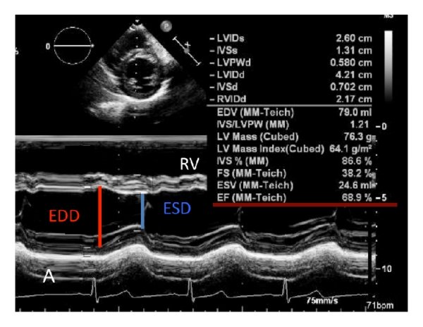
4.Echo is likely to show more of a Global hypokinesia rather than specific coronary territory

5.Mechanical complication, though less common in NSTEMI , Ischemic MR especially with LCX- NSTEMI can be problematic and much commoner than we think.

6. A subset of NSTEMI precipitated by acute severe HT and flash pulmonary edema has excellent prognosis if BP is reduced promptly. (This can be simply a equivalent of HT, with no true supply side ischemia with LVF with global ST depression )

Management

*More or less similar to STEMI with aggressive opening of culprit lesions with few differences. (unlike STEMI, CULPRIT SHOCK trial doesn’t apply here )

*May require CABG more often

*Mechanical circulatory support will be needed in many

*Finally, and importantly, there is more likelihood of systemic factors like sepsis, Anemia, or renal or kidney failure contributing to the CS in NSTEMI than STEMI. In fact, we have observed pre-existing HFpEF can be a contributory factor.

Outcome

There are differing data about prognosis of CS in STEMI vs NSTEMI. Early mortality is higher with STEMI; but, late mortality converges. Ironically, in many patients of CS in NSTEMI, the outcome can be worse than STEMI, as there is no single culprit and myocardial salvage does not appear to be a primary issue. (Ref 2)

What does SCAI guideline say about CS in NSTEMI?

Nothing, yes it is true. Are you surprised ? A search for the word NSTEMI in both these document drew a blank. May I kindly request SCAI team to look in this, CS in NSTEMI deserve better recognition in their guidelines at least in their next edition (Ref 3,4)

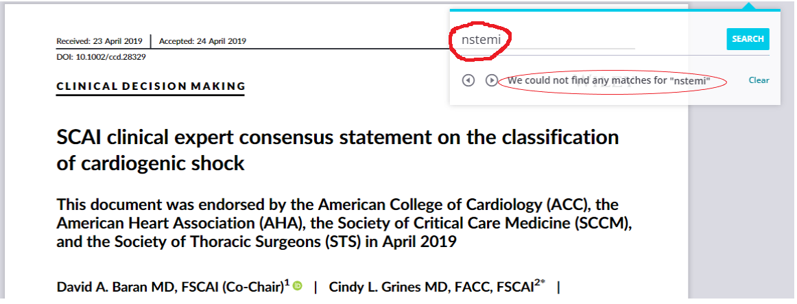
I am not sure why SCAI classification didn’t address CS in NSTEMI as a separate entity.

Final message

Surprisingly , CS in NSTEMI is not a well researched entity in cardiology literature. Fellows are requested to analyse the GRACE registry once again or create their own institutional experience.

Reference

1.Martínez MJ, Rueda F, Labata C, Oliveras . Non-STEMI vs. STEMI Cardiogenic Shock: Clinical Profile and Long-Term Outcomes. J Clin Med. 2022 Jun 20;11(12):3558. doi: 10.3390/jcm11123558. PMID: 35743628; PMCID: PMC9224589.

2.Anderson ML, Peterson ED, Peng SA, . Differences in the profile, treatment, and prognosis of patients with cardiogenic shock by myocardial infarction classification: A report from NCDR. Circ Cardiovasc Qual Outcomes. 2013 Nov;6(6):708-15. doi: 10.1161/CIRCOUTCOMES.113.000262. Epub 2013 Nov 12. PMID: 24221834.

3.SCAI 2019 Catheter Cardiovasc Interv.2019;94:29–37

4.SCAI 2022 consensus update.

Read Full Post »

Read Full Post »

       ”It looks like, science oftentimes struggles at the hands of scientists

  The weakest link in medical research is framing the right research question. How will you explain the drama of research on myocardial viability studies that have been going on full steam for more than 3 decades,? Which has become junk now.

This truth came out from a secondary analysis of the famous REVIVED-BCIS2 trial. For the busy guys, the conclusion is given in the box below.

JAMA Cardiol. 2023;8(12):1154-1161.

Final message

We realize, in the game of myocardial revascularisation the quantum of dead tissue determines the outcome of revascularization, not the amount of living tissues. This same question was asked & answered more than a decade ago (2009) on this site, of course without that damning evidence.

Common myths in cardiology: The presence of viable myocardium does not mean one must do a revascularisation procedure following myocardial infarction!

March 15, 2009 by dr s venkatesan 

Many times, we don’t require large-scale RCTs, years of toil, and efforts wasting resources to prove an apparent sense of thinking.

Read Full Post »

Conservative management conveys two tangentially opposite meaning.

For the superior “physicians & Interventional cardiologists” it would mean

For the “Inferior genre of physicians” it will sound like this

Reference

This is a partial repost from a 2008 article . https://drsvenkatesan.com/2008/11/30/what-do-we-mean-by-conservative-management-why-it-is-often-considered-as-an-inferior-form-of-treatment-in-medicine/

Read Full Post »

Which point in ECG is taken as reference for End systolic dimension for LV function assessment?

Marking the end diastolic point in ECG is quiet straight forward. Peak of R wave.(or Q)*

But, what about the reference point for end systole.

  1. Descending slope of T wave
  2. Peak of T wave
  3. End of T wave
  4. If T is absent or not clear , I will be confused
  5. To be realistic, ECG has no reliable reference point for end systole.

Answer: If I say the answer is 5, no one is going to agree. Please note, the relationship between the T wave and the peak systolic phase in echocardiography is weak. Is there any relation at all ? Then, how to measure LV function in echocardiography? There is a electro-mechanical delay in every segment and sequence of cardiac contraction and relaxation, with former piping the later (electricity beats).

Surprisingly (illogically as well), we take the point of maximum thickness in M-mode as end-systolic, which, in fact, corresponds to peak mechanical systole. This point has no consistent relationship with any part of the T wave. We must realize, the clinical cardiac cycle is defined based on sounds, i.e. S1 and S2, while the biomechanical cardiac cycle is different. Similarly, echocardiographic systole is not the same as clinical systole. 2D echo eliminates this uncertainty to a large extent. This is one of the reasons , we are advised and encouraged, not to measure LV dimensions in M mode.(A very tough advice to follow though)

Final message

We are not completely clear yet, in the “ECG vs Echo” time correlation of hemodynamic events. The errors may be in only milli-seconds, still, when planning for Interventions like re-synchronisations, and in the follow up of DCM patients this could matter much.

Reference

1.Mada RO, Lysyansky P, Daraban AM, Duchenne J, Voigt JU. How to define end-diastole and end-systole?: Impact of timing on strain measurements. JACC Cardiovasc Imaging. 2015 Feb;8(2):148-57. doi: 10.1016/j.jcmg.2014.10.010. Epub 2015 Jan 7. PMID: 25577447.

2.Gill H, Hoffmann A. The timing of onset of mechanical systole and diastole in reference to the QRS-T complex: a study to determine performance criteria for a non-invasive diastolic timed vibration massage system in treatment of potentially unstable cardiac disorders. Cardiovasc Eng. 2010 Dec;10(4):235-45. doi: 10.1007/s10558-010-9108-x. PMID: 21113800.

Read Full Post »

Learning resources in the web, has made the traditional methods of learning & teaching almost redundant.

American society of Echocardiography (ASE) has produced this 20 page PDF document, which covers the entire field of echocardiography , in an Illustrative format.

Take a print out, of this and stick in your lab (If you have space) for an Immersive experience. Hope, sharing this document here doesn’t violate any copyright issues.

Read Full Post »

Answer : Is the coronary dissection really painful ? As in most situations of scientific medicine, the answer to the question is, It “may or may not”. But, for some strange reason, it is more often painless . Mind you, even if it occurs, it is atypical, continuous, non-anginal if flow is unaffected, and not relieved by nitro-glycerine. This has important clinical significance , as many successfully lysed STEMI patient might have minimal segments of dissection/deep plaque fissures. , may be misdiagnosed as post infarct angina.

Spontaneous coronary dissection vs Iatrogenic dissection

SCAD is a rare , different entity , enjoys a popular space in the patho-physiology of CAD. it is a disorder more common in women, especially in pregnancy where hormones tend to soften the connective tissue in vascular EC matrix. An important therapeutic caution is PCI is contraindicated in SCAD except in critical locations such as left main.

Image courtesy : An amazing learning resource by Charles Bruen, MD at http://www.rhesusreview.com

Reference

A best review article on SCAD from NEJM Esther S.H. Kim N Engl J Med 2020; 383:2358-2370

Next query

What is the difference between plaque fissure and coronary arterial dissection? Is plaque fissure painful ?

Answer could not be synthesised with the available cardiac literature.

Read Full Post »

Is it a physiological molecule ?

1.Yes, It is a physiological molecule.

2.No, Amyloid is always pathological.

Where does it gets deposited ?

A .Extracellular*

B. Intracellular

C. Both

Answer : Q 1: A / Q 2: C. It is indeed a physiological molecule in small amounts that help carry hormones across the blood. In pathology, it accumulates in huge amounts. It is a disorder of protein folding, making them thick, stiff , sheets of peptide, hence mis-behaving with adjacent cells, injuring them in the process. This is responsible for the systemic nature of disorder right from the brain to peripheral nerves, Heart, kidneys, liver, spleen, etc. (Ref 1)

(*It should be stressed , majority of deposition occurs in extracellular space.They clog the interstitial space, also can invade the cell, especially in neurons in Alzheimer’s disease . Now we have evidence that Aβ (A beta) get into the myocytes as well. (Troncone L, J Am Coll Cardiol. 2016) Fellows, better say amyloid is primarily extracellular, but, by pressure effect and injury of cell membrane it results in cell death.)

Ultrastructural Organization of Amyloid Fibrils byAtomic Force Microscopy Image source Chamberlain AK, . Biophys J. 2000 Dec;79(6):3282-93.

Can we dissolve it or get rid of it ?

Till now, these disorders has been termed as a degenerative disease with no viable options. Now, we have made a breakthrough. A group of drugs genetically created work by unfolding the proteins by interfering with RNA (SiRNA) that home in on the host liver and help clear these systemic proteostasis, the key pathology in Amyloidosis (TTR).

Patisiran is approved by FDA for peripheral neuropathy and used in cardiac amyloidosis. Patisiran is a siRNA encapsulated within a lipid nanoparticle delivery system for targeted delivery to the liver, the primary source of serum TTR . Once in the liver, siRNA binds to the untranslated region of TTR mRNA and degrades it, inhibiting TTR synthesis. ( Adams D et al NEJM 2018)

Tafamidis is another similar drug. (N Engl J Med 2018; 379:1007-1016)

Final message

We are moving in the right direction in tackling the cell aging, degeneration and cell fibrosis that is behind majority of chronic systemic disorders.(Anti-fibrotic drugs is the next big target Ref: Front. Pharmacol., 31 May 2017 )

Reference

1.Greenwald J, Riek R. Biology of amyloid: structure, function, and regulation. Structure. 2010 Oct 13;18(10):1244-60.

2.Adams D, Gonzalez-Duarte A, O’Riordan WD, Yang CC, Patisiran, an RNAi Therapeutic, for Hereditary Transthyretin Amyloidosis. N Engl J Med. 2018 Jul 5;379(1):11-21.

Read Full Post »