Feeds:
Posts
Comments

Posts Tagged ‘amyloidosis’

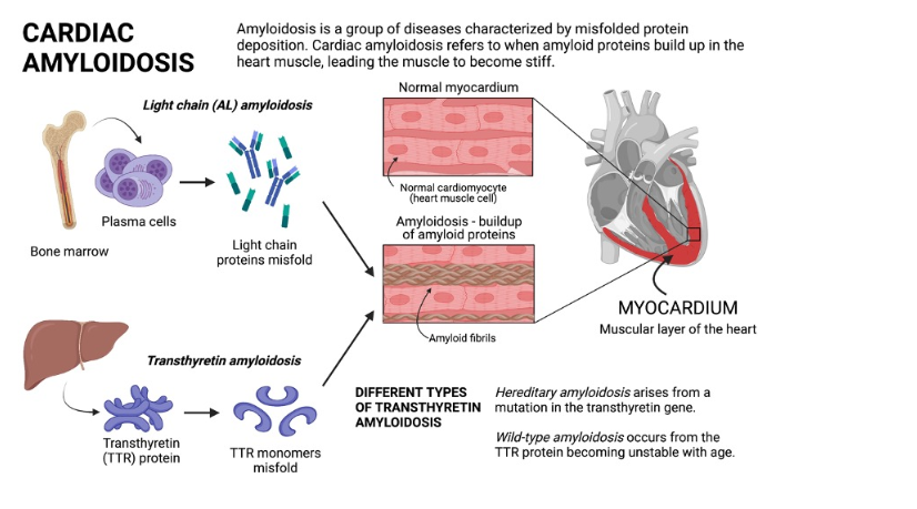
Amyloidosis is a proteostatic disease , meaning a progressive disorder of extracellular deposition of insoluble protein fibrils in tissues.It affects , brain, kidney, nerves and almost any organ.Crucially, amyloid is an interstitial disease, accumulating strictly in the spaces between cells rather than inside them. This interstitial buildup exerts physical pressure, alters tissue architecture, and finally invading the cell and hence the organ function.

The primary culprit is a normal transport protein   transthyretin (TTR) (same as prealbumin) It is synthesized in the liver as a stable, four-part tetramer structure that safely circulates in the blood. Under stressful or aging conditions, this tetramer destabilizes unstable monomer pieces. These monomers undergo misfolding, polymerizing into rigid cross-beta-sheet amyloid fibrils. These abnormal ATTR fibrils escape cellular degradation.

Cardiac amyloidosis

Cardiac amyloidosis , presents as mainly as restrictive cardiomyopathy, but can be seen in any chronic heart failure associated with LVH, especially in Aortic stenosis, where it can involve both the valve and myocardium.There are multiple reasons why we diagnose amyloidosis more often than before. But , one thing is sure, it is not due to increasing incidence in the population , but more of a high awareness and availability of advanced cardiac imaging .
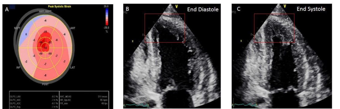
Image source : Agha AM,. Role of cardiovascular imaging for the diagnosis and prognosis of cardiac amyloidosis. Open Heart. 2018;5:

What is the confirmatory test for Amyloidosis ?

It was classically diagnosed by tissue biopsy with Congo red staining, showing “apple-green” birefringence under polarized light microscope. Mass Spectrometry, Immunofluorescence is then used to decode the protein composition. Currently , with typical patterns of cardiac imaging we can diagnose with confidence without biopsy. While echocardiography is good enough , MRI and technetium-99m pyrophosphate scans are crucial.

What are the specific treatment option for amyloidosis ?

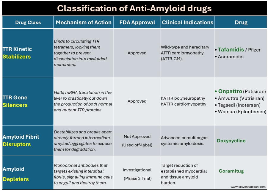
Until recently, Amyloidosis, has been considered as irreversible degenerative disease .Now we have strategies to reduce progression or even regress the Amyloid deposition. Many drugs are still being studied with varying success.

Following table summarise the different pharmacological modalities to tackle Amyloidosis.
Final message

A limitation of currently approved anti-amyloid medication such as Tafamidis or Onpattro , is that it cannot be reversed and only be prevented.Emerging technologies like monoclonal antibody depleters hold promise to actively clear out existing deposits, potentially turning it to a truly reversible condition. Chronic fibrotic pathways from triggered fibroblasts are closely linked to Amyloidosis. Hence, antifibrotic drug research should go hand in hand with Amyloidosis.

Read Full Post »

Is it a physiological molecule ?

1.Yes, It is a physiological molecule.

2.No, Amyloid is always pathological.

Where does it gets deposited ?

A .Extracellular*

B. Intracellular

C. Both

Answer : Q 1: A / Q 2: C. It is indeed a physiological molecule in small amounts that help carry hormones across the blood. In pathology, it accumulates in huge amounts. It is a disorder of protein folding, making them thick, stiff , sheets of peptide, hence mis-behaving with adjacent cells, injuring them in the process. This is responsible for the systemic nature of disorder right from the brain to peripheral nerves, Heart, kidneys, liver, spleen, etc. (Ref 1)

(*It should be stressed , majority of deposition occurs in extracellular space.They clog the interstitial space, also can invade the cell, especially in neurons in Alzheimer’s disease . Now we have evidence that Aβ (A beta) get into the myocytes as well. (Troncone L, J Am Coll Cardiol. 2016) Fellows, better say amyloid is primarily extracellular, but, by pressure effect and injury of cell membrane it results in cell death.)

Ultrastructural Organization of Amyloid Fibrils byAtomic Force Microscopy Image source Chamberlain AK, . Biophys J. 2000 Dec;79(6):3282-93.

Can we dissolve it or get rid of it ?

Till now, these disorders has been termed as a degenerative disease with no viable options. Now, we have made a breakthrough. A group of drugs genetically created work by unfolding the proteins by interfering with RNA (SiRNA) that home in on the host liver and help clear these systemic proteostasis, the key pathology in Amyloidosis (TTR).

Patisiran is approved by FDA for peripheral neuropathy and used in cardiac amyloidosis. Patisiran is a siRNA encapsulated within a lipid nanoparticle delivery system for targeted delivery to the liver, the primary source of serum TTR . Once in the liver, siRNA binds to the untranslated region of TTR mRNA and degrades it, inhibiting TTR synthesis. ( Adams D et al NEJM 2018)

Tafamidis is another similar drug. (N Engl J Med 2018; 379:1007-1016)

Final message

We are moving in the right direction in tackling the cell aging, degeneration and cell fibrosis that is behind majority of chronic systemic disorders.(Anti-fibrotic drugs is the next big target Ref: Front. Pharmacol., 31 May 2017 )

Reference

1.Greenwald J, Riek R. Biology of amyloid: structure, function, and regulation. Structure. 2010 Oct 13;18(10):1244-60.

2.Adams D, Gonzalez-Duarte A, O’Riordan WD, Yang CC, Patisiran, an RNAi Therapeutic, for Hereditary Transthyretin Amyloidosis. N Engl J Med. 2018 Jul 5;379(1):11-21.

Read Full Post »

y

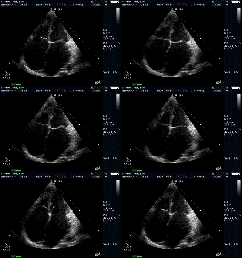
A classical restrictive cardiomyopathy . Both atria balloon out as they face stiff resistance from the ventricles

Restrictive cardiomyopathy(RCM) is a common form of myocardial disease which was difficult to recognise  in the past.It was also commonly confused with constrictive pericarditis. Today , one can easily recognise this entity.A simple clue is bi- atrial enlargement  with relatively normal ventricle size.The above case is a classical form of RCM.

In late stages of RCM ,both LV, RV begins to dilate and can mimic a dilated cardiomyopathy. Doppler filling pattern  , and tissue Doppler motion of mitral annulus are recently validated methods to identify RCM. Still ,  2D features  are  very useful .This implies , the anatomical changes in the chamber size  are as important as physiological AV filling profiles. It is generally believed , physiological impairment precedes anatomical defects.( But not proven concept yet ! )

Note: The ventricles are not dilated and retain good systolic function

In the above patient  , etiology could not be confirmed and was labeled as idiopathic RCM  as tests for amyloid and eosinophilic infiltrations were negative.

M -Mode of the same patient confirms good systolic function

One would  call  for doppler mitral filling profile here ,  to confirm restrictive physiology ( A short DT , Short IVRT  . A reversal in  pulmonary veins , E/E’ ratio etc etc ) But all these are redundant here.

How is RCM different from non dilated cardiomyopathy ?

A new entity is being recognised in the cardiac  muscle disease.This is often referred to as NDCM (Non dilated cardiomyopathy)  .Global  systolic LV dysfunction  with normal  LV dimension.This a similar to the terminology MDCM (Minimally dilated  cardiomyopathy  where LV dimension increases  not more than  15 % of basal size ) .

This is seen in CKD and diabetic individuals.Atria may be enlarged .Diastolic dysfunction may co exist.  It is no surprise,  this entity closely mimics RCM. But in RCM LV systolic function  is not greatly compromised till late stages , while NDCM it begins with systolic dysfunction. This is the only difference .There can be overlaps .

Many strongly believe ,  both  RCM  and NDCM are one and the same entities ,  that present in different time frames in their natural history.

Final message

Diagnosing RCM is no longer difficult in established cases* . The message from  this article is , 2 D echo can  strongly suggest  the possibility of RCM (or even clinch it)  .  Never ever  diagnose RCM with normal 2 D echo. Doppler filling profiles are useful  additional tools . We  can not diagnose RCM with doppler features alone , but we can  be fairly certain about RCM when we encounter  typical bi-atrial enlargement and a normal LV by  2 D echo.

Caution : Patients  with longstanding atrial fibrillation of any cause , can dilate their atria and could mimic RCM .They can be some compromise in LV function due to chronic tachycardia .

* Recognizing RCM ,  very early in the course is still a problem . Here the  newer modalities like Phase  MRI, tissue doppler, speckle tracking, and velocity vector imaging may be useful.

Read Full Post »
