Feeds:
Posts
Comments

Archive for April, 2021

This question might squeeze the collective coronary knowledge of any cardiologist. (At least, it does for me !)

What is an intermediate coronary lesion? (ICL) 

Traditionally it is an “angio-ocular reflex” measurement of coronary arterial diameter stenosis that lies between 40 to 70% (Mind you, 70 diameter stenosis is 90% area. So, we must be clear what we really mean in any revascularisation debate).

Above one is the simplest expression of ICL. (* While 70% cutoff is fairly constant, the lower limit 40% is still not a settled issue. It can be 30 or even 50 %. I think we haven’t yet named the lesions 1 to 49 %. It is the spectrum that contains  Coronary erosions, ulcers, luminal irregularity, or the evasive term minimal CAD  )

Many sub-classes exist in ICL.

  1. Should ICL definition be different in proximal LAD? (A 40% PDA or OM2 lesion is not the same as 50% LAD right.Maybe we need to artery specific redefinition, left main we did it already)
  2. It can be de nova chronic (most common ) Acute  /subacute, acute recanalization (Each has a different management strategy)
  3. What about ICL with good TIMI 3 flow. Mostly safe and can be ignored?
  4. Should we bother to know the content of ICL? It could be a minor plaque or just thickened and narrowed arterial wall or even layered thrombus.
  5. Is it isolated ICL?  When ICL occurs in isolation it gets more attention is natural to ignore if ICLs are noted along with other critical lesions nearby. The risk of ignoring or risk of including ICL in the final angiogram reports is unquantified. 
  6. When two ICLs lie by next to each other (Tandem ICL) will you add the stenosis resistance in series? Does the length matter.(Can we measure net FFR ?) 
  7. Is it symptomatic vs asymptomatic? (very difficult query )In stable non-Infarct CAD Internedaite lesions do not obstruct flow, but Post ACS it is the distal microvasculature that determines the epicardial flow. so even intermediate lesion resist flow.
  8. ICLs in ecstatic segments pose a special issue. Adding to this Galovian positive remodeling mask the true plaque burden(Currently liberal use of OAC like warfarin are used in ectatic vessels with ICLs)
  9. By the way, is it true, ICLs are more prone for  ACS?  We believe it based on small studies and sort of biased teaching. Of course, there is some truth in it, but in a larger sense, it is not correct thinking. ICLs by sheer number overtake the critical lesions in terms of Incidence. So more ICLs present as ACS. But in, pure pathological terms flow-limiting lesions do carry more risk for ACS. (Of course, calcification might stabilize a few of them, and convert them to CCS) . For argument’s sake, if we agree ICLs are more prone for ACS, we should first fix these lesions than the more tighter ones.(Any guidelines forthcoming ?)
  10. Finally, the most important query Is the ICL vulnerable, or is it flow-limiting? (read below)

Imaging and physiology

CAG is just a shadow of contrast luminogram. Further, the contrast flowing across a lesion cannot be equated with the true velocity of blood flow. So, what shall we do? How do we overcome the limitations of CAG shadow? We need to go after more glamorous shadows like IVUS and OCT. They do suffer from myopia and hypermetropia respectively. Still, they are good enough to reveal important info like the content of lesions like calcium thrombus with acceptable precision, etc. The thickness of the fibrous cap (TCFA) is a current marker of vulnerability. This thickness is dynamic as do plaque liquefaction. We are looking ahead to the days of virtual histology and plaque metabolism by NIR spectroscopy. Decisions based on a single one-time snapshot from intermediate lesions would largely be meaningless. 

What about physiology? FFR, iFR,(Adenosine free)  QFR (Based on TIMI frame count) offer a more scientific assessment of flow across the lesion. Still, it is not clear. An elegantly made study is available that depicts the relation between stenosis and FFR.

Realtionship between diameter stenois snd FFR. Note even a 30% lesion has low FFR and wide variation a 70% lesion show on either side of cut off .8Does Routine Pressure Wire Assessment Influence Management Strategy at Coronary Angiography for Diagnosis of Chest Pain? The RIPCORD Study Nick Curzen circulation cardio vascular Interventions 2014.

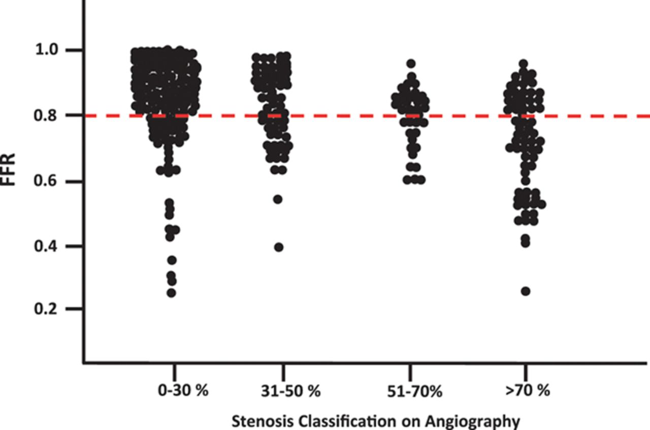
Relationship between diameter stenosis snd FFR. Note, even a 30% lesion can have a low FFR, and a 70% lesion show the FFR to scatter on either side of the cut-off value .8 . So, what does it mean?  We have simply shifted our ocular bias to objective flow bias. Does Routine Pressure Wire Assessment Influence Management Strategy at Coronary Angiography for Diagnosis of Chest Pain? The RIPCORD Study Nick Curzen circulation cardiovascular Interventions 2014.

What is the effect of statin on ICL?

There is no specific large-scale study that looked into this. Plaque regression and stabilization are expected in most ICL with intensive statin regimens. (Seung-JungPark et al  JACC 2016) It reduces new-onset TCFA. Will it increase the cap thickness? It can be assessed by the OCT study. (Maybe it is already available will search for it ). PCSK & Inclisiran should do it if not a statin. 

Final message

Coming to the title question, the term ICL means nothing without the clinical background and the angiographic setting it is detected. Realize, the intermediate lesions don’t Imply intermediate risk. We can’t do  IVUS or OCT in all intermediate lesions. Even if we detect vulnerability in a 50% lesion, treatment will remain mostly intensive medical management. (There is absolutely no good evidence to show stents stabilize vulnerable plaque that does not limit flow ) 

So, the best approach to all those billions of ubiquitous ICLs scattered across the human coronary landscape is to stabilize it OMT( Open-minded medical therapy), lifestyle modification (taking style out of life), reassurance, and propagation of peace that will passively the plaques. Imaging and FFR can do wonders in an elite minority population at a considerable cost. (However, for the sake of demystifying atherosclerosis we should continue research with such modalities, sparingly though )  

Reference


Read Full Post »

It was the final case on weekend Echocardiogram review day, I asked my fellow for a brief summary of the patient. 

A 5 -minute conversation

“Yes, sir, he is a 62-year-old male retired govt officer. He has a severely stenosed aortic valve, with a peak gradient of 90 mmHg and a mean gradient that comes to almost 50 mmHg. LV  EF is 58%, GLS is 18, LVH is obvious. LA is not dilated (Didn’t measure volume though), but DT is short. Valve orifice is hovering around 1cm, mild calcium noted in LCC  I am not sure whether it’s bi or tricuspid still. The annulus is 22mm. The mitral valve is perfect, no calcium spill over to the mitral curtain and the rest of the annulus”.

“That is ok, what for he has come”?

“A GP from Tambaram has referred him after he detected a murmur over the chest”.

“Oh Ok. What are his symptoms”?

“He is denying any symptoms”.  

“Are you sure? did you ask him specifically about it during exertion”? 

“Yes, he says he can climb 3 flights of stairs. (In fact, he was sort of amused when I told him to be frank in his expression,since  he has a potentially serious obstruction in the main valve that connects his heart and body.”

“I agree, but his reaction was not inappropriate I thought, after all, he didn’t feel any symptoms right”. “So what shall we do for him?  TAVR? SAVR? or Leave him alone? Shall we put him on the treadmill? to document symptoms? Is it that risky”? 

“But , he says he can walk for a mile or two every day” 

“That’s fine. Can you really predict when his ventricle will fail and he may land up in a semi-emergency surgery?

“I think we can’t,  but why is he is so asymptomatic sir”?

“Wow, that’s more than a million-dollar question. You need to address that query to the vascular Goddess. I don’t know the answer.It is all about the ability of the heart to perfectly balance the ventricle and aorta in spite of severe obstruction. It is something like TIMI 3 flow and good FFR  in a patient with 90% occlusion.) My guess is, the LV does this by modulation of systemic pressure &  resistance in such a way , it neither feels the strain nor does it reduce the stroke volume much. By the way,  have you heard about this ? Z- Va score. I would like you to read about that. It will help you understand the hemodynamic nuances of severe AS and how the ventricle manages to serially couple the afterload of the vascular system”. 

“Make a pardon sir, I haven’t heard about it. What is Zva? 

“Never mind. It is not a new index. Was first introduced 16-years ago by Martin Briand et al from Quebec, heart Institute Canada  J Am Coll Cardiol 2005 Jul 19;46(2):291-8.  Z Va score(Valvulo-Arterial) is the collective flow impedance of the aortic valve and the entire aorta. It is more attractively defined as the cost of blood pressure in mmHg for pushing one ml of blood per body square meter area

Formula for Z va : (Systolic BP × Mean gradient)/ Stroke volume Index

Unit : mmhg /ml/m²

Normal value:  < 3.5 to 4.5 (Actually no normality, rather it must be acceptable value .It is still being defined )  if the cost is more than 5mmhg it suggests significant Aortic stenosis) A high value > 4.5 is a definite index of poor outcome. In a well-compensated heart, Zva is maintained far less than 5 and many such patients are asymptomatic as well. Zva has specific clinical value in all critical AS especially so if they are asymptomatic. It is no longer a research topic, has an important role in the bedside too. Here is an excellent resource on Z Va score from ESC.


Final message 

The timing of AVR in aortic stenosis is very critical. All symptomatic severe AS must be immediately intervened. Currently, with surgical risk falling rapidly ( & the option of TAVR looming large) even many of the asymptomatic AS need to be considered for valve intervention at the earliest before or at the onset of LV dysfunction. Zva’s score will definitely add more light to our  limited hemodynamic wisdom in aortic stenosis(Zeineb Hachicha  JACC 2019) 

Read Full Post »

ASD device closure has become a de-facto modality for most ostium secundum defects(<35mm). The stupendous success of this procedure is attributed to careful pre and Intra-procedural Imaging, new generation hardware and of course the ever-improving expertise among Interventional cardiologists.

Still, there is one issue that is bothersome. It is the late complications of this device and the need for follow-up (Unlike surgery where close and forget option seems real and confer lot of comforts) The delayed mechanical complications are now extremely rare still  follow up of these patients is advised.

What is the mechanism of Aortic erosion in ASD device closure ?

The IAS is a dynamic structure. (Ask any echoc’ardiographer ,how ASD size varies with cardiac cycle.) The device should sit right across all rims including the  Aortic rim . If the device if larger , and if the Aortic rim is less it has on other option but to splay over the Aorta . Enthusiastic young cardiologists should be aware this splaying is not in our control at all. Not all splaying are good and safe as well. If its not smooth and if the septum is mal-aligned there could be friction Injury to Aorta. A very early manifestation of device dislodgement and later a trickle of  pericardial effusion. This should be watched for. (Please be reminded a early pericardial minimal effusion due to sudden shrinkage of RA, RV and due to some unknown hypersensitivity response ? can confuse us )

Link between deficient Aortic rim and Erosion : An unsettled Issue (But , we settled it ! )

One issue that is poorly understood is, many Interventional cardiologists believe strongly that the length (and even quality) of Aortic rim is the least important and need not to be respected. I am still not clear on what basis this piece of Interventional literature came in. This is exactly is the reason even novices take liberty and large devices are implanted casually encroaching the Aorta. Though most cardiologists shrug of this risk of Aortic rim deficiency and subsequent erosion,  at least one study clearly showed a serious link between the two. I feel the issue is not yet settled and demands re-scrutiny.

This presentation was made in Tamil Nadu Interventional (TIC)council meet at held recently


This image has an empty alt attribute; its file name is aortic-erosion-2-1.gif

Link to the PPT presentation aortic erosion 2

Final message 

After going through all relevant literature as on 2019 , the incidence of aortic erosion is rare but the fear is real (Many feel it is paranoid and largely unfounded ) I won’t agree though.The message must be, a good quality Aortic rim is important too.

However, a properly sized device, perfectly delivered with good Image assistance by a trained cardiologist in a high volume center (? >25/year) shall prevail over surgery in most patients with ASD. 

Reference


Further issues : Stroke risk with ASD device 
 

One more Issue with ASD closure device is delayed embolic episodes from thrombus attached to device. This is prevented by  routine anti platelet drugs practiced by certain Institutions .The new generation devices (Occlutech Germany) has modified the LA side of the disc (No Hub) to reduce this risk

Read Full Post »

Some of the questions  addressed  in this presentation

1.What happens to fetal blood pressure during maternal hypotension how good is fetal autoregulation?

2.Why is LSCS increasingly preferred mode of delivery in heart disease complicating pregnancy challenging the traditional scientific concept?

3.What is likely hood of patients with moderate mitral stenosis developing pulmonary edema during prolonged 2nd stage of labor?

3.What is the missing link between PIH and PPCM? How prepartum cardiomyopathy differs from postpartum?

4.Is Eisenemneger really an absolute contraindication for pregnancy?

5. How can we continue VKAs warfarin or Acitrom throughout pregnancy? What are the potential problems of double switching one at 6th week from VKA to Heparin and again from heparin to VKA  at  12th week?

Hope, the man-made hematological bridge in pregnancy has been finally liberated from confusion (Who is saying not yet?)


6. On what evidence base the safety margin of 5mg cutoff for Warfarin and 3mg for Acitorm was decided?

7. Who is insisting on us to do Anti-Xa monitoring for LMWH in pregnancy? Is it really needed? What does the American society of hematology say?  (ASH guidelines for VTE in pregancy 2018) Why we don’t insist on Xa estimation in acute coronary syndrome?

8. What is the inflection point of at which risk of termination is almost at equipoise with continuing pregnancy in various heart diseases.

A GIF run-through of the presentation.

PDF & video version will be posted


The ultimate reference 

Read Full Post »