Archive for March, 2023
AI, is not risk free in medical science !
Posted in Uncategorized on March 25, 2023|
A hemodynamics quiz: What is the effect of AF on mean LA pressure?
Posted in Uncategorized on March 25, 2023|
ERS pattern in ECG : “Iatrogenic panic” is unwarranted !
Posted in Uncategorized, tagged ers pattern in ecg, ERS syndrome on March 23, 2023|
This is an ECG of a 25-year-old, recorded in master health check-up.
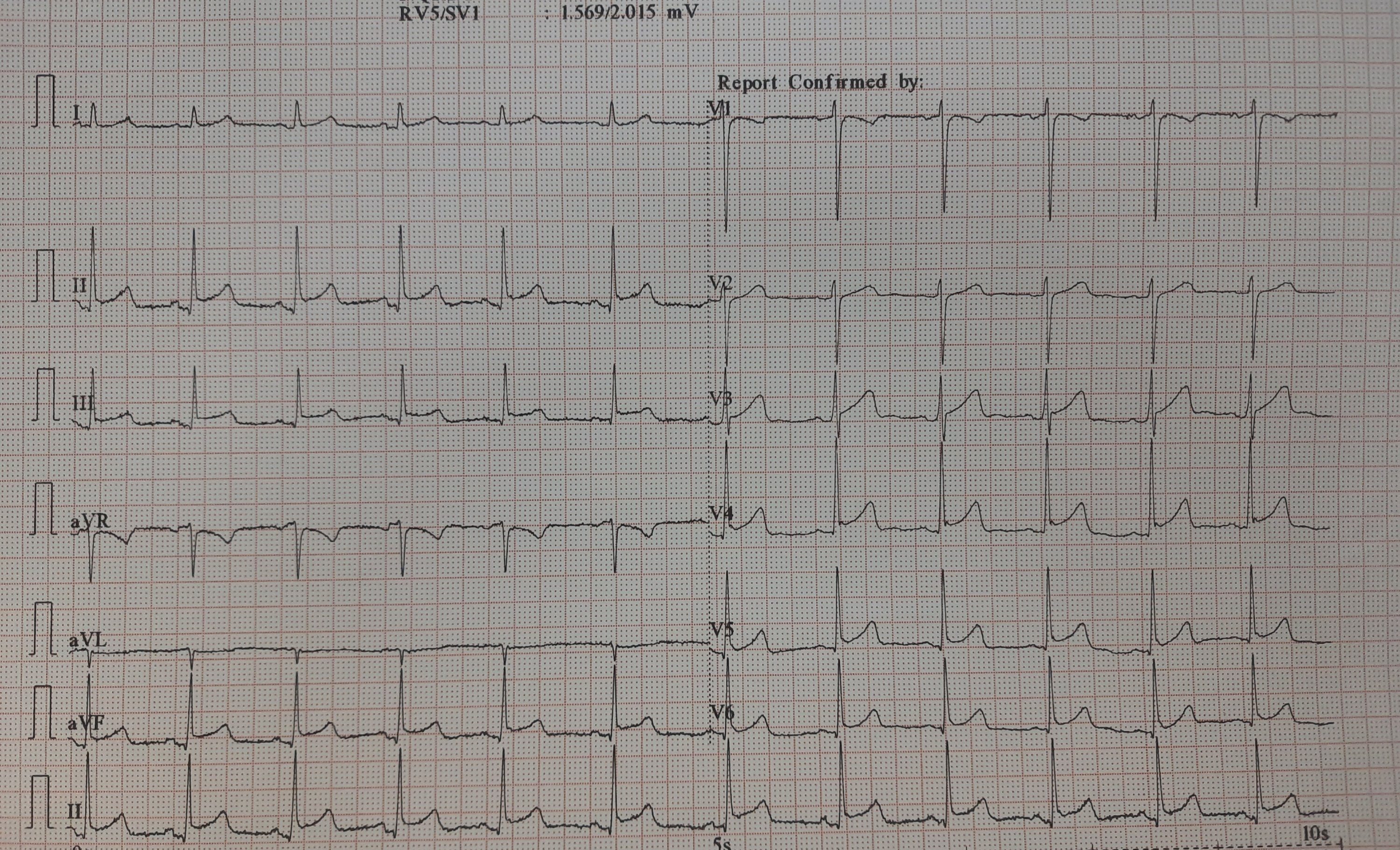
It would be mind-boggling to know the prevalence of such ERS patterns in the general population. One estimate suggests it could be anywhere between 3 to 13 % depending upon the criteria used. Let us assume the mean as 5 %. Then, it would be 30 crores of human beings in our habitat show this ECG pattern. If applied, in my city Chennai alone 5 lakh people could carry this tag.
While it is true, some forms of ERS and J wave syndrome can be markers of serious ventricular arrhythmias, either spontaneous or at times of Ischemia. Currently, It has become a fad, in cardiology academic circles*, to propagate the idea that ERS is no longer a benign condition. This is not acceptable at any degree of cognition. This happened mainly after few studies in powerful journals created some alarmist views. (*Maybe there is a bit of truth there. I still have doubts about whether we interpreted the Michel Haïssaguerre study properly)
Final message
ERS is a widely prevalent normal ECG variation with a minuscule risk. High-risk subsets need to be screened only if the J waves encroach and spill dangerously into the ST segment as well. Of course, this pattern is of serious concern if there is a family history of young SCDs has occurred.
Reference
Here is a good review of this topic by
Learning targeted IAS puncture in 20 minutes
Posted in Anatomy of heart, Uncategorized, tagged anatomy of heart, ias puncture, right vs left atrial anatomy on March 10, 2023|
The main reason for all those jitters, we cardiologists, get every time we puncture the IAS is not due to a lack of expertise and experience perse. There are two more reasons. First and foremost, it is still largely a blind* procedure. (Even in this era, where drones with HD vision shoot one-meter targets from a 1000 KM range ) *TEE and ICE are there, but they rarely give enough confidence.
The second reason is more important and is rectifiable. It is the perception error in our anatomical cognition, that is fed to us from first-year medical school. We are made to believe (at least to people like me ) The right atrium is aligned like a perfect box on the right side, sharing a wall called IAS, and the left atrium is obediently placed left of the right atrium. Please realize the heart is such a complex twisted single tubular organ, the venous end, in a stunning backward loop brings the LA most superior and posterior to the right atrium overriding the left-right relationship.)
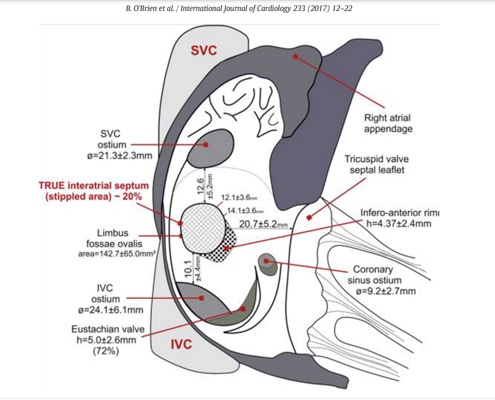
The right atrial terrain and IAS with multiple bumps and holes. Note the true IAS constitutes only 20% . This is where our punctures need to be.
Development of IAS
IAS development and the number of layers it sandwiches, the tortuous tracts of PFOs, the fossas, and its variable limbus is a big topic. Further, It is worth recalling, the true IAS hardly forms 20 % of the area of the interatrial contact surface.
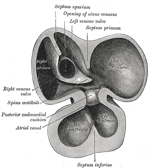
(the differential regression of sinus venous, along with infolding of the roof and along with curious septum spurium , the ubiquitous septum secundum make the texture, area & shape of IAS, a fascinating creation, though troublesome for the cardiologists ! ) Whoever named that part of vanishing IAS as spurious, (I think it is Henrry Grey ) has much fore-vision.
Forget about all this. Tell me how to cross this difficult terrain
Coming to the real world of interventions, we need to do targeted punctures in different spots of IAS in various interventions.(Mitra clips, LAA device, PTMC, PV abaltions, Mitral paravalvular leaks , TMVR etc) This has made this task even more tricky. Experts are always there to help us out. Like swimming, it can never be learned in books.
This 19-minute clip from. Seoul, South Korea is an excellent resource. Thanks to Dr. Sang Weon Park
Along with sound anatomical knowledge, improved hardware, and imaging like deflectable sheaths, TEEs, and ICE (intracardiac echo ), let us hope, it will soon become an easier task for everyone.
Final message
Understanding “attitudinal cardiac anatomy” with fluoroscopic overlay is the key. Again, it needs to be stressed, “Right is not right, and left is not left” when it comes to true atrial geo position. LA is equally posterior, superior, and of course to the left of RA. Some of my colleagues are blessed with a special 3-dimensional skillset (Inherited ?) I failed miserably to understand this, till very late. I am sure, Dr. Park’s video will help all our youngsters to cross the difficult gateway to the left side of the heart.
Reference
One more good read
B. O’Brien et al. / International Journal of Cardiology 233 (2017) 12–22
P wave spotting in AF is not forbidden
Posted in Uncategorized, tagged cardiology research topics for fellows, causes of absent p wabes, p vs f waves in af, p waves in atrial fibrillation, research topics in atrial fibrillation on March 8, 2023|
Fibrillation is a continuous, chaotic muscular activity. In AF, atrial muscle is expected to lose all coordinated contractions with fibrillatory waves replacing P waves. Have you ever spotted a suspicious P wave in a strip of otherwise explicit AF? If not, this write-up is not for you.
An evolving rare theme in Atrial fibrillation
Have a look at this ECG
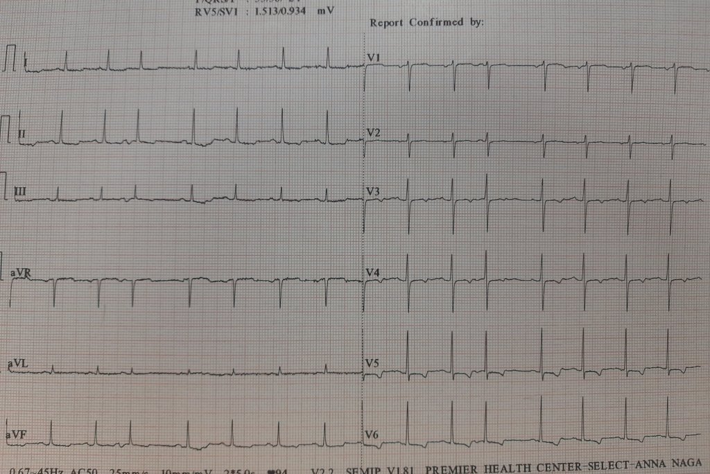
Here is an ECG, that was reported as AF, multiple APDs, or Possible AF, Pre AF. I suggested the term AF in transition. While few agreed, many said it is a straightforward SR with APDs, making it appear irregular RR.
But, the fact of the matter is, ECGs are insensitive to pick all fibrillatory wavelets. It can selectively pick a few coarse F waves and make them appear as P. I think, in this era, we should not diagnose AF by proxy, ie absent P waves. Rather, we need to look actively for fibrillatory wavelets. (Imagine all sinus arrests will qualify for f fine AF with a slow ventricular rate is it not ?)
The semantics of AF nomenclature is long. Intermittent AF, and paroxysmal AF, are well-known entities. It is now clear, AF can occur for a few seconds and vanish too. It seems we need to play some more linguistics with the most common cardiac arrhythmia. (Non-sustained AF, evanescent AF, etc )
Some thoughts on this hide & seek P waves
- Apart from the conventional list of absent P waves, one more example is repetitive APD can stun the atrial muscle for a few moments or minutes.
- Then, we always have the issue( eluded to earlier) of sinus node paralysis, with irregular junctional escape mimicking AF.
- Amiodarone can reduce fibrillatory rate, and (AF cycle length ?) Coarse F waves slow and stabilize it to mimic an organized P wave
- P on Ta waves (Like R on T ) can trigger a nonsustained AF for a few moments in a functional manner without real pathology in atria.
A funny memory brings back an EP truth
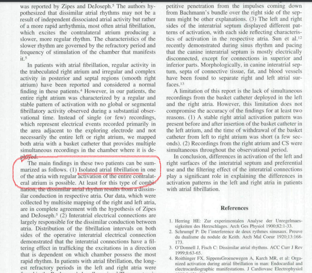
During our student days, my Professor used to trap us with this question, Which atria would fibrillate in mitral stenosis? Many of us blinked, and few had no hesitation to say, it is the LA that fibrillates. Now, after 50 years we realize, how fascinating the secrets AF has unfolded. Some organized activities are often in the right atrium, even as LA begins the process of AF. It is possible it may take variable time for the left atrial chaos to spill over to RA*. During these electrical uncertain times, some of the right atrial P wave activity refuses to die down. Even more dramatic one Atrium alone can permanently fibrillate and others completely insulated by blocking the signal in the Interatrial pathways. (Ref 1 ) Ndrepepa’s paper in the JCE 2000)
Final message.
True scientists rarely bother about questioning a dictum. The concept of non-uniform AF was first thought of by (Schrmp et al Ref 2) 100 years ago in 1920, and Zipes(Ref 3) hypothesized this in 1973. now, in the Year 2000, Ndrepepa confirmed it with EP studies. The spotting of occasional P waves is not forbidden in AF if the contralateral atria decide to block the incoming AF signals and keep generating their own P waves
Young EP guys, now that you are equipped with the sophisticated intracardiac GPS, please pursue this provocation in AF. One more piece of evidence we observed in the echo lab. Try to look at Tricuspid doppler A velocity waves in full-blown AF patients. You can see the surprise for yourself. This is very good research work to do. This is one of the ideas I gave to my fellows at MMC. Now, it is free for all to pursue whoever wants to do this. The clinical implication* will follow.
* A lingering query, how common is RAA clot in mitral stenosis with AF and the possible threat of pulmonary embolism?
Reference
2.Zipes DP. DeJoseph RL: Dissimilar atrial rhythms in man and dog. AmJCardioi I973;32:6l8-628.
3.Schrumpf P: De 1’interference de deux rythmes sinusaux. Preuve du dualisme du nodule de Keilh. Arch Mai Qwur 1920;l3:168-173
Postamble
The snapshot from Ref 1 . The term Isolated AF confined to one atrium could be a rare event, but, no one can deny we have plenty to learn from them
Categories
-

-
The contents of the this blog is being published as Kindle E book , as per the request of many of the readers. Every article will continue to be open source in this site. Again I shall reiterate the book format is not aimed at any commercial intent. It is only to facilitate learning in a single book format Here is the link to book
https://amzn.in/d/euhL5vu Archives
- July 2026 (4)
- June 2026 (9)
- May 2026 (6)
- April 2026 (11)
- March 2026 (10)
- February 2026 (8)
- January 2026 (8)
- December 2025 (11)
- November 2025 (7)
- October 2025 (8)
- September 2025 (7)
- August 2025 (9)
- July 2025 (10)
- June 2025 (8)
- May 2025 (9)
- April 2025 (7)
- March 2025 (10)
- February 2025 (4)
- January 2025 (9)
- December 2024 (11)
- November 2024 (8)
- October 2024 (10)
- September 2024 (5)
- August 2024 (5)
- July 2024 (6)
- June 2024 (5)
- May 2024 (4)
- April 2024 (7)
- March 2024 (4)
- February 2024 (8)
- January 2024 (6)
- December 2023 (8)
- November 2023 (13)
- October 2023 (14)
- September 2023 (5)
- August 2023 (6)
- July 2023 (10)
- June 2023 (5)
- May 2023 (5)
- April 2023 (4)
- March 2023 (5)
- February 2023 (2)
- January 2023 (7)
- December 2022 (3)
- November 2022 (5)
- October 2022 (5)
- September 2022 (4)
- August 2022 (3)
- July 2022 (9)
- June 2022 (2)
- May 2022 (1)
- April 2022 (2)
- March 2022 (1)
- February 2022 (3)
- January 2022 (7)
- December 2021 (3)
- November 2021 (5)
- October 2021 (8)
- September 2021 (4)
- August 2021 (6)
- July 2021 (6)
- June 2021 (7)
- May 2021 (5)
- April 2021 (4)
- March 2021 (3)
- February 2021 (6)
- January 2021 (8)
- December 2020 (4)
- November 2020 (5)
- October 2020 (7)
- September 2020 (7)
- August 2020 (10)
- July 2020 (6)
- June 2020 (9)
- May 2020 (9)
- April 2020 (5)
- March 2020 (7)
- February 2020 (3)
- January 2020 (4)
- December 2019 (4)
- November 2019 (6)
- October 2019 (3)
- September 2019 (6)
- August 2019 (3)
- July 2019 (1)
- June 2019 (3)
- May 2019 (2)
- April 2019 (2)
- March 2019 (2)
- February 2019 (4)
- January 2019 (2)
- December 2018 (2)
- November 2018 (2)
- October 2018 (2)
- September 2018 (1)
- August 2018 (2)
- July 2018 (3)
- June 2018 (1)
- May 2018 (3)
- April 2018 (1)
- March 2018 (3)
- February 2018 (3)
- January 2018 (1)
- December 2017 (3)
- November 2017 (3)
- October 2017 (3)
- September 2017 (2)
- August 2017 (2)
- July 2017 (2)
- June 2017 (2)
- May 2017 (4)
- April 2017 (3)
- March 2017 (3)
- February 2017 (5)
- January 2017 (3)
- December 2016 (2)
- November 2016 (5)
- October 2016 (4)
- September 2016 (3)
- August 2016 (5)
- July 2016 (3)
- June 2016 (4)
- May 2016 (3)
- April 2016 (6)
- March 2016 (4)
- February 2016 (3)
- January 2016 (5)
- December 2015 (6)
- November 2015 (5)
- October 2015 (8)
- September 2015 (2)
- August 2015 (5)
- July 2015 (7)
- June 2015 (4)
- May 2015 (6)
- April 2015 (5)
- March 2015 (7)
- February 2015 (15)
- January 2015 (8)
- December 2014 (5)
- November 2014 (9)
- October 2014 (7)
- September 2014 (9)
- August 2014 (5)
- July 2014 (11)
- June 2014 (5)
- May 2014 (4)
- April 2014 (5)
- March 2014 (8)
- February 2014 (8)
- January 2014 (5)
- December 2013 (7)
- November 2013 (7)
- October 2013 (14)
- September 2013 (12)
- August 2013 (15)
- July 2013 (15)
- June 2013 (15)
- May 2013 (15)
- April 2013 (15)
- March 2013 (15)
- February 2013 (15)
- January 2013 (15)
- December 2012 (15)
- November 2012 (15)
- October 2012 (15)
- September 2012 (15)
- August 2012 (15)
- July 2012 (15)
- June 2012 (15)
- May 2012 (15)
- April 2012 (15)
- March 2012 (15)
- February 2012 (15)
- January 2012 (15)
- December 2011 (15)
- November 2011 (17)
- October 2011 (17)
- September 2011 (17)
- August 2011 (21)
- July 2011 (20)
- June 2011 (17)
- May 2011 (15)
- April 2011 (17)
- March 2011 (25)
- February 2011 (20)
- January 2011 (20)
- December 2010 (18)
- November 2010 (21)
- October 2010 (21)
- September 2010 (25)
- August 2010 (20)
- July 2010 (10)
- June 2010 (11)
- May 2010 (19)
- April 2010 (16)
- March 2010 (14)
- February 2010 (22)
- January 2010 (18)
- December 2009 (20)
- November 2009 (20)
- October 2009 (3)
- September 2009 (21)
- August 2009 (19)
- July 2009 (12)
- June 2009 (12)
- May 2009 (11)
- April 2009 (15)
- March 2009 (21)
- February 2009 (4)
- January 2009 (12)
- December 2008 (13)
- November 2008 (9)
- October 2008 (22)
- September 2008 (20)
- August 2008 (16)
- July 2008 (14)
- June 2008 (7)
Blog Stats
- 6,703,400 hits
Please give your feed back .
Click below to see who is watching this website live !

- This site will never aim for profit. Still ,this donation link is added at the request of few visitors who wanted to contribute and of-course that will help make it sustainable .

Please Note

