Just roll over the virtual marker along the coronary lesion to get the underlying flow ratio. Blue is an absolute normal segment. Green is ok, orange and red slow-moving coronary traffic jam zones. it’s just like drawing a google map showing life traffic. No wire, no adenosine FFR comes inbuilt in every angio shot. Looks great Isn’t it? This is called QFR. Quantitative flow ratio derived from routine coronary angiograms. It can also guide us to find the optimal sites of both proximal and distal stent landing zone in the best physiological manner.
Which company makes this ?
Any studies done with QFR ?
FAVOR 2 study was reported in TCT. This modality is expected to evolve.
Final message
Whenever possible every anatomical lesion in the coronary should be substantiated by physiological parameter and possibly coronary Imaging to know plaque morphology and vulnerability. Though it is wishful thinking, still for all logistic reasons, most of the real world stenting will be based only on the blind anatomical luminogram.
At this point, please let me utter a non-academic hyperbole. Even a casual query to your beloved patients about their true symptoms and exercise capacity shall make these ultra-modern coronary physiology studies redundant in many. A well-performed and well-interpreted stress test is a good, objective, non-invasive indicator of coronary flow across lesions. It is wise to keep this as a basic clinical foundation in the evaluation of CAD,even as we continue to learn and forget half evolved modalities with rapid expiry dates like FFR, IFR, CT-FFR. QFR shows some promise though. Please watch for next in line coronary physiology – OFR, Optical flow ratio from OCT run through.
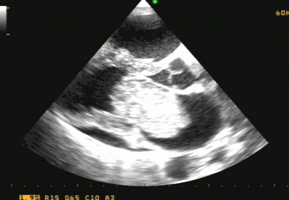
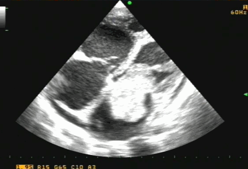
Bicaval view is an Important TEE view to visualize, the LA, IAS, and right atrium. I used to have some trouble getting oriented to this view. Hence this post. It is obtained in the 90-120 degree view at the mid esophageal position. Imagine the patient is lying on his left side and the probe comes from above down between the spine and heart to the LA from within the esophagus. This is the best view to see IAS in the profile.(Subcostal TTE can also do it) Note how the LA hugs the right atrium which is actually an ill-defined (In TEE I mean) common meeting point of both IVC and SVC. Also important is the relationship of RUPV with SVC & the horizontally running RPA sitting right over the top of LA.
The relationship between RUPV and SVC is crucial in device closure of large ASD, especially in sinus venous defect.
Clinical Importance of this view
Very useful in ASD rim morphology especially in the posterosuperior rim.
Delineates clearly the defect boundaries in SVC ASD.
Sinus venosus defect: Image source not known. Thanks to the creator.
This view doesn’t miss even the smallest PFO (With Contrast )
Can be used to guide IAS puncture in structural heart Interventions.
IVC /SVC mass extension into RA well visualized.
RA myxoma attached to septum: Image source -Michael Essandoh from Research gate
Final message
Getting oriented to TEE planes and images is so useful in structural heart interventions, like TAVRs, mitral clip, LAA occluder, tandem heart, valve in valves, etc. It is indeed a tough exerciseand requires re-learning of cardiac anatomy with fluoroscopic overlay*.I wish, I go back and sit with first-year medical school students and start all over again.
*Current hybrid cath labs do provide Echo/Fluro co-registration, still it demands core 3D anatomical Imagination.
Cardiac myxoma is the most common primary tumor of the heart that presents as mitral inflow obstruction/ regurgitation often with a systemic presentation. It can be either familial, syndromic, or sporadic. Excellent imaging is possible and diagnosis has become straightforward. Surgery is the specific treatment,
What information do the Surgeons need?
Size, attachment to surrounding structures is the key. The myxoma origins most often in IAS and defining its attachment is crucial. Mitral leaflet distortion, Injury ( and even attachment) is possible. It is helpful for the surgeons if we let them know the mechanism of mitral regurgitation prior to surgery. Echocardiogram including TEE is sufficient in most. MRI may add some more info. The aim of surgery is to remove the tumor mass completely.
Is myxoma a completely benign tumor?
Another issue is our poor understanding of the recurrence of myxoma. Why should a benign tumor be recurrent? If recurrence is a feature to be counted as a sign of malignancy, myxoma can be definitely a suspect. There seems to be a catch. It is invasive, locally recurrent, still not malignant. (Whether sarcomatoid degeneration happens is not known. Most pathologists deny this) The problem is, still we are not clear about the cell of origin of this tumor. All that we know is its origin mesenchymal stem cell.
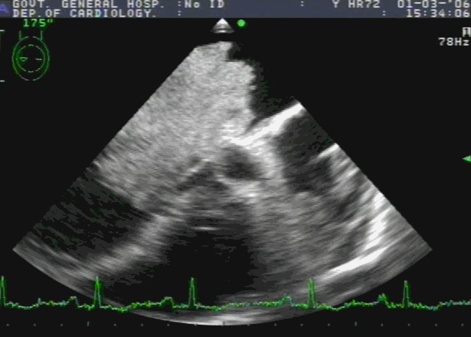
Note 50 % tumor mass enters the left ventricle with diastole. No wonder, as the tumor plops with diastolic cardiac cycle a high-pitched sound simulating opening snap followed by an MDM perfectly mimics rheumatic mitral stenosis. An MR murmur is equally common.
Common sites of recurrence
Interatrial septum
The atrial surface of anterior mitral leaflet
From contra-lateral atrium rarely
The mechanism of recurrence is either due to incomplete resection or due to its multifocal origin.
4 chamber view showing what appears to be a small narrow pedicle attaching to IAS. Please note echo imaging can be deceiving. Surgeons must inspect the mass in toto before taking the decision to excise IAS or not
What the surgeon needs to do?
The aim of surgery is to remove the tumor completely. It’s painful to diagnose recurrence and subject the patient to another surgery. (We encountered a sorry situation recently) So, when we remove the tumor we should ensure sufficient clearance with normal tissue.Biatrial approach is preferred by some surgeons.( Ahmet Yüksel Braz. J. Cardiovasc. Surg. vol.31 no.4 July/Sept. 2016)
If the tumor is not well delineated, it is better to remove a significant area of IAS along with tumor massand subsequent patch closure. Recurrences in AML and contralateral atria is unfortunate and can’t be predicted.
Further, the mitral valve is to be inspected and functionally tested. Minimal repair work or even rarely replacement might be necessary.
TEE imaging of LA myxoma. Note how fragile the edges of the tumor looks. It explains the high incidence of tumor embolus in this condition. Also, to be noted is the forceful impact of the tumor mass on AML that predisposes chronic mitral valve damage.
Final message
Referring a patient to a cardiac surgeon is not a bland ritual. (Have seen many single line referrals such as triple vessel disease referred for CABG) A well-informed interaction by the cardiologist with the surgical team and a possible per-operative echo consult, especially in rare surgeries will bring the best for the patient.
PCI is effective in relieving angina, what does it do to LV dysfunction?
This is a fundamental query in the principles of revascularisation of CAD . The term LV dysfunction can convey a bizarre meaning.It can constitute any of the combinations of the following.Cell death, necrosis, scarring, fully dead, partially dead, partially viable, apoptotic cells that are clustered across various myocardial segments. These cells are interwoven with fibrotic interstitium. Microvascular integrity is also altered.
Cells stretch, slip and slide with one another. Contractile architecture is lost. This is referred to as remodeling.In the process, the ventricle gets dilated. Wall stress increases, LVEDP raises. Patient may go for progressive failure.The whole concept of chronic myocyte loss is due to the process called programmed cell death.
Does PCI cancel this pre-planned program?
The answer seems to be a clear ” No” (Of course few studies do show some improvement ) It is becoming clear, chronic ischemic juggernaut moves on. The mechanical spiral effect on the myocardium will go unabated whether you rectify the small residual ischemia or not), However, tissue engineering, anti-fibrotic drugs, cell repair molecules, stem cell assistance are attractive approaches to prevent or treat ischemic cardiomyopathy in the future.
If PCI can’t do it what about CABG ?
Read the STICH trial in Ref 2
Point of clarification
Revascularisation does have a role in salvaging the myocardium and improves LV function when done before irreversible damage has happened. When does it happen? To be precise, within 24 hrs of IRA occlusion. This is all about knowing the science of myocardial viability. Of course, In (un)real world this 24 h deadline is the least respected time window because cath lab viability directly competes with myocardial.
The contents of the this blog is being published as Kindle E book , as per the request of many of the readers. Every article will continue to be open source in this site. Again I shall reiterate the book format is not aimed at any commercial intent. It is only to facilitate learning in a single book format Here is the link to book https://amzn.in/d/euhL5vu
Click below to see who is watching this website live !
This site will never aim for profit. Still ,this donation link is added at the request of few visitors who wanted to contribute and of-course that will help make it sustainable .
Please Note
The author acknowledges all the queries posted by the readers and wishes to answer them .Due to logistic reasons only few could be responded. Inconvenience caused is regretted.