Feeds:
Posts
Comments

Archive for May, 2017

A 45 year old man came with  recent onset breathlessness.His left ventricle was dilated along with left atrial enlargement.The LV EF was 42% (By current definition mid range preserved systolic function( Circ Heart Fail. 2016 Apr;9(4))

But, he was severely symptomatic because of combined  systolic and  diastolic dysfunction.Diagnosing and grading diastolic dysfunction has been extensively done in last decade.Now , we realise without significant diastolic dysfunction symptoms of pulmonary congestion can never occur in patients with DCM.

We don’t require complex tissue Doppler parameters to diagnose high-grade LV diastolic function.Just have a look at LA dimension,  concentrate the E to A ratio. A tall E that humbles the A by more than 2 to 3 times is clear evidence for  LA mean Pressure exceed  18 to 20 mmhg or so.

This , in combination with dilated LA is a marker of chronic severe diastolic dysfunction.The fact that A is diminutive in no way takes the Importance of Atrial contribution to LV filing at this critically compromised LV status.

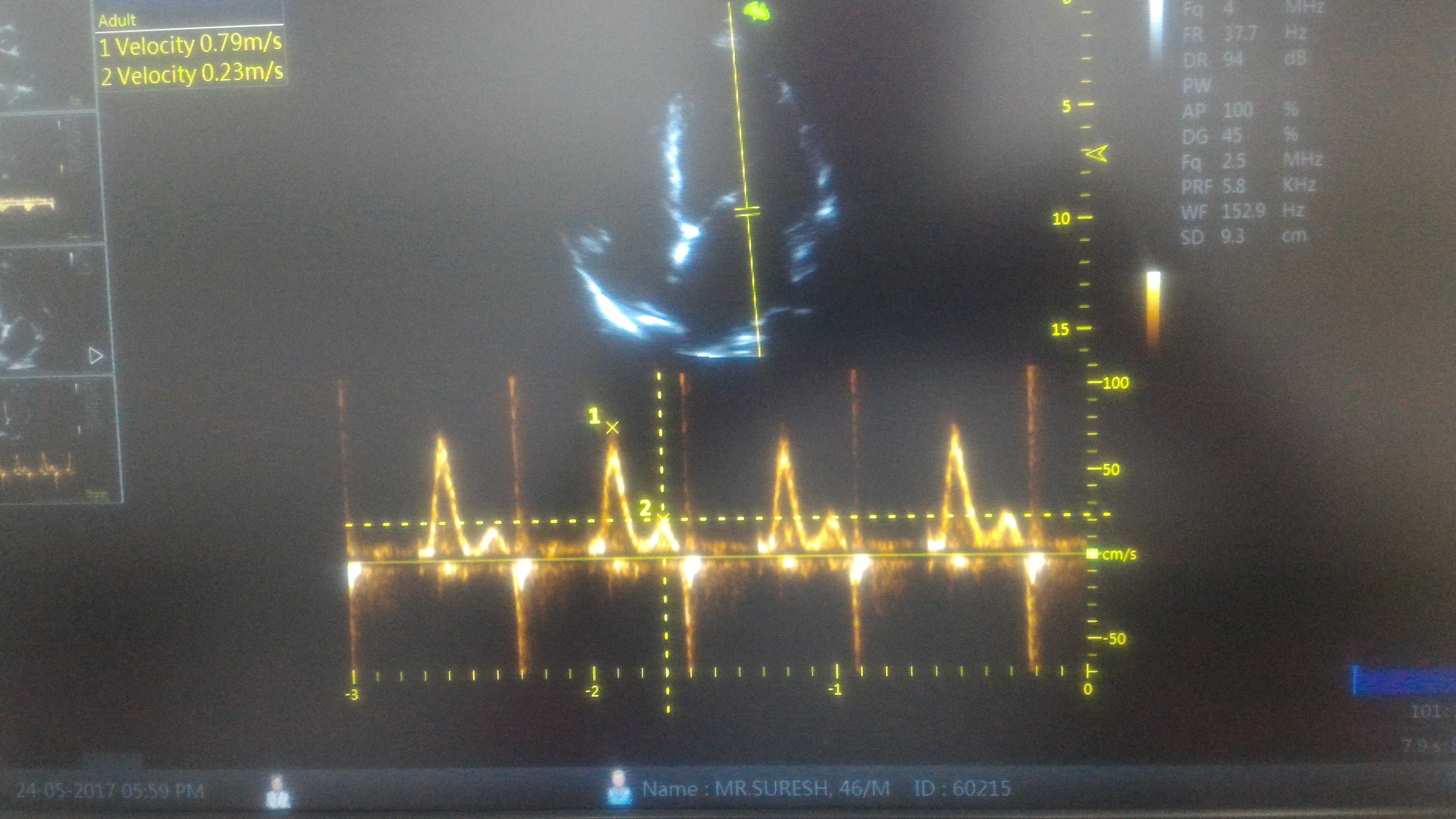
Note E:A ratio is 3:1 .This simply means the early (and mid to a certain extent ) diastolic pressure in LA is high and most of the filling takes place before Atrial contraction .There is one more reason for diminutive A . Atrial contractility fails to prevail over E in late diastole as LV end diastolic pressure is significantly high in these patients with diastolic dysfunction.

A dilated left atrium is an Independent marker of significant LV diastolic dysfunction (In the absence of MR) .When does LA begin to enlarge in diastolic dysfunction ? There is uniform rule.Generally LA size more than 4.5cm indicate grade 3 or 4 LV diastolic dysfunction.

LA size and Pulmonary congestion 

It’s a paradox , a roomy  LA dampens the LA pressure curve and A reversal into lungs may not happen.

*AF irony on A reversal

Logic might suggest , loss of atrial contraction might attenuate A reversal and less blood flooding into pulmonary veins.No, It doesn’t happen that way.If  AF is precipitated for any reason its going to be “switch on”  for acute pulmonary edema.

What is the relation between systolic and diastolic dysfunction in DCM ?

We find about 30 % of DCM has documented resting diastolic dysfunction.This is actually a underestimation of true diastolic dysfunction as it can very well manifest only during exertion.

Though generally , there is good correlation of grade of diastolic and systolic dysfunction in terms of severity , some of the patients show severe diastolic dysfunction out of proportion with systolic dysfunction.

Note : In the above patient it’s actually a fairly preserved systolic function but still has advanced diastolic dysfunction.

Grading of diastolic dysfunction .Image courtesy MM Redfield et al: JAMA 289:194, 2003. Note E:A >1.5 is

Final message

Relying on E:A ratio to diagnose diastolic dysfunction  may appear  amateurish for some of us .The rampant reporting of E>A for grade 1 diastolic dysfunction has made this parameter a “Doppler cliché”. But , the fact of the matter is,  it does help us confirm severe (Grade 4) diastolic dysfunction when E stands  tall and towering over an almost dwarfed A.

Clinical Implication

Please realise ,In patients with DCM  when you find an  A that is too diminutive in combination with  a menacingly tall E , it may be prudent to raise diuretic dosage. It’s a sure signal for impending pulmonary edema.

Queued queries 

Can DT and IVRT normalise with progressive diastolic dysfunction ?

(more…)

Read Full Post »

Technology is a great equalizer.Never in my dreams, I would  have thought as I drive through the dense Nilgiris forest , a satellite  located 36000 Km up in the sky would guide  me through  every turn and bend most accurately.

The curvy roads are coded with  live traffic  flow in Red ,orange & green . That’s “Google map”  for you. (By the way, proud to note Google runs with an Indian CEO who hails from my city Chennai !)

Now , coming to academics , . .Some one thought,  if the traffic in the entire globe can be monitored with few clicks,  How about  adding live traffic data to the otherwise  dumb anatomical coronary angiogram images we get in a non Invasive  CT scan ?  We can even color code the different segments of coronary artery based on the velocity profile and pressure drop. That is CT- FFR . Now technology is  available to get online live FFR as well. (Siemens )

Live coronary traffic blood flow 

Fractional_Flow_Reserve_Coronary_CTA (1)

Heart flow the newest technology in coronary Imaging and  non invasive Quantitative assessment is possible .It provides direct information about how to navigate the coronaries and intervene only the reddish areas  leaving the greens untouched.

Principle

Its called computational fluid dynamics .A super computer calculates live FFR for the entire segment by measuring the drop in  CT density data in Hounsfield units and translates into pressure equivalents and hence non invasive FFR.This modality has been approved by FDA.The heart -flow and Siemens has come out with onsite CT FFR.

Reality check :Have we conquered the coronary physiology ?

Trying to  understand coronary flow with a engineering mind-set is Insulting the complexities of biology. Be reminded , Invasive FFR is assumed as a gold standard, Inspite of the fact that , its blessed with flaws in concept , techniques ,(Hyperemia vs no hyperimia) and lesional variation . Now ,what is the big deal , a non invasive CT -FFR  is compared Impure gold standard  and claiming a breakthrough ?

Of course,logic would suggest,if both FFRs are flawed why not use  a less invasive one that is CT -FFR. It can atleast save time, cost, and potential procedure related issues.May be ideal in ACS situations were catheter FFR can destabilise the patient.Further, it can provide  continuous live information in a hybrid lab , hence post procedure FFR is readily assessed . (Converting Red coronary into Green ones  would become cardiologists new moto!)

Final message

The point of contention for the modern day  cardiologist is ,they have realized (Not all ofcourse !) in a harsh way that , they must use a physiological confirmation of a lesion severity before indulging on fixing it with a metal. Whether CT-FFR will increase the number of angioplasties  or reduce will remain a mystery . Whatever it does , it should do it for appropriate reasons . We know any technology has a shelf life and If MRI can provide the MRI-FFR (Journal of Cardiovascular Magnetic Resonance January 2014, 16:O55)  , CT will be pushed back for obvious reasons (Prohibitive radiation hazard)

Reference

Status of MRI based FFR 

Read Full Post »

Brugada syndrome is  as an  Inherited sodium Ion channel defect leading to loss of /or reduced sodium channel function.This specifically causes RV epicardial Imbalance of In-flowing(depolarising)  and out-flowing (repolarising)current , potentially triggering ventricular arrhythmia. This happens either spontaneously or during electrical stress times which include, fever, various drugs , adverse autonomic fluxes etc. So far,  we have been thinking it as primary electrical disorder with no macroscopic/ histopathologic  defects.

Newer Insights are emerging

But, how is this primary electrical disease , harbor a well demarcated  RV epicardial phenotypic substrate ? .  . . ablation of which eliminates the VT.

Zone of probable structural defect over RV epicardium (Pink zone) amplified by infusion of Ajmaline. Note the ECG showing typical ST elvation lead V1 to V3 .(Image courtesy Carlo Pappone et all  )

A recent study from Italy from the original founders  (Brugada team Ref 1 ) has confirmed RF ablation of RV epicardial tissue  is indeed feasible in many and should be considered in high risk Brugada syndrome. (Then should we suggest , ICD is no longer a choice in Brugada ?)

MRI findings in Brugada has shown some structural defects .(Ref 3,5) .It seems  Brugada is an Inherited electrical cardiomyopathy with a structural defect. (The overlap between ARVD and Brugada syndrome appear more real than we thought before ! (Ref 7 )

Final message

Still , Brugada is more of a electrical disorder,  but soon we may refer it as structural heart disease.

Reference

4.Catalano O,Antonaci S,Moro G,Magnetic resonance investigations in Brugada syndrome reveal unexpectedly high rate of structural abnormalities. Eur Heart J. 2009;30:22412248Abstract/FREE Full Text/Google Scholar
A best review comparing Brugada vs ARVC

Read Full Post »

An event that happened recently  that shook my country’s  collective conscience .It was, loss of  hugely popular and beloved  President of India , Dr Abdul Kalam on 27-07-2015. He was 84.Death came in a most dramatic way when he fell down midway  during his lecture to students of Indian institute of management ,Shillong in the state of Megalaya.

image

Indian President Kalam addressing the students snapped moments before he dropped down dead due to cardiac arrest

What is the implication of this VVIP’s death for  cardiac Academic ?

I believe , there is lot .The presumed cause of death was cardiac arrest . As we know , it must have been an instant electrical death as the local medical personnel  couldn’t  revive him after an Initial  CPR and later shifting him to state of the art facility . The ex-president was known to have a good health record and the heart should have been normal until prior to the cardiac arrest.

Now coming to the key question what is the chances of survival of cardiac  arrest ?

While there have been many survivors  of cardiac arrest within hospital premises  and coronary care units . . . still ,  life cannot be  guaranteed even if prompt CPR is initiated .

It’s the height of  Heisenberg  irony,  some lives can be saved  even when cardiac arrest happens out of hospital , while it’s also a fact deaths due to cardiac arrest happen right inside the cath lab where all emergency strategies are in place .

How much delay is permissible in resuscitating cardiac arrest ?

Cardiac arrest is nothing but activation of  the switch of death . Evey second is important . Experience suggest if reversed within first 2 minutes maximum survival is expected .Beyond 5 minutes and within 10-15 minutes most deaths will ensue.Up to 20 minutes survival is possible though with a risk of brain permanent brain damage.

A  recent study  from Sweden which addressed this  issueand  confirmed a  dismal fact  that even in the presence of best emergency care system,  survival is meager  4 -8% . However , the positive outcome was , If the bystander does some form of CPR till the arrival of  emergency service reaches the spot it can go up to 10.5 %.

cpr cardiac arrest

It is very clear , surviving an unexpected cardiac arrest is in the domain  of  God , still  we must encourage lay persons  to know and learn the techniques of CPR. The protocols has been simplified now , to include chest compression alone as the Initial measure.

Coming back to the question of death of our president,  as some one asked me direct question if the death could have been prevented ? , I said , yes if God willing !

Post-ample

Note : Cardiac arrest in patients who are at high risk with underlying heart disease (structural or genetic ) will require implantable cardiovertor  defibrillator ( ICD ) that can save thousands of life every day across the globe .


Read Full Post »