Feeds:
Posts
Comments

Archive for August, 2014

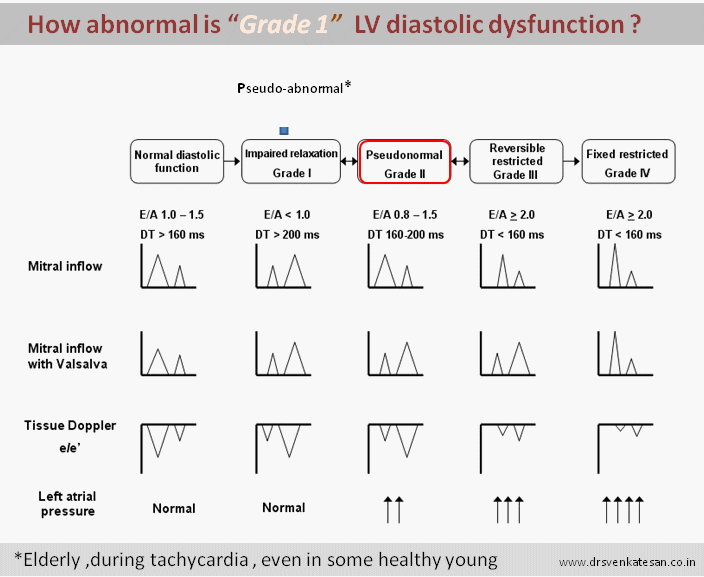
The correct  answer could be any of the above , depending  upon the level of your knowledge.

Ever since Herrick reported coronary thrombosis as a cause for MI and Davies documented it by angiogram many decades later (1980) ,the fate of thrombus  and the mechanism of its dissolution is the key to our understanding of ACS.

Even though we are now able to take on this thrombus in a direct fight  by aspiration techniques ,still the hematological  aftermath  and the aberrant coronary behavior  can fool us at any time ! The major lesson learnt  in recent times  is the  success of pPCI  is not in clearing the thrombus but ensure it never accumulates again  at the site  in the future .This is why there is whole big industry working on post PCI anti coagulation and anti platelet strategies .

Clinical correlates of poor  perfusion in micro circulation.

Plugging of micro circulation is the most under-recognised  issue.This results in no reflow in acute fashion or LV  dysfunction and micro-vascular angina in long term . Late recovery of LV function is attributed to late clearance of thrombotic debri.

RCA vs LCA thrombus load.

*One interesting observation is RCA thrombus clears more slowly as it has no well formed venous circuits .most RCA blood drains through thebesian veins which traverses  RV  myocardium .this can be hemodynamic hurdle unlike the LCA venous drainage

Read Full Post »

As the medical care advances  human care  has taken the back seat. It is said super specialists  read more and more about less and less ! In the  process they  fail to see the  patients  as a single biological unit  instead as collection of organs .

While  organs in turn are looked as  pile of data.Hence the  treatment they provide lack the soul !

In the prevailing circumstances , how do we ensure  modern medicine  does not interfere with  these vulnerable souls,either to live in peace or leave in peace ?

Medical Ethics

Image : Source and Courtesy of http://illuminationstudios.com

It appears doctors are not at fault . The system is  biased towards raw science .Highly trained  doctors are tied down by  both  true and pseudo  scientific Intellect .Often  times they are compelled to do some procedure or interventions  just to  justify  the  premier status of the hospital  .While few do it  to show off  their expertise or  to impress  their peers   others are simply bound by rigid and obsessive  protocols and guidelines . Few others do it  for the burning  desire  of  scientific accomplishment .

One can offer hundred reasons for doing a procedure . . . but we always struggle to justify  with a valid reason for not doing a investigation or  procedure !

In fact , the  concept of appropriateness  criteria came out with good intention .But , it had failed miserably.

The irony is  . . . we need to indulge in something to avoid something.

Example 1 If homocystiene and  hsCRP vanish from the CAD screening industry   Adiponectin and Vitamin D3 comes in with a thunderous applause like a new Hollywood movie  !

Example 2: In cath lab  for leaving alone an insignificant  coronary stenosis , we have to do  another procedure  called FFR to satisfy  scientific ego ! (I know one senior doctor , who left a 80% LAD  lesion for medical management without FFR ( with all his clinical acumen )  was ridiculed for being unscientific !)

Here is a recent perspective article NEJM has discussed  this  important issue that plague us

Why should big  Tertiary  teaching hospital  are  flooded  with super specialists  which by default shun basic human care ?

Read this article*

Super specialist tertiary care hospital NEJM

*The article I have quoted  may  not  be completely relevant here  . . . It  answers  few of the queries raised!

Read Full Post »

The diastolic mitral filling pattern has been  named and  graded  umpteen times in the last  decade. We believe it has  reached some semblance of clarity.I beg to differ.

pseudo abnormal relaxation grade 1 003

Image template taken  from  http://www.learntheheart.com

There need to be one more  grade between Grade 1 and grade 2 .Grade 1  is defined as A velocity > E velocity . This is the  commonest abnormal pattern and is often  man made.We can’t help it . We have to report it  anyway. Significant number of elderly show this pattern  without any pathology. It simply represents augmented atrial contribution  at times of apparent ventricular stress .

I wish a good chunk  of  grade 1  pattern ,  especially  in elderly or during tachycardia should be labelled  as physiological  grade 1 pattern  (or simply as  normal variant ) . However I would prefer it to be named as  pseudo abnormal pattern* !

* In my experience , currently medicine is taught in a complex manner .Facts that are told  in simple terms are rejected  straightaway . It would seem,too much clarity is not good for  science So,let us get confused one more  time  for the sake of our patients !

Read Full Post »

Coronary artery disease (CAD)  is man-kind’s  greatest threat in modern times.CAD ,diabetes ,Hypertension, obesity, mental illness  has become an epidemic  even among the young !


Lifestyle diseases cad risk smoking alcohol

There is a simple solution for  lifestyle diseases !

Just  . . .  Remove style from your life !

lifestyle diseases coroanry cardiology medical ethics inappropriate stents over treatment excess medical care , bio ethics,

Instead . . . try to live like these  Tibetian villagers

life purpose of living

Final message

One study which researched all lives who crossed 100 Years  concluded something like this !

“To live a longer and healthy life* ,Get up early  , have a purposeful daily chore that must include a physical component , work with conscience ,love every one sync with the nature and  lastly and most importantly remove style from your life !

Choose  your life . . . It is simply there in your hand for grabs !

Post-amble.

* Please note , Doctors  are never listed in the top with relevance to health of mankind  ! They simply cure some illness !

Read Full Post »

Answer  is question is wrong : RAA clot do occur in AF and severe right heart failure.It is less often recognised , since echo views are difficult and clinical events are silent.

RAA right atrial appendage clot tee echocardiographyBrief account of RAA clot formation

  • RAA is broad flat ,thin ,  chamber comparable to elephant’s ear.The ostium is not that distinct as the body as it  blends  with crista  terminalis .
  • Rough pectinate muscles  should make it prone for thrombus.Further , RAA has more sluggish flow than LAA  increasing the propensity for thrombus.However , the flat nature of the chamber , absence of tortuous tracts , constant  SVC flow which is abutting the  RAA can counteract this.
  • RAA clots are  less recognised as echo views are difficult .TEE is often required.
  • Overall RAA clot is 50% less common than LAA.
  • RAA clot should be specifically looked  for  in chronic AF and any severe right heart failure. (Unlike MR jet TR jet has less efficiency in flushing the  Right atrium )
  • Finally,clinical events from RAA clot are less conspicuous as the emboli reaches the pulmonary  bed silently.Unlike its colleague on the left side it  neither triggers TIA nor a stroke !

Reference

right atrial appendage clot raa clot in af atrial fibrillation

1. Buğan B, Baysan O, Demirkol S, Güngör M, Yokuşoğlu M. Right atrial appendage thrombus in a heart failure patient with sinus rhythm. Gulhane Med J. 2011; 53(3): 214-215.


2.Subramaniam B, Riley MF, Panzica PJ, Manning WJ. Transesophageal echocardiographic assessment of right atrial appendage anatomy and function: comparison with the left atrial appendage and implications for local thrombus formation. J Am Soc Echocardiogr.; 2006; 19(4):429-33.

3.Sahin T, Ural D, Kilic T, Bildirici U, Kozdag G, Agacdiken A, Ural E. Right atrial appendage function in different etiologies of permanent atrial fibrillation: a transesophageal echocardiography and tissue Doppler imaging study. Echocardiography;2010; 27(4):384-93

4 .Ozer O, Sari I, Davutoglu V. Right atrial appendage: forgotten part of the heart in atrial fibrillation. Clin Appl Thromb Hemost; 2010; 16(2): 218-20


Read Full Post »