Feeds:
Posts
Comments

Archive for May, 2020

The mainstream (& the sidestream) medical media have already named one culprit ie hypertension (HT) as a significant comorbid entity in the current Corona pandemic. But, If we ask one direct question, how Corona and HT are linked ? , the answer is not forthcoming fluently.

.

Some of the thought process about the Issue.

1. Does Coronavirus relish high blood pressure on its journey to attack lungs ?

Funny question you may think. The virus primarily lives and tracts through the respiratory tract to reach lungs. Viral load in the bloodstream is nil or minuscule till late stages. BP is nothing to do with what is happening in pulmonocytes . BP doesn’t influence the viremia in any significant way.

2. Does HT related pulmonary endothelial dysfunction facilitate and accelerate viral injury?

We don’t know. If at all,  this is true,  we have to worry about pulmonary hypertension (PH) and not SHT . PAH and SHT have absolutely no relation as far as I know.

3. Is concomitant anti-HT drugs cause relative hypotension and cause morbidity?

Maybe. We don’t know. Hypotension should be avoided at all costs.

4. Is there a conflict between Antihypertemive agents and Corona?  

We don’t know.(Much has been written about the ACEI genetic susceptibility Here is a current reference South, A.M., Tomlinson, L., Edmonston, D. et al. Controversies of renin–angiotensin system inhibition during the COVID-19 pandemic. Nat Rev Nephrol 16, 305–307 (2020).)

5. Does Corona precipitate  LV dysfunction in patients with corona infection?

Possible. Very rare (We haven’t seen much of uncontrollable HT causing afterload mismatch in corona setting as of now)

6. Do silent or manifest Coronary artery disease (CAD) and diabetes (The close buddy of HT) add up the risk?

Yes.definitely is possible. (No doubt, DM confers definite additional risk in Corona) 

7. Is preexisting HT cause CKD and argument the morbidity?

Yes. This happens only if HT has damaged the target organs sufficiently. Here the outcome of Corona infection might have an aggressive course.

Final message

So what exactly is the relationship between HT and Corona?

(Actually, this question was asked not in a cardiology academic meet. It was a casual query from one of my studious patients. I think nowadays we learn cardiology more from them than textbooks)

I guess, there is no direct, meaningful link in most people who just have isolated  HT without any target organ damage. It is more of perceived risk, and views are speculative and conjectural (Including what is written here) Corona affects the elderly and HT is also common in the elderly (30% of all elders) that’s it. 

The estimated 300 million people who are labeled as hypertension around the globe need not panic. Just because you are having high BP, corona doesn’t have any special attraction to you.   

Read Full Post »

We know, LV dysfunction of any etiology can cause VPDs.This must be differentiated from VPD induced LV dysfunction. Mind you, this is not an easy job at all.

When do you suspect excess VPDs are the cause (or might cause) of LV dysfunction?

  • In young, otherwise healthy persons with “VPDs and LV dysfunction”  would suggest chronic abnormal electrical activity is the cause for subsequent LV dysfunction. (An expression of electromechanical remodeling)
  • Monomorphic VPDs more often suggest primary electrical pathology(Like OTVT, Fascicular VT)
  • VPD count more than >10000 in 24 hrs in Holter will probably Indicate an electrical defect that requires  Intervention.
  • VPD burden (>10% and usually >20%),
  • Frequent NSVT,
  • Retrograde P-wave after the PVCs,(Chronic AV desynchrony)
  • Lastly one may argue its myth as well. VVI (& mode switched DDD) is a perfect example of how our heart tackles undesired VPDS. (We see hearts living comfortably with only with VPDs from the right ventricle (Pacemaker rhythm) without any troubling LVD.

Ref 

1.Baman TS, Lange DC, Ilg KJ, et al. Relationship between burden of premature ventricular complexes and left ventricular function. Heart
Rhythm. 2010;7:865–9.
2.Ban JE, Park HC, Park JS, et al. Electrocardiographic and electrophysiological characteristics of premature ventricular complexes associated with left ventricular dysfunction in patients without structural heart disease. Europace. 2013;15:735–41

When do you suspect VPDs are because of LV dysfunction?

  • History , clinical examination is the key. If there is a known cause for myocardial pathology.

  • Here, the VPDs are more often multifocal (Rarely monomorphic)
  • QRS complex other than VPDs may show slurring or fractured abnormality 
  • More severe forms of LV dysfunction  
  • Severely scarred ventricle obviously would Indicate primary structural disease that causes VPDs

Clinical implication

  • If VPDs are the cause of LV dysfunction we may try to suppress or abolish it.
  • No point in ablating otherwise asymptomatic VPDs in cardiomyopathy. Here we have to identify the cause for LV dysfunction(CAD, Myopathy etc) 
  • Beta-blockers can be useful in both subsets.
  • The over-enthusiasm of ablating all forms of VPDs in any structurally abnormal heart is to be restricted. (Of course, Indication for RF ablation/ ICD may be appropriate in malignant forms )
  • The beenifits of CRT therapy can be negated with frequent VPDs
  • The relationship between the risk of SCD with the number of VPDs,  is never found to be linear. (We have learnt in a hard way, that It is the degree of LV dysfunction that writes the script for SCD in a given patient)

What is the mechanism of VPD induced LV dysfunction?

VPDs alter the way ventricle contracts by inducing wall motion defect. In fact, it is intermittent cardiac desynchrony. Generally, LV tolerates this well. When the number exceeds a critical level LV size, shape and contractility is affected.

We need to differentiate chronic tachycardia mediated LV dysfunction (Tachycardic cardiomyopathy) with VPDs per se. This differentiation we can only guess. 

* Some how cardiologists have not,  implicated RV dysfunction induced VPDS. My guess is, it is equally important. Logically,at least 30% of VPD in end-stage DCM must be attributable( and arise from) RV dysfunction.

How to confirm is it a cause or bcoz ?

The only way is to prove LV function improves to normal or near-normal with ablation of VPDs .This can happen only with primary electrical disorders.

Final message 

The link between VPDs and LV dysfunction is stuck in a complex two-way affair. Though a cause and effect component might be quantified to a certain extent, both can be coupled sequentially . “VPD promoting  LVD” &  “LVD begetting VPD”  is always a possibility. This is the reason, we are tempted to take on any VPDs,  which of course, is definitely not warranted.

  Two resources 

The HRS guidelines must-read for all fellows


This is a real state of the art lecture on VPDs straight from a world-renowned Dr Gacia  Courtesy : Methodist, Houston 

Read Full Post »

Surprised to see many of my colleagues, physicians and fellows are beaming with new pride even in this troubled corona times. Paradoxically, could see some fresh clinical sense in their approach to problems as well.

Each one of them has a story to tell

  • Sir, I could suddenly diagnose heart failure for the first time with my eyes and ears without NT- Pro BNP or E/E’ . I agree with you sir, textbooks seem to be right. There was indeed basal rales and JVP was elevated. I was astonished I could diagnose CHF clinically!
  • I feel proud, that I have acquired the rare expertise of giving fitness to an emergency appendectomy just by ECG.Its unbelievable, I had the courage of not asking for a pre-op echocardiogram.
  • Oh yes, it was a real flash of bedside brilliance. I could rule out Infective endocarditis, in a patient with prolonged fever, without caring to call for a bed side screening echo for vegetation. I am really proud of my acumen! I realised, Duke criteria is far more deep than our urge to have a glimpse on vegetation.
  • I can’t believe myself, yesterday, I was able to Ignore a 90% LAD lesion, first time in my life, by clinical means without FFR and QFR stuff.
  • This one is again from the echo lab. I refered a patient with aortic stenosis, for AVR, without bothering about all those low flow and high gradient conundrum. I was sure it was severe AS. The dense calcium and LVH were good enough to tell the complete story.

Finally, one of my senior colleagues, who lives half his awake time in cath lab, confessed to me. “Yes,Venkat, it’s all happening right in front of my eyes. Miracles based on absolute truths . I have since learnt the ultimate lesson in cardiology. How to treat, many of my CAD patients, without knowing coronary anatomy, that too without any major adversaries”

Postamble

After listening to these sobering stories , I got into a mid-afternoon nap, wherein, my good old professor came in my dream. He blessed me with his famous smile and hug for practicing and propagating clinical cardiology, as he taught to me.

But sir, I blinked, sorry sir, I don’t deserve your compliments. This newfound sense is going to vanish along with this dream. Blame it on the terrible corona times, which has forced us to deliver this low-quality care based on the antique clinical methods.

My mentor’s happiness was short-lived, as he realised these guys will soon be consumed again, by the glamor machines that runs medical science. He left silently, still pleading us to try our best.

Read Full Post »

This post was originally written in 2013.

A middle-aged man with STEMI  came to our CCU.  It is just another case of STEMI and asked my fellow to lyse.

Anterior STEM ecg

But it was not the case . He, told me, Sir, the patient had a syncope following chest pain and he has injured his face and Jaw. He was actively bleeding. When I saw this face, it was indeed  frightening.Strptokinase induced bleeding

What shall we do ? When a patient  with STEMI presents with bleeding facial Injury

  1. Rush for Immediate PCI (Which was  of course not possible in our place as it happened out of office hours! )
  2. Take that ultimate risk and thrombolysis
  3. Give only heparin ( Many times it is as good as  lysis )

We took a (bold ? ) decision to thrombolyse with streptokinase.(After  a CT scan which ruled out any Intracranial bleed like hematoma etc) Clopidogrel was also given.

absolute contrindication for thrombolysis facial trauma

Patient continued to bleed in the initial 3 hours and was oozing in the next 12 hours. Blood transfusion was contemplated, but it was not required. Dental surgeon opinion was sought, his teeth were pulled and a compressive bandage was applied.It arrested the bleeding.The ECG settled down.LV function was almost normal with minimal wall motion defect. He is posted for a coronary angiogram later.

Final message

 There may not be anything called “Absolute contraindication” everything appears relative

I presented this in the weekly clinical meet,  with a tag line of  How to save a patient, apparently by violating a standard guideline. Not surprisingly, It evoked laughter amusement from learned physicians. I wasn’t. Guidelines are meant to guide us agreed.They can not command us. They are not legally binding documents as well! Many lives can be saved if only we have the courage to overrule when it’s required.

Afterthought

Had this patient has bled to death during lysis what would have happened to the treating doctor? (or )If the patient has died due to MI, because of deferred thrombolysis, what would be the line of argument?

2020 update.

This case scenario is a non-issue as of today. With so much experience, we straight away do PCI . Just manage the oral bleeding if any.


Read Full Post »

What is the incidence of Isolated systolic pulmonary arterial hypertension (ISPAH) and its Implication? We attempted to answer this question and found some interesting answers. It was published in the Indian heart journal  December 2007 Abstract issue. More than a decade gone. I think this issue is still largely misunderstood. Fellows may pursue this. One more parameter that can be explored is pulmonary artery pulse pressure and effect on progressive pulmonary vascular disease and PVR. Mean while  PAH definition and classification has changed many times, ISPAH definitely requires a place in the new scheme of things.

The abstract

ISOLATED SYSTOLIC PULMONARY ARTERIAL HYPERTENSION
S.Venkatesan ,G.Gnanavelu,V.Jaganathan , Madras Medical College. Chennai

Pulmonary circulation is a classical example of a low-pressure low impedance circulation. It is generally presumed high output states generally do not increase the systolic blood pressure in the pulmonary circulation. In systemic circulation, there can be divergence of systolic and diastolic blood pressure depending upon the cardiac output and peripheral vascular resistance. This has resulted in separate clinical entity -Isolated systolic hypertension.(ISH). It has been our observation many of the patients with PAH during echocardiographic and cath study were found to have an elevation of systolic pulmonary artery pressure(PAP) with normal diastolic PAP . In this context, this study was undertaken to specifically identify whether there is an entity of Isolated systolic PAH
( ISPAH ) and it’s the incidence in various clinical situations.
We analyzed the echocardiographic data of patients who were referred to our echo lab retrospectively. A total of 4000 echocardiograms over a period of 6 months were reviewed. Majority of these patients were referred for routine screening echo from our OPD. Data from patients who were assessed to have PAH were thoroughly scrutinised. They constituted shunt lesions, RHD,PPH, COPD, pregnancy, and patients with unexplained dyspnea for evaluation Those Patients who had both TR and PR jet were only considered for analysis .The Systolic PAP was estimated with TR jet and diastolic PAP with End diastolic PR Jet. ISPAH was diagnosed when the calculated systolic PAP was more than 30mmhg. And the diastolic PAP was less than 16mmhg  Antenatal women formed 2 % of the study population. A total 72 patients fulfilled the criteria of ISPAH Among the shunt lesions it was most common in large VSD( 4/10), followed by ASD(14/35) and PDA( 1/3) . In patients with RHD it was observed in 12%(15/110) , COPD 10%(15/150), in pregnancy and general population it was 5%(23/450). None of the patient with PPH had ISPAH.The mean Systolic PAP was 38mmhg(R 32- 74) The mean diastolic PAP was 14mmhg(R 8-15).The highest systolic PAP was 74mmhg recorded in patient with large VSD.
It is often presumed hyperkinetic states elevate systolic PAP and reactive elevates diastolic PAP .But it is clear from our study the rule is not that simple. Surprisingly many of the RHD patients had only the systolic PAP raised.It is important to recognize systolic PAP was very high in some of the shunt lesions. Taking this alone as an index of severe PAH is fraught with the risk of declining corrective surgeries in these patients.
Perhaps the most important observation from the study is the incidence of PAH in apparently healthy individuals, which is very significant as it could be the marker of continuously increasing chronic lung disorders due to the worsening environment of the 21st century.


A PowerPoint presentation of the paper is available with the author and may be requested.

Read Full Post »

I think it is an Invalid question. Whether you like it or not , medical science and philosophy are always bonded together and its relationship is eternal. It doesn’t make sense to separate them. I think we have misunderstood the meaning of philosophy. While science is presumed truths, philosophy is trying to believe in unknown truths. Philosophical truths are built-into every decision a medical professional takes.

If the expected natural history of any disease is science, unexpected deviations are philosophy. (RT PCR testing for diagnosing  Corona is science, why 90% of them are not infective and don’t transform disease is philosophy) When something is not seen or quantifiable like human immunity, it is a perfect example of concealed science or manifest philosophy.

Taking about what we think we know is science, Talking about what we really don’t know is philosophy. The term Idiopathic syndrome finds a  proud of the place in every specialty in medicine, Isn’t? 

 What will be your answer when your patient wants an assurance that a stent, you had just implanted will not get occluded in the next 6 months or so.“I don’t know, I cant assure you about that”  will be your most likely answer. (Though, we do it in style, hiding behind  the scientific hyperbole decorated with numbers,  also referred to as statistics) Please realize, this is the expression of medical philosophy in the finest form.

Final message 

My Impression is, philosophical truths should be liberally used in a regular fashion right from the first-year medical school to advanced specialty teaching. This seems essential as science in the current times suffers from too much sanctity. This has spilled over to the doctor population as well, and make them appear invincible. 

If only we realize science often trails behind the philosophical truths at least by a few decades, our patients will not be injured inappropriately and prematurely. Mixing science with philosophy in the right composition ( a perfect academic cocktail ) will bring out the best from the noble profession.   

Postamble

Can anyone guess, why scientists are given a doctorate in Philosophy degree  (PhD ) ?

Read Full Post »

Fortunately, indications for DC cardioversion in pregnancy is rare. A literature search suggests only about 50 cases are reported. I haven’t shocked electively in pregnancy but occasionally have come closer to it. In this current corona lockdown period, we had a call for a potential shock in pregnant mother with fast AF, which was again avoided by the optional  rate control measures.

Let us see, how often DC cardioversion might be necessary during pregnancy and few tips for its safety.

General principles

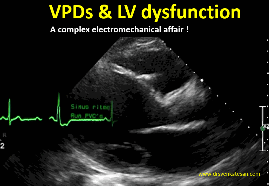
We know pregnancy can be pro arrhythmogenic. Most arrhythmias are non-sustained VPDs and APDs.They can be ignored if there is no structural heart disease, or at least postponed till delivery.

Drugs remain the mainstay.

The most common sustained arrhythmia in the young reproductive age group is SVT (AVNRT/AVRT) that can be managed with drugs like  Adenosine and beta-blockers. (Flecainide/Sotalol are found to be safe in pregnancy) . Though IV Verapamil is very effective, it has with some concern for fetus, so better avoided. Please, note many of the SVTs can be reverted with simple vagal maneuvres and oral beta-blocker /Verapamil. IV Digoxin has been widely used in RHD population for AF during pregnancy.

Mind you, even Injections Adenosine, Esmolol do have bradycardic potential and need to be given in monitored setting (No surprise, they are called medical cardioversion with the attendant risk) 

The universal antiarrhythmic drug Amiodarone still might come in handy in any refractory arrhythmia (Including AF) though it comes under the list of contraindication.(Safety of amiodarone in pregnancy) 

One important suggestion to make. Magnesium is a wonder antiarrhythmic drug, a membrane stabilizing agent through its indirect Ca + and K + blocking properties can be a powerful antiarrhythmic agent, especially in VT. This has a unique safety profile in pregnancy.We use it in eclampsia liberally  with the same action to suppress brain convulsions. (Cerebral tachycardia). Please consider IV magnesium  prior to considering  shock in VTs with dysfunctional ventricles as in peripartum cardiomyopathies et(Dose  to 2 g in 10mL of D5W over 1 to 2 minutes)

Consider DC shock only if there is hemodynamic instability.

Hemodynamic instability demands DC version. One practical issue is , what defines hemodynamic instability? In pregnancy, the systolic BP is already in lower normal due to systemic vasodilatory state. An HR>150 makes it further fall to around 90mmhg. This tempts us to label it as unstable. In this situation, we have to rely on patients’ symptoms to define hemodynamic instability. Never try to shock a comfortable pregnant women in whatever tachycardia she is in . (Including some VTs especially from outflow, fascicular, etc  ) Try to use drugs and get an expert opinion. to rule out subsets like cardiomyopathy, documented CAD, LV dysfunction.

When to shift to a cardiac facility?

This question crop up often. It is mainly logistic. May be in peripartum cardiomyopathy /Suspected ACS  with VT require special care.

 DC Shock checklist  & Precautions 

  • Biphasic shocks with energy levels 100 joules ( up to 200). Ideal to give single shock, ok to err on high energy  
  • Pads should be well away from the abdomen. 
  • Synchronized with QRS complex (Machine does this) 
  • In an unusual event of VF and cardiac arrest Defibrillation with 300/320 J (Here unsynchronised) 
  • Check the crash cart ready with essential drugs.
  • Keep cardiologist either on-call (Even a junior resident in labor room give immense confidence) 
  • Rule out  LV dysfunction or significant valve disease by echo (CAD can’t be ruled out though) If echo machine is not available ask the radiology or cardiology fellow to use the abdominal USG probe to document good LV contractility and gross EF% estimate.
  • If intramural thrombus is not convincingly excluded and there is AF and valvular heart disease, better to heparinse  and shock to avoid embolic events.
  • Temporary pacemaker support (Some of the cardioverters has transcutaneous pacing to tide over transient bradycardia that might occur post-shock) 
  • CPR readiness ( Extreme precaution !)
  • Fetal heart monitoring and Emergency cesarian readiness. 
  • Finally, most important consent with patient and family.

Is electrical Insulation of baby necessary or is it possible? 

 It’s not required. Fetus inherently tolerates stress better. Even if,  few joules reach the fetal heart inadvertently it may not mean much. What is, to be worried is maternal hypotension or bradycardia post-DC shock.

Impact on the fetus: Evidence?

The impact on fetal blood flow is not significant. This report from Taiwan  reassures there is no adverse effect by measuring umbilical artery flow (Yu-Chi Wang European Journal of Obstetrics & Gynecology and Reproductive Biology 126 (2006) 268–274)

While we consider DC shock during pregnancy is safe for the fetus, still, shock pads close to the abdomen, amniotic fluid being a good conductor of electricity at least one mother showed a sustained contraction of the uterus and fetal distress. This was possibly attributable to DC shock  Eleanor J. Barnes BJOG 2003 https://doi.org/10.1046/j.1471-0528.2002.02113.

Final message 

Most cardiac arrhythmias in pregnancy are carefully managed by non-electrical means. Of course, emergencies can’t afford to wait. Though two lives are at stake, it’s the mother’s heart that prevails over in drug selection and risk estimation. After all, it is her loving heart, that keeps the fetus alive.

I have seen Obstetrician anxiety (which spills over to attending cardiologist too!) can be extreme in such situations. I must admit, Obstetricians, are truly sincere warriors fighting at odd hours to protect the two delicate lives. After all, taking  responsibility brings the anxiety. Cardiologists must understand this and help them out in their difficult times.(without any super specialty ego !)

Reference

  1. Crijns HJ. Electrical cardioversion in healthy pregnant women: safe yes, but needed?. Neth Heart J. 2011;19(3):105‐106. doi:10.1007/s12471-011-0079-3


2.Finlay AY, Edmunds V. D.C. cardioversion in pregnancy. Br J Clin Pract. 1979;3:88–94

3.Oktay C, Kesapli M, Altekin E Wide-QRS complex tachycardia during pregnancy: treatment with cardioversion and review. Am J Emerg Med. 2002 Sep;20(5):492-3.

Which drugs are safe?

From BMJ 

Click to access pregnancy_heart_disease_v28_web.pdf


Further frontiers 

The DC shock from ICD experience

There have been a good number of women who got ICD for various indications (Commonly HOCM, long QT, ) who subsequently became pregnant, successfully managed during pregnancy.

(One rare study Andrea Natale et ll Circulation. 1997;96:2808–2812 documents at least 10 shock episodes documented in large series of 44 patients without any consequences)


Read Full Post »


A throbbing query …for so long

Got into this amazing lecture, that Infuses knowledge and wisdom about values in life, in just 14 minutes .Probably, It has more treaures than what we may get, in our entire life time as we search for truths.

I wonder, such thoughts can come only from God’s special messengers.Devdutt Pattanaik seems to be one. 


Read Full Post »

LA volume is one of the critical parameters that define (as well as determines the symptoms) both diastolic and systolic LV dysfunction.Still, we are tentative in the true estimates about the normal range of LA volume. (Upper limit 40ml/m²) .We are surprised to note, the difference between MRI derived and Echo measured LA volume showed a disturbing variation nearing 80 %.

How to measure  LA volume?

While the timing of LA volume measurement has not much controversy (end-systole), and the shape errors are largely eliminated by 3D echo, still why this variation?

It is a telltale error, of either including (or excluding ) the pulmonary vein ostium and complete blindness to LAA during LA border tracing. We know, LAA is physiologically, pathologically, and electrically is a critical accessory of LA. Still, we have so for excluded it from routine LA volume calculations. Is that a right-thinking in the overall evaluation of LA volume?

Normal LAA volume

Though the LAA volume is directly related to its size, shape much great confounder, since it precludes in arriving any mathematical calculation of volume from the area. Direct casts ofLAA and 3D echo to a certain extent will help measure LAA volume. MRI may also do the same.The normal LAA volume is calculated to be up to 20 ± 9 ml. Whatever be the  LAA  volume, one estimate suggests it will reach 25 % of total LA volume. This is very important to know. In fact, In significant LV dysfunction, LAA  is expected to stretch, efface, and dilate and contribute more to LA volume. Both static and dynamic LAA volume status also gives us an idea about potential thrombus formation risk.

(Ref : Measurement of Left Atrial Appendage Size by Transesophageal Echocardiography  Kazuko Yoshimoto et al. J Med Ultrason (2001). 2012 Jan.)

Are we justified to Ignore the LAA volume during routine LA function assesment ?

I don’t know. It may be wise to routinely add LAA volume to LA chamber volume to truly assess  1.Overall LA function, 2.Estimate the risk of thrombus formation and  3.Risk of developing AF. Meanwhile, its found LAA appendage volume might even approach that of  LA volume when it’s pathologically enlarged. (Left Atrial Appendage Volume as a New Predictor of Atrial Fibrillation Recurrence After Catheter AblationPedro Pinto Teixeira et al. J Interv Card Electrophysiol. 2017 August) 

LAA volume is Important for one more reason 

We are getting new data about dynamic LAA volume status during LAA closure.In fact , this particular study (JACC: Cardiovascular Interventions Volume 8, Issue 15, December 2015)  documented how LAA appendage balloons out during volume loading of LA.This study suggests we have to be careful about the hidden potential of LAA to expand and if ignored the device is likely to get dislodged with volume overloading. These observations make it clear we can’t isolate LAA volume when calculating LA volume. 

Final message

There is a strong case for measuring  LAA size & volume separately and preferably be added in the net LA volume Index. We can’t simply Ignore this vital and inherent part of LA , just because its called as an appendage. Of course, even a novice will rank LAA first,  as the pathological hot spot within the entire LA.

Reference

Everything about LAA

Giuseppe PattiVittorio PengoRossella Marcucci,The left atrial appendage: from embryology to prevention of thromboembolism  European Heart Journal, Volume 38, Issue 12, 21 March 2017, Pages 877–887, https://doi.org/10.1093/eurheartj/ehw159


Read Full Post »