Feeds:
Posts
Comments

Archive for the ‘cardiology- coronary care’ Category

Yes, it is a triple vessel disease, with one tight lesion and at least two other significant lesions. One of them appears diffuse as well. 

Representative Image: Source courtesy DOI: 10.14740/cr548w LicenseCC BY-NC 4.0

“What to do next?. Is he symptomatic?  Yes. Definitely has significant angina” but LV function is normal.

“Ok then. If you are daring enough, ask this question”.

Which lesion is causing angina?

No easy answer at all. Try looking for some clues right from history, ECG, stress ECHO, meticulous assessment of individual lesions. Realize, even sophisticated imaging like SPECT, PET functional MR, may not help much either.

Oftentimes, we need to use the lean resources of collective common sense and clinical acumen. 

  • If it is post ACS status,  consider residual ischemia in the culprit artery is the cause for angina.
  • Second, consider the tightest lesion as angina-related.
  • Or the complex, eccentric, thrombotic lesion is responsible.
  • Next, consider LAD as default lesion as  angina related artery (Statistically right 75%, prognostically perfect decision) 
  • Watch for ECG changes during chest pain (ST depression usually don’t localize, but experience tell us V5 /V6 ST depression is more likely to be LAD ischemia )
  • Echo wall motion defect either during rest or (more usefully) in stress can really help. (It needs some effort to look for Wall motion mapping with coronary lesion subtending segment)
  • What about balloon inflation test during PTCA ? . Prompt angina when a lesion is occluded may give a direct clue.

Want to get more confused?

  • Ask your colleagues for an opinion either online or offline.
  • Do FFR/QFR/IFR  and OCT and look for intracoronary pressure-flow data and plaque burden. We are entitled to get excited about fibrous cap thickness, and hunt for vulnerable lesions and decide thereupon.  

Finally some easy options. 

Which lesion is causing angina? Never entertain that troubling question at all. (Need not  squeeze your coronary intellect you know ) 

Consider every lesion as important 

  • Get ready to stent all three or more lesions.(Many times forbidden though !)
  • (or) More convenient, refer to CABG. (Surgeons will welcome for sure )

Final message

Which lesion is causing angina? is indeed an important query one should raise. This paves way for selective focussed PCI in deserving lesions alone. However, when dealing with complex lesions subsets. the most pragmatic way as of today is to educate the patient and include them in the decision-making process (Never forget to offer medical management as a permanent option, especially if there is no critical LAD disease, and say thanks to  ISCHEMIA/COURAGE/ BARI 2D.)

Read Full Post »

This post was originally written in 2013.

A middle-aged man with STEMI  came to our CCU.  It is just another case of STEMI and asked my fellow to lyse.

Anterior STEM ecg

But it was not the case . He, told me, Sir, the patient had a syncope following chest pain and he has injured his face and Jaw. He was actively bleeding. When I saw this face, it was indeed  frightening.Strptokinase induced bleeding

What shall we do ? When a patient  with STEMI presents with bleeding facial Injury

  1. Rush for Immediate PCI (Which was  of course not possible in our place as it happened out of office hours! )
  2. Take that ultimate risk and thrombolysis
  3. Give only heparin ( Many times it is as good as  lysis )

We took a (bold ? ) decision to thrombolyse with streptokinase.(After  a CT scan which ruled out any Intracranial bleed like hematoma etc) Clopidogrel was also given.

absolute contrindication for thrombolysis facial trauma

Patient continued to bleed in the initial 3 hours and was oozing in the next 12 hours. Blood transfusion was contemplated, but it was not required. Dental surgeon opinion was sought, his teeth were pulled and a compressive bandage was applied.It arrested the bleeding.The ECG settled down.LV function was almost normal with minimal wall motion defect. He is posted for a coronary angiogram later.

Final message

 There may not be anything called “Absolute contraindication” everything appears relative

I presented this in the weekly clinical meet,  with a tag line of  How to save a patient, apparently by violating a standard guideline. Not surprisingly, It evoked laughter amusement from learned physicians. I wasn’t. Guidelines are meant to guide us agreed.They can not command us. They are not legally binding documents as well! Many lives can be saved if only we have the courage to overrule when it’s required.

Afterthought

Had this patient has bled to death during lysis what would have happened to the treating doctor? (or )If the patient has died due to MI, because of deferred thrombolysis, what would be the line of argument?

2020 update.

This case scenario is a non-issue as of today. With so much experience, we straight away do PCI . Just manage the oral bleeding if any.


Read Full Post »


Professional competence is defined as doing things, always in the Interest of patients. It’s generally believed small hospitals are not competent enough to treat cardiac emergencies . . .Do you agree with that ? No, Its largely a myth . Do you know there is a absolute lack of proficiency threatening to plague our country’s coronary care system. ? It’s the professional Incompetence by the space age, star hospitals (mis)managed by masters of the noble business. None (am I right ?) of this hospitals either monitor or publish the outcome of their treatment.

Backed by pseudo scientific data , amplified by unrealistic expectations of ill Informed patients , some hospitals are avoiding Initial emergency treatment of acute MI , instead they waste time ( load DAPT ofcourse !) in securing the finance for the costly Invasive procedures or refer them out of their premises if they can’t afford for it.In the ensuing emotional and financial melee many of the ill-fated patients lose vital time window of thrombolysis as well ! and carry risk of fatality or damaged myocardium.

Every stake holder in the current coronary care system simply assume the enforced modality must be far superior because they administer the most modern and costly treatment suggested by few high intensity cared clinical trials originating from west. The wisemen who run the corporate hospitals never realise medical competence and outcome is not entirely defined by science. Their primitive cognition wouldn’t allow to think beyond business equations either.

Please believe me, time and again, I have witnessed patients reaching Government hospitals after being shunned away by big (Some times even medium sized ) hospitals who boast themself only as PCI enabled care. Even if they want to lyse they stock only the Tenekteplace .

I think tragedy is a lesser word to describe the scenario , where a distressed family is trying to arrange for a Rs30,000 shot of Tenekteplace when thirty times cheaper still equally efficacious (Rs 1000 Streptokinase) is concealed from their visibility .The Govt should urgently look into instances of large private hospitals avoiding Govt insurance scheme patients even in cardiac emergencies ! To label our poor patients as unaffordable ones is a outright misnomer, rather its the rich hospitals that are “not affordable” to lose profit and treat our countrymen , in a cost effective manner is the reality !

Who is Poor ? You decide.

Two forbidden things in coronary care

1.Cajoling and manoeuvring a distressed family for a primary PCI as a routine treatment hyping its beneficial effect and underplaying the true advantages of thrombolysis in largely technical jargons is the current norm in most coronary care units.

2.Another issue is , after confused confabulations with the duty medical officer, if a rare patient family choose the option of thrombolysis , comes the next googly*. Many noble minded hospitals do not stock the low-cost and equally efficacious thrombolytic agent and offering only the costly option to the anxious families when the myocardium is on fire.

Hospitals that practice these two coronary protocols need to be shamed and labeled as “Coronary Incompetent ” In spite of having 24/7 cath labs. (Realise , they are just like any remote rural hospitals , at least the later can’t be faulted as they don’t withhold a reperfusion strategy !)

Final message

I think , mindless proliferation of cath lab based cardiac care , which follow this theme , ie “Thrombolysis incapable but PCI capable “ are biggest threat to coronary care in our country ! For the best coronary care for any country ,what we need is efficient prehospital thrombolysis team .We have conveniently forgotten the great study of CAPTIM wherein the ambulance drivers replicated the same effect of primary PCI performed by highly trained cardiologists in modern labs.

In India, primary health centers which is within few km reach of entire population can be designated as static ambulance equivalents with basic resuscitation facility . If a multipurpose health worker can be trained to lyse, with remote supervision that will accomplish 90 % of what the cathlab guys can achieve ! Selective shifting is suffice.

Postamble : Ofcourse, not doing pPCI for high risk or complicated STEMI is unscientific and we need to have proper consenting and referring frame-work for such patients.

Counter point : One of my colleagues asked me ? Why do I enjoy attacking the established scientific practices ? May be I have a problem , yes, but I think in a true medical democracy we have right to debate anything , absolute truth is a ongoing journey !

*Googly: An unplayable ball delivered to a batsman in the game of cricket.

Read Full Post »

When a culprit thrombus keep the  myocardium as hostage . . . don’t storm the coronary artery  indiscriminately   !

When a single gun men  keeps 100 innocent people as hostages , threatening their  lives, rescue mission should start .No can can afford to wait. But, without knowing  the  culprit’s true nature the process of rescue mission is always going to be tricky .There are so many instances Newton’s third law  was reversed , when reactions  evoke more chaos  than the index action.

In the recent world terrorist events ,  the  rescue missions  were so delicate and  it was very  unfortunate we  lost  many   innocent hostages !  The reasoning is ,there  is no way we can avoid these. I wonder is it really true ? !

rescue missionNot all culprit lesions  are true ones.They simply threaten  our myocardium with  thrombus and plaques  in various forms .Don’t show aggression to pseudo threats  you may  ultimately end up with more damage.(What I call as crazy culprits!)

(  Read here , why unstable angina even though thrombus is sitting right inside the coronary artery attempting to lyse it causes more  damage !)

After thought

Iam sure ,bulk of  the Interventionists wouldn’t agree with this thought . They would decry , watching a person  silently when the myocardium  is on  fire is a serious crime !

But . . . we  need to  remember the process of extinguishing  the fire  with some more fire arms is a delicate game played in undefined  philosophical turf.

The only way to introspect  such events in life is , to accept any eventuality    arising out of “not pursuing”  a  presumed rescue mission with vigor. No need to be guilty about that,after all , it can be a myth !

Modern human cognition , growing with a staple  scientific  feed  on a 24/7  basis  is  unlikely to realise , restraint can be an effective tool  even in critical moments !

Oh,is all that I have  scribbled so far  is just a repetition  of 1000 year concept of  “Primum non nocere”

Read Full Post »

When a patient comes with angina at rest , it could mean two things .Either a  STEMI or an NSTEMI .This , we can diagnose only after seeing the ECG .

Can we differentiate these two by the  character of chest pain alone ?

Very  tough task isn’t  ? But there are some definite clues .

Infarct  pain

  • Is mostly sudden .
  • Likely to be crescendo , lasts more than 20-30 minutes .
  • Fails to get relived by rest or even  Nitrites.
  • Sweating due to sympathetic activation is more pronounced.

Unstable angina

  • Is rarely  sudden .Often has a pro-drome.
  • UA is  mostly precipitated by an increased demand situation or a stress.
  • It has  a typical waxing and waning  pattern . Rarely assume a true  crescendo character  as myocytes  does not necrose (Just threaten to die !)
  • The chest pain radiation   to  shoulder is less  conspicuous , instead it  tends to  reach  the  jaw area .(* An observation,Is it something to do with multi-vessel CAD in UA ?)

Mechanism of the difference : Epicardial vs Endocardial angina

The pain of UA is   due to subtotal occlusion and  endocardial ischemia , while STEMI is  sudden total occlusion  and the resultant  transmural  ischemia . In STEMI  epicardial  surface is always involved (Which lifts the ST segment in ECG .).We know epicardium  is same as  visceral layer of pericardium which is well innervated .Hence  pain  of STEMI   acquires  more of somatic character  than a  predominately visceral type pain  that occurs with  UA/NSTEMI where epicardial ischemia is absent.

Clinical importance

The demarcation between unstable angina and Infarct pain becomes vital when we calculate the time window for thrombolysing STEMI .Many of them have a phase of pre infarction angina which is a type of unstable angina. If we mistake it for Infarct pain then one may falsely calculate a prolonged time window and deny re-perfusion therapy.

Post -amble

It is tricky issue  to differentiate the  chest pain of  STEMI and NSTEMI  .A significant overlap can occur  in  real coronary care scenario . We know   chest pain  that occurs in both   pre and post infarct  phase  is considered  as unstable angina .(With infarct pain sandwiched between them!) Hence differentiating  them may even be termed as futile.

Still,clinical cardiology  can be  made  fascinating by indulging in such exercise !


Read Full Post »

We know  streptokinase  is a non fibrin specific   agent that   results in systemic lytic state and hence more chance of bleeding.

TPA is fibrin specific  and it  will act only on fibrin  bound to clot , hence systemic bleeding risk should be less.

However , in real world , it is well  documented  stroke risk with TPA is consistently more than streptokinase .(It varies between .0.3-.5% with streptokinase , 0.7-to 1%  with TPA)

How do you explain this apparent  paradox ?

Possible explanations.

  1. The fibrin selectivity pf TPA is not absolute* .
  2. The lytic power of  TPA is more hence stroke is more likely.
  3. The FDP* released by TPA can trigger a systemic lytic state
  4. In the  post TPA protocol   heparin  is  mandatory and  this  contribute to stroke risk.

*What happens o fibrin degradation products (FDP) levels after TPA ?

FDP levels do increase after TPA  .This peaks at 1 hour after lysis.it Correlates well with risk of stroke.(Ho CH, Wang infarction.Thrombosis Research ).

Reference

This is an excellent review with analysis from 14 studies with total of 142 907 patients with thrombolysis

A meta  analysis of thrombolytic agents streptokinase vs tpa tnktpa  stroke risk fibrin slectivity

Ho CH, Wang SP Serial thrombolysis-related changes after thrombolytic therapy with TPA in patients with acute myocardial infarction.Thrombosis Research

Read Full Post »

Is “Non-flow limiting coronary  lesions  more prone for ACS ?

  • If  your  answer  is “No”, you can skip this article.
  • If your  answer is “Yes” , you need to read this article.

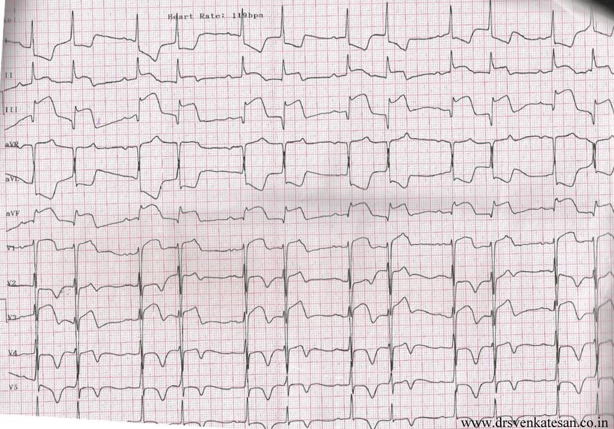
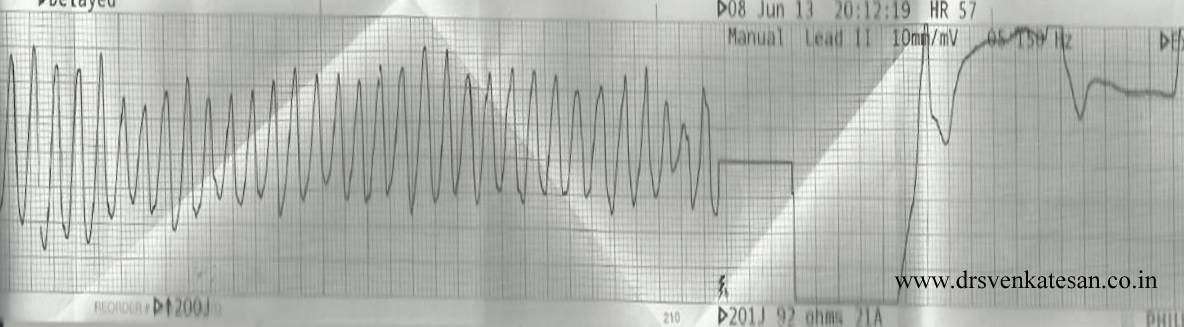
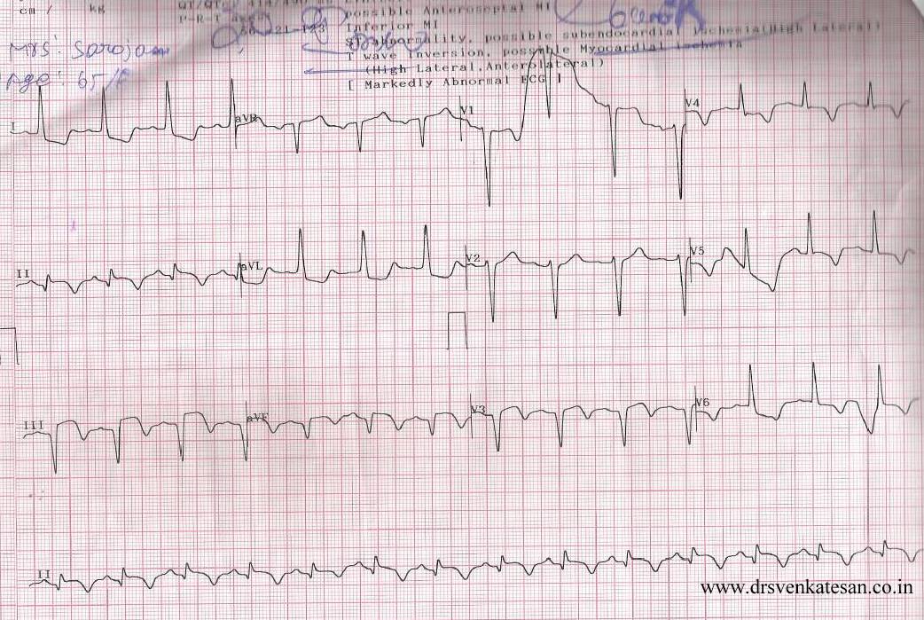
ACS is the commonest cardiac emergency .Thousands of patients are treated every day.Millions of dollars are spent.Bulk of the cardiologist’s life revolves around this entity.

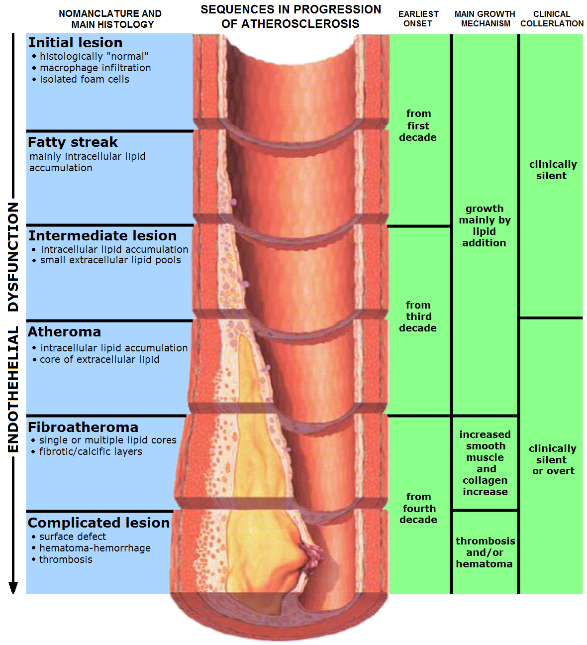
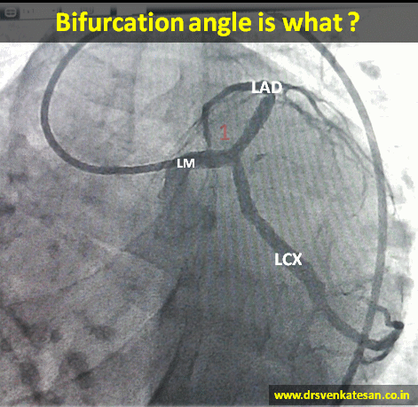
Scattered atherosclerotic plaques in coronary artery lead to ACS either in a random fashion or in a predictable manner .

Still, we are  highly  uncertain about  which lesions are likely to result in ACS ! Some time in the beginning of  21st century, the main stream cardiology media were abuzz with the concept, that non obstructive , non-flow limiting lesions are more prone for ACS rather than more tight  stenosis.

atherosclerosis flow limiting lesion glagov plaque rupture vulnerable erosion fissure vs dissection

I fail to understand how a tight lesion is less  prone for ACS. Tighter lesions are  bigger and must be  prone for more complications . Image courtesy :http://upload.wikimedia.org/wikipedia/commons/9/9a/Endo_dysfunction_Athero.PNG

This reasoning was based on few studies, that lacked  solid scientific proof . In fact the initial  observation was  not made in living coronary arteries rather by autopsy observations .(Later live virtual histological studies came ,  but didn’t confirm this !)

Surprisingly the degree of  anatomical narrowing was conferred  vulnerability  , when we know plaque compositions , morphology and hemo-rheological  factors are many fold important in precipitating ACS . (Lipid content , fibrin cap  thickness, eccentricity , etc)

So where is the truth hidden?

Is it really possible, lesser the stenosis more  is the propensity for rupture ?

 We need to introspect .

“In all probability,  it is a meager statistical illusion”

For every tight lesion there are as many minor lesions scattered around in a given a coronary artery. These can progress into ACS  later.

It is basically wrong to assume non-flow limiting lesions are more prone for ACS than non-flow limiting lesions.To believe so , seriously underestimates  the  culpability of big lesions .It appears a coronary mockery to me  !

At best , we can conclude  non-flow limiting lesions  are not benign and can be an important source of ACS.

An unscientific chain reaction !

If we start believing non flow limiting (say  30%  stenosis ) is more prone for ACS , why we are not stenting all  those lesions ?

If the above concept  is  is applied in cath lab  routinely , the principle of  FFR   which relies solely on hemodynamic impact  will  crash into the dustbin !

Some  more truths

However , It is indeed true  when a plaque is hardened by severe sclerotic process or calcification it is less prone for  rupture and clinical ACS  but can be a source for stable angina.

Is it  justified to assume , larger the plaque the harder  would be it’s content  that  resists ACS ?

Meanwhile , we also know there need not be any lesion at all to cause an ACS.( In a young  smoker ,  100 % thrombotic STEMI  is possible  over an area of coronary erosion caused by endothelial dysfunction ! So , where do we go from here !)

Let us be clear

Are you confused more !   . . . after  reading this article, let us clear it by two-line summary !

As on 2014 ,

  • Symptomatic flow limiting lesion   are tackled by stents.
  • All non-flow limiting lesions  are treated by  high dose Statins  and vigorous medical management.

Final message

Contrary to popular  perception, tight lesions are  more complex, eccentric , soft and are at immediate risk of ACS.

Non flow limiting lesions remain static in most,  regress in many , still  carries  distinct  risk of progression into full blown ACS , at any time if conditions are favorable.

Fixed concepts and ideas in medical science do not help us  taking medicine forward. Especially so, when these are based on assumptions and approximations. If only we redo these studies with the currently available technology (FFR/OCT/NIR the conclusions would be dramatically different. !

Waiting for someone to nullify such false concepts in a more scientific way !

Reference

2.Glagov S, Weisenberg E, Zarins C, Stankunavicius R, Kolletis G. Compensatory enlargement of human atherosclerotic coronary arteries. N Engl J Med. 1987; 316: 371–375.

3.Fuster V, Lewis A. Conner Memorial Lecture. Mechanisms leading to myocardial infarction: insights from studies of vascular biology. Circulation 1994;90:2126-2146.

4.Ambrose JA, Weinrauch M. Thrombosis in ischemic heart disease. Arch Int Med 1996;156:1382-1394

Postample 
This post was written in 2014. Happy to find a scientific proof to this concept in 2018.
Source :  PROSPECT study

Retrospective angiographic studies and the prospective PROSPECT (Providing Regional Observations to Study  Predictors of Events in the Coronary Tree) study have shown that plaques with severe stenosis carry a higher per-plaque risk for producing clinical events than plaques that cause no or non severe stenosis.

However, such lesions are few, and overall, most ACS are precipitated by plaques  without significant stenosis on an antecedent angiography
weeks or months before. This epidemiology is consistent with the distribution of TCFAs, as shown by a combined angiography and optical coherence tomographic imaging study of nonculprit lesions.

Lesions that caused severe stenosis were twice as likely to be
TCFAs than lesions with only non severe stenosis, but the total number of TCFAs with nonsevere stenosis was three times higher than those with severe stenosis. The mild pre-existent stenosis of most TCFAs and ruptured plaques is explained by expansive remodeling, because such lesions are, on average, large.

The long-held notion that mild to moderate obstructive coronary lesions are responsible for the majority of MIs has been challenged by studies that described significant narrowing in the days preceding MI. However, significant narrowing shortly before MI may be a result of (rather than a precursor) for rupture.


Read Full Post »

Only fools will   manage unstable angina  medically !
 
That was exactly the statement ,  one popular Interventional  cardiologist   told a small gathering  in one of the weekly meet .
 
Do you agree ? 


Answer
 
We can’t  make a blanket  statement like that . We have clear  guidelines (Of course as licensed and certified cardiology  practitioner  you have  every right to violate it !)  .
 
UA is risk stratified in Low , Intermediate and High risk  categories .Only high risk group  require emergency Intervention .Even in high risk group there are some reservation.(ICTUS  study )
 
There are some very mild forms of  UA (High grade stable angina precipitated by an emotional stress will exactly mimic UA. Similarly most  secondary UA due to tachyardia , Anemia  etc should not  cause an alarm .)
 
 
*Please note  , currently coronary angiogram is included in medical  investigation  in most  patients with UA . The  confusion in interpreting  such statements  is partly because many physicians/ cardiologists consider  doing a coronary angiogram by itself an  Interventional  management
 
 
Reference

Read Full Post »

This  is the story of a 55 year old  women ,  who was received  in our CCU  with a  dramatic STEMI (ECG looked like an action potential ) ,  LV  S 3  and  hypotension.    It was impending cardiogenic shock.Since we do not have full fledged primary PCI  program  , thrombolysis was planned. She had  cardiac arrest   immediately after  starting streptokinase infusion . She  was  promptly shocked  and  revived .  The ECG changes rapidly  reversed(ECG -3) . Every other  hemodynamic parameter got stabilised as well . To our surprise   ( few hours later ) this patient  was  so comfortable , sat up on her bed ,  demanded a discharge . (Which was refused of course !)  One week  later coronary angiogram was done, a near complete recannalisation of RCA was documented.

ECG 1 on arrival
Inferior MI 2  

ECG -2 Developed cardiac arrest  10 minutes  after  starting the Streptokinase Infusion

primary VF 2

ECG -3 .Taken few minutes following   the VF

inferior MI evolved 2


Acute myocardial infarction (STEMI)  kills more than a million life every year . Majority of death  happens within an hour of onset of symptoms. Ventricular fibrillation  is the arrhythmia of death. Why this occurs  only in  few , while  many are  immune to it ?

God keeps  this secret  close to his chest ,  how and why  he selects   candidates for this arrhythmia !

Scientists are still  far away  in finding the truth . But , one thing  is obvious .The  moment   coronary artery is totally occluded  , the heart begins a fight  and try  to  get rid of this obstruction . In the process ,  it  goes into convulsion (VF)  with a foolish belief  , it  can shrug of the thrombotic insult . Death often   ensues if  not intervened . (Very rarely  VF can be a non sustained one  and patient survives cardiac arrest !)

VF  as  a electrical  response  to  reperfusion injury .

Often times ,  we witness patients  to  go  for  VF  very early following thrombolysis . The  thrombus in situ is an irritant , it  triggers the inherent fibrinolytic system (Natural TPA included) If it is successful  it opens the occlusion ( atleast partially )  and salvages the myocardium .If the fate is against  the patient , very early reperfusion of IRA triggers  VF  .  If this occurs at home   survival  is  low .If  the VF occur at hospital the probability of survival is near 100 % .

               The  intensity of  natural lytic mechanism  is the major determinant  of   early reperfusion . Ironically  the same  factor   determines  occurrence of the deadly  VF .

I would believe  , the STEMI patients  who die early (even before reaching  the hospital ) are (un) blessed with a  fighting  heart  ! Ironically , the lazy hearts  reach the hospital  alive ! (slow &  steady win the race !) .  Of course , reperfusion  injury is not the only mechanism of VF . Other common suspect is  left main STEMI .

Link to related video “Ignorance based  cardiology ”

https://www.youtube.com/watch?v=J9DH6Vr04es

Final message

While , VF  is  referred  to as arrhythmia  of death , it may  in-fact , represent  a common form  of  reperfusion arrhythmia in  the setting of  STEMI !  .  . .  Hence , it can  Initiate  a new lease of life in  many   lucky ones !  I hope the title of this article  makes sense  !

Read Full Post »

bifurcation angle

  • At any branch point three angles are possible .True bifurcation angle is formed between LAD and LCX .
  • The angle between LM and LAD or LM and LCX can also be important in specific situations ,especially when we encounter short left mains and Medina 1,1,0 lesions .
  • Major bifurcation angle can  occur in mid  segments  as well ,  between LAD / major Diagonal  , LCX and OM.
  • Logic would tell us the  left main  bifurcation  angle is relatively fixed by the anatomical AV and IV grooves. Still early course of LAD and LCX can be out of grooves.
  • Further ,the bifurcation angle is imparted some amount of dynamism by cardiac cycle . It can vary between 80 -120 degrees (LAD/LCX).
  • Most importantly various  angiographic views can alter the true angle (by illusion ) in dramatic fashion . RAO caudal view appear ideal to measure it. (LAO caudal make every bifurcation angle obtuse !)
  • Acute angled bifurcations are prone for stent related mechanical issues both during deployment and in the long term outcome . (When two stent technique is used) This is because ,  acute  angled bifurcations has a tendency to drift the carina , and  encroach  the lumen  which can create new  turbulence . Of course final kissing balloon is expected to reduce this hemodynamic side effect at least on paper !

Read Full Post »

Older Posts »