Feeds:
Posts
Comments

Archive for May, 2009

Thrombolytic therapy ,  has been  the specific treatment  for STEMI for  many decades. Primary PCI*  is  shown to be  superior  than  thrombolysis  if   performed   early  by an experienced  team in a dedicated facility. (*Conditions apply). It is estimated ,   currently only a  a fraction  STEMI  population get primary PCI (<5%) in ideal conditions . Another fraction , get  primary PCI by inexperienced cardiologists  in low volume centres.

So , thrombolysis   remains, and  would continue to remain ,   the    primary  mode of therapy for STEMI  in the  present and near  future !

How do you assess the successful  thrombolysis ?

It should be recognised ,  there is a fundametal flaw in this  question !

The aim of thrombolytic therapy is  not  to   lyse  the thrombus  , but also  to restore the coronary blood flow to the  myocardium – also called reperfusion . One may wonder , why the term ,  thrombolysis  should ‘t be  used interchangeably with reperfusion. 

A successful thrombolysis  never guarantees  a good reperfusion , for the simple reason ,  distal blood flow in an  obstructed coronary artery  is dependent on ,  many factors  other than relief of obstruction.

Apart from the potency of drug,     other   important factors  that determine  successful  lysis &  reperfusion are  . . .

  • Timing of opening of artery , if the thrombolysis is delayed  ,  the distal myocardium is dead , and   it won’t allow blood flow to enter the mycardium.
  • Microvascular integrity is as vital as epicardial vessels.
  • Distal microvascualture  plugging by the thrombotic debri . This is called”no reflow “

So , we should  primarily assess myocardial reperfusion rather than epicardial thrombolyis ! following thrombolysis .

What are the parameters available to assess successful reperfusion /thrombolyis?

  1. Clinical : Relief from chest  pain. Angina relief  , though subjective is an indication for adequate reperfusion of ischemic myocardium.
  2. ECG-ST segment regression > 50%
  3. Cardiac enzymes: Early flushing of  intra myocytic CPK into systemic circulation and hence early peaking of CPK MB (<1ohours instead of 24h)
  4. Reperfusion arrhythmias(AIVR-Less specific) .Primary VF is now thought to be reperfusion related.
  5. Infract related artery(IRA) patency by coronary angiogram
  6. Distal TIMI flow/ myocardial blush score/ TIMI frame count

ECG ST regression ,  is a direct indicator  myocardial reperfusion   as the ST segment shifts  towards baseline ,  implies  of infarct current of injury . ST regression almost always correlate with good  recovery of LV function  in STEMI .

IRA patency , is an epicardial index , it  does not give information about myocardial blood flow . But ,  a good  distal TIMI flow generally indicates good reperfusion.This  again ,  is  not a fool proof  index,  as even many of the TIMI 3 flow patients  have severely damaged myocardium by echocardiography .

Final message

For the above reasons, one should always  make a distinction between successful lysis and successful reperfusion . Surprisingly ,  ECG  is  the gold standard for assessing successful reperfusion of myocardium ,  while CAG tell us  about epicardial patency and possibly reperfusion also.

Read Full Post »

Apart from  acute  coronary syndrome,    cardiac  failure is   the most common clinical  presentation of  CAD. Cardiac failure ,  classically present with dyspnea on rest or on exertion , while angina is the dominant presentation in ACS.  

What if  ,  both these  occur together in an acute fashion ?

Yesif it occurs  together it is called ischemic cardiac failure . Fortunately , this is quiet uncommon . It has   an adverse outcome,  especially if it occurs  as a companion of NSTEMI . Let us see how . . .(  Most of the episodes of cardiac failure  in CAD  means only  LV failure )

For cardiac failure to occur , there need to be a mechanical contractile dysfunction or defect . In CAD population , this can  occur in  one of the following way.

  • Loss of LV muscle (Acute  Myocardial infarction as in STEMI)
  • Mechanical defects (Mitral regurgitation/VSR etc)
  • An arrhythmia (Commonly VT or AF / CHB )  can precipitate  cardiac failure

Apart from these three , there is  an important mechanism of acute LVF, namely ischemic stunning of major part of LV resulting in severe mechanical dysfucntion.This is a dangerous form of cardiac failure (Pathologivcclaly it is thought to represent  contraction  band necrosis !) this occurs in global ischemic situations manifested as gross global ST depression.

So,  there are two types of  ischemic LVF  .  STEMI   occuring due to infarct( ± ischemia ) Other  one (NSTEMI)entirely due to ischemia.

Logically ,  one  may n’t   refer  STEMI related LVF as  ischemic LVF at all  , as infarct has already occured. While , NSTEMI related LV could be the ” True ischemic LVF “


What are the differences between cardiac failure that occur in  STEMI and NSTEMI ?


lvf in nstemi stemi

Is post infarct failure  ( The commonly used terminology  , now out of vogue ! )  a type of ischemic LVF ?

In the strict sense , it is not . Here the dead myocardium , is responsible  for the   failure .To label a  LVF , as  ischemic , ongoing ischemia must  be  documented and further it  should  be shown to  contribute   for the  mechanical dysfunction .

This is of vital importance ,   if you wrongly attribute ischemia  as a cause for  the LVF , the patient may be taken up for emergency  revascularisation .It is not going to help much (Infact , it may  worsen !) as  this cardiac failure is not going to be corrected  .What we require ,  here is an  aggressive medical management  protocol .


Read Full Post »

Failed thrombolysis is an important clinical  issue  in STEMI   as  successful thrombolysis  occurs  only in  about 50-60%  of pateints . The typical criteria to define failed thrombolysis is  the  regression  of less than 50% of sum total( or maximum)  ST elevation in infarct leads.

So what do you do for these patients with failed thrombolysis ?

It depends upon the patient’s symptom, hemodynamic stability, LV dysfunction .

They  should  get one of the following .

  1. Conservative medical management  with /without CAG
  2. Repeat thrombolysis
  3. Rescue PCI
  4. CABG

Medical management is  thought to be  too inferior a  management,  many of the interventional cardiologists  do  not want to talk about . But  , there is  an important  group of patients (Not often addressed in cardiology literature)  who  technically fulfill the criteria  of failed thrombolysis  , but   still  very  comfortable , asymtomatic  and in  class 1. These patients ,  have  a strong option for continuing the conservative management .

Repeat thrombolysis does not have a consistent effect but can  be  tried in some  stable patients. CABG  can be a genuine option in few

Rescue PCI

This terminology  has become  the  glamorous one since the  catchy word  rescue is tagged in the title  itself. For most of the cardiac physicians ,  this has become the default treatment modality.This is an unfortunate perception . What  one should realise   here is  , we are  tying to rescue  the myocardium and  the patient ,   not the patient’s coronary artery !

Opening up a coronary obstruction is not synonymous with rescue .

For rescue PCI ,  to be effective it should be done within the same time window as that for thrombolysis (ie within 6 or at the most  12 hours) .This timing  is  of vital importance  for the simple reason , there will be nothing to rescue after 12 hours as most of the muscle  would be  dead. Reperfusing a dead myocardium has been shown to be hazardous in some ,  as it converts a simple  infarct into a hemorrhagic  infarct.This softens the core of the infarct and  carry a risk of rupture. Further,   doing a complex emergency  PCI  ,  in  a thrombotic milieu with   presumed  long term  benefit ,  is  a  perfect recipe for a potential  disaster.

While the above statement may be seen as pessimistic view , the optimistic cardiologist would vouch for the“Curious  open artery hypothesis” .This theory simply states , whatever be the status  of the distal myocardium ( dead or alive !)   opening an obstruction in the concerened coronary artery  will benefit the patient !

It is  huge surprise , this concept   continues to  be alive even after  repeatedly shot dead by number of very good clinical trials (TOAT, CTO limb of COURAGE etc ).

The REACT study (2004) concluded undisputed benefit of rescue PCI for failed thrombolysis  , only if the rescue was done  within  5-10 hours after the onset of symptoms.The mean time for  pain-to-rescue PCI was 414 minutes (6.5hours)

Final  message

It is fashionable to talk about time window for thrombolyis but not for PCI  .The time window for rescue PCI is an redundant issue  for many  cardiologists ! . But ,  the fact of the matter is ,  it is not . . .

The concept of time window in rescue PCI  , is as important as ,   that of  thrombolysis. Please , think twice or thrice !  if some body suggest you to do a rescue PCI in a stable patient  ,  12hours after the index event .

Important note : This rule   does not (  or need  not  ) apply for patients in cardiogenic shock  or patient ‘s with ongoing iscemia and angina.

Read Full Post »

NSTEMI constitutes a very heterogeneous population .The cardiac risk can vary between very low to very high . In contrast , STEMI patients carry a high risk for electro mechanical complication including sudden death .They all need immediate treatment either with thrombolysis or PCI to open up the blood vessel and salvage the myocardium.

The above concept , may be true in many situations , but what we fail to recognize is that , STEMI also is a heterogeneous clinico pathological with varying risks and outcome !
Let us see briefly , why this is very important in the management of STEMI

Management of STEMI has undergone great change over the past 50 years and it is the standing example of evidence based coronary care in the modern era ! The mortality , in the early era was around 30-40% . The advent of coronary care units, defibrillators, reduced the mortality to around 10-15% in 1960 /70s . Early use of heparin , aspirin further improved the outcome .The inhospital mortality was greatly reduced to a level of 7-8% in the thrombolytic era. And , then came the interventional approach, namely primary PCI , which is now considered the best form of reperfusion when done early by an experienced team.

Inspite of this wealth of evidence for the superiority of PCI , it is only a fraction of STEMI patients get primary PCI even in some of the well equipped centers ( Could be as low as 15 %)

Why ? this paradox

Primary PCI has struggled to establish itself as a global therapeutic concept for STEMI , even after 20 years of it’s introduction (PAMI trial) . If we attribute , lack of infrastructure , expertise are responsible for this low utility of primary PCI , we are mistaken ! There are so many institutions , at least in developing world , reluctant to do primary PCI for varied reasons.( Affordability , support system , odd hours ,and finally perceived fear of untoward complication !)

Primary PCI may be a great treatment modality , but it comes with a inherent risk related to the procedure.

In fact the early hazard could exceed the potential benefit in many of the low risk STEMI patients !

All STEMI’s are not same , so all does not require same treatment !

Common sense and logic would tell us any medical condition should be risk stratified before applying the management protocol. This will enable us to avoid applying “high risk – high benefit” treatments in low risk patients . It is a great surprise, the cardiology community has extensively researched to risk stratify NSTEMI/UA , it has rarely considered risk stratification of STEMI before starting the treatment.

In this context , it should be emphasized most of the clinical trails on primary PCI do not address the clinical relevance and the differential outcomes in various subsets of STEMI .

Consider the following two cases.

Two young men with STEMI , both present within 3 hours after onset of symptoms

  1. ST elevation in V1 -V6 , 1 , AVL , Low blood pressure , with severe chest pain.
  2. ST elevation in 2 ,3, AVF , hemodynamically stable , with minimal or no discomfort .

In the above example, a small inferior MI by a distal RCA occlusion , and a proximal LAD lesion jeopardising entire anterior wall , both are categorized as STEMI !
Do you want to advocate same treatment for both ? or Will you risk stratify the STEMI and treat individually ? (As we do in NSTEMI !)

Current guidelines , would suggest PCI for both situations. But , logistic , and real world experience would clearly favor thrombolysis for the second patient .
Does that mean, the second patient is getting an inferior modality of treatment ?

Not at all . In fact there is a strong case for PCI being inferior in these patients as the risk of the procedure may far outweigh the benefit especially if it is done on a random basis by not so well experienced cath lab team.
(Note : Streptokinase or TPA does not vary it’s action , whether given by an ambulance drive or a staff nurse or even a cardiologist ! .In contrast , the infrastructure and expertise have the greatest impact on the success and failure of PCI )
Final message

So , it is argued the world cardiology societies(ACC/ESC etc) need to risk stratify STEMI (Like we do in NSTEMI ) into low risk, intermediate risk and high risk categories and advice primary PCI only for high risk patients.

Reference

https://jamanetwork.com/journals/jamainternalmedicine/fullarticle/226907

Read Full Post »

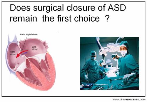
Atrial septal defect is one among the commonest congenital heart disease .After years of controversy, there is consensus  now , all significant ASDs  need to be closed ,  at whatever age it is detected.

This rule does not apply to small ASDs without chamber  right atrial and right ventricular dilatation. These defects and PFOs need not be closed .

Over the years , the  controversy  has shifted  from   Should we close ?  to  How to close ?

There are two options available : Device closure , Surgical closure

asd closure device www.drsvenkatesan.com

asd closure www.drsvenkatesan.com

The following table compares the both treatment  modalities

( Personal perspective )

asd device closure 4

Final message

Device closure is a complex, costly, often  difficult  and  error prone   cardiac procedure .It needs long term follow up and may  carry a life long risk of major cardiac complication.It is useful only in selected subset of ASD patients. Surgical closure prevails over device closure in most situations.

Is this article  has biased view against this  emerging pediatric  interventional procedure of ASD closure ?

It may appear so . But that is the reality as on 2009 !.May we hope technology evolves further and take our surgeons head on .

2012 update on ASD device closure .

The   hard-ware  as well as the  expertise has   improved a lot and it is on right track to become a real challenge to surgery.

The only issue again is the availability of  rims to mount the device . Another  realistic and sensitive issue  which  have I come across is  , many interventionist cardiologist do feel awkward  when they experience  unexpected rim shortage on table.  They should realise it is not their  fault.

Always be ready to abandon the procedure and refer to the surgeon , according to your  true conscience 

After all , improperly delivered device is  a life long pain for the patient .He has come to you with a  great belief  isn’t !

2014 update

Device closure for most ASDs in both children and adult is  now possible with high degree of success. We have crossed about 50 patient experience. And  I am truly amazed  , how within a short period the device closure is about to conquer the crown from the surgeons ! (Exciting new data are coming from   my colleague Dr Gnanavelu from  the new Super specialty hospital of Government of Tamil Nadu Chennai. )


Reference

Aortic erosion following ASD closure

http://content.onlinejacc.org/cgi/content/full/45/8/1213

Read Full Post »

Acute coronary syndrome is the commonest cardiac emergency. STEMI and NSTEMI are the two clinical limbs of ACS. Generally they have distinct clinical, ECG, angiographic features.(Ofcourse, with some degree of overlap) . It is  a  mystery , both clinical presentations differ so much inspite of the common denominator  , namely ,  an injured plaque with add on thrombus  within the coronary artery. The primary difference between these two entities is, in  STEMI the occlusion occurs sudden and complete and in NSTEMI it occurs slow and incomplete

Cardiac arrhythmias in ACS

It is a  much published factoid  for  many decades, that  only one third of STEMI patients  reach the hospital alive ! The reason being , STEMI  is very much prone for primary VF.  Contrary  to this ,  most pateints with NSTEMI reach the hospital alive ! How ?

Both are ACS, if ischemia is a powerful trigger for dangerous ventricular  arrhythmia’s , NSTEMI should also behave  similarly .So what protects against arrhythmias in NSTEMI ?

  • We realise ,  by observational experience (Not EBM !)  It is the suddenness and totality of ischemia that trigger dangerous form of arrhythmia  .
  • Further, a balanced  ischemia in two contralateral segments (or global  ischemia) some how protects against development of ventricular  fibrillation .This may be due to preservation  of  electrical homogeneity  , and the spherical VT spiral waves are not sustainable.
  • In contrast , STEMI has a sudden  focal , ischemic  zone that initiates the VT and    ischemia free  contralateral segment  welcoming  and sustaining the  reentrant wavelet.
  • The observation of primarily single vessel disese in STEMI and multivessel disease in NSTEMI also give credence to this concept.
  • Further , ischemic preconditioning can exert an important anti arrhythmic  effect in NSTEMI as  patients with unstable angina have   slow, repetitive episodes of ischemia prior to the index event .
  • Post MI scar mediated VT/VF is independent of degree of overall ischemia
  • It is also established ,  a sub group of  STEMI pateints  who  had  preinfarction angina(  ie . a brief  period of UA/NSTEMI) have very low risk of SCD  supporting the concept of sensitising the myocardium against ventricular arrhythmias.

Final message

Even though , there is a convincing concept  of  Ischemia induced  cardiac arrhythmia in literature ,in real patients it is very difficult to link the two in many situations..UA/NSTEMI is the most common  acute ischemic event but the incidence of VT/VF here,  is far less than one would expect.In ACS , focal , total  ischemia is more likely to precipitate a VT/VF than multifocal and global ischemia.

Read Full Post »

Can we advice CABG for single vessel disease  ?

Yes, CABG  may be indicated  in

  • Critical , proximal , complex  LAD disease   with or without  ostium involvement.
  • Many of the bifurcation lesions with large and significant  side branch
  • Small caliber LAD with diffuse disease .

When these occur  in diabetic  subjects , the  indication for CABG is more certain .

* Present generation cardiologists  would feel  every  lesion  is  stentable and should not be referred to the surgeon .But it should be emphasized here,   technical feasibility alone  ,  does not  imply  PCI is superior and ideal in all coronary interventions.

Can we do a CABG  in  single vessel disease  with  normal  LAD ?

CABG is  very rarely  indicated   for isolated RCA or LCX disease. It should be consciously avoided in this patient population.

This is because the at risk myocardium  supplied by these vessels are far less than that of LAD. PCI  is  preferred    in these vessels .(Ofcourse , after considering medical management  ) .

CABG is  ,  too traumatic a  surgery , to  offer  in this  low  risk  coronary  lesions.

Exceptions

CABG  can still be done in following situations  for non LAD single vessel disease.

  • Left dominant circulation  with  complex lesions in LCX /OMs.
  • It is common to see diffuse , long segment  and severe disease of RCA with normal LAD /LCX system .PCI is not feasible in this subset.
  • Failed PCI
  • Recurrent instent restenosis.
  • Bail out CABG after a acute complication during PCI

One should remember ,  inability to do a PCI  does not  mean ,  the patient  should   land in surgeon’s table .We should recall , from our memory medical management is an effective and established form of treatment in single vessel disease ( Mainly for non LAD , and some cases of LAD also !)


Read Full Post »

04_16

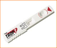
How good is Troponin T or I  to rule out acute coronary syndrome in the emergency room  when a  patient presents  within two to three hours after the onset of symptoms ?

  1. Very useful
  2. Useful
  3. Rarely useful
  4. Not useful
  5. Not at all useful

The answer is  5 , can be 3 or  4 , never 1 or 2 !

If you are surprised with the answer

Findout why , read further

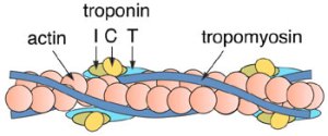
troponin-i-troponin-t1

19_trop-t-sen1

troponin-i-troponin-t-2Final message

Troponin has a definite diagnostic  and prognostic value in  STEMI or NSTEMI  but relying on a single normal troponin level very early after an ACS can be . . . futile.

Realis,   diagnosis of ACS , especially  STEMI , is primarily by ECG and clinical features . Even in NSTEMI biomarkers help primarily to risk stratify the event. Bio markers come into picture only in borderline  ECGs and in baseline ECG defect like LBBB/Pacing rhythm .

It should be recognised , the major draw back of cardiac markers is , it  does not represent real time cardiac myocyte  events. (But the good old ECG has this unique property !) .The myocyte secretion & release  kinetics , the effect of  native (and pharmocological ) reperfusion make it a unreliable  marker.Apart from the time lag  , the  laboratory methods to detect these  molecule needs further refinement.

For the current day cardiologists ,  it is  required to finish off the entire treatment  of MI  within 6  hours by doing a primary PCI . It is an irony , troponin begins to appear only by  then to be detected in the blood !

Further reading

A .All about troponin

http://www.annals.org/cgi/content/full/142/9/786

B.Troponin In aortic dissection

http://www.ncbi.nlm.nih.gov/pubmed/15887472

Read Full Post »

Ventricular remodeling  follows large myocardial infarction .This term denotes to  change in size , shape  and function  of the ventricle   due to altered  myocyte geometry .It is now believed  , this  process begins to occur very early  following a STEMI.(less than 24hours)

lv-remodeling

In which MI remodeling is more common ?

Any MI of large size , especially  anterior  and lateral MI.  Inferior and posterior MI are less affected by adverse remodeling.The incidence is up to 20% of all myocardial infarction ,  if left untreated. Ventricular aneurysm formation and dyskinetic segments can be termed as the worst form of remodeling. The old terminologies of infarct extension and expansion could by synonymous with ventrilar   remodeling .(Note : Every patient with STEMI undergoes some form of physiological remodeling that should not be confused with progressive pathological remodeling , we are discussing  here ! )

What is the clinical impact of remodeling ? How to prevent it ?

Progressive cardiac failure and a  poor outcome .  It may provoke ventricular arrhythmias. ACE inhibitors (CONSENSUS study 1992 )  has since revolutionized  the pharmacological  prevention of adverse remodeling.

How to recognise left ventricular remodeling ?

Many methods are available .

  • 2 D Echocardiography
  • Tissue doppler
  • LV angiogram
  • MRI

These imaging methods diagnose remodeling  only after it manifest* .We know remodeling is a cellular and molecular process .The earliest trigger for remodeling is the mechanical stretch and wall stress on the ventricles.Large areas of necrosed myocardium and  the adjacent normal myocardium sets a perfect stage for eccentric pulling of myocardial segments and unregulated slippage of myocytes.

* Diagnosing fully established  ventricular remodeling  serves ,  no great   purpose as it is very difficult to reverse it by pharmacological methods.it requires complex surgery.

What is the effect of mechanical stretch on cellular function ?

It is well known  myocyte granules secrete Type B  -Naturetic peptide  in response to stretch. It could be a very early sign of adverse remodeling. So monitoring of  BNP may give us an opportunity to intensively treat those patients who are likely to land in progressive cardiac failure.

A baseline level of NT-proBNP >120 pmol/L identified patients  prone for adverse remodeling .Serial measurements showed further increase. It is possible to identify adverse remodeling of LV by documenting fresh elevation of BNP following MI .

Reference :

1 )Nilsson JC, Groenning BA, Nielsen G, et al. Left ventricular remodeling in the first year after acute myocardial infarction and the predictive value of N-terminal pro brain natriuretic peptide. Am Heart J 2002;143:696-702.

2)http://content.nejm.org/cgi/content/full/352/7/666#R24

Read Full Post »

pci-ptca-ebm-stent

Scientifically ,  the  indication for coronary revascularisation   should be  based on following

  1. Patient’s  symptom ( more specifically angina , dyspnea is less important !)
  2. Prov0kable  ischemia  ( A significantly positive stress test )
  3. Signifcant LV dysfunction with  documented  viable myocardium &  residual ischemia
  4. A revascularisation eligible coronary anatomy * TVD/Left main/Proximal LAD etc ( *Either 1, 2 or 3 should be  present  in addition )
  5. All emergency PCI during STEMI /High risk NSTEMI

Practically ,

A CAD  patient  may fulfill  “Any of the above 5 or  “None of the above 5” ,  but ,  if   a coronary obstruction  was  revealed  by coronary angiogram  and if he  fulfils The 6th criteria , he becomes  eligible for  revascualrisation

6th criteria

If the patient has  enough monetary   resources (by self  ) or by  an  insurance company  to take care of PCI /CABG *

*The sixth  criteria overrides all other criteria in many of the cath labs .Of course , there are few genuine ones still  fighting hard , to keep the commerce out ,  from contaminating cardiology !

Read Full Post »

Older Posts »