Feeds:
Posts
Comments

Archive for August, 2017

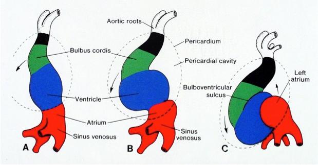
Left main bifurcates into two , that’s  the classical anatomical behavior of LCA. (Or it trifurcates) When left main divides , it tends to share its diameter between its two siblings LAD and LCX with considerable  whims and fancies.(Though Finet* et all thought it has a working rule !) * From  Biomedical Engineering, Cardiovascular Hospital and Claude Bernard University France

Now , have a look at this , its a rare example of  how a left main might Ignore the rule of bifurcation just like that !

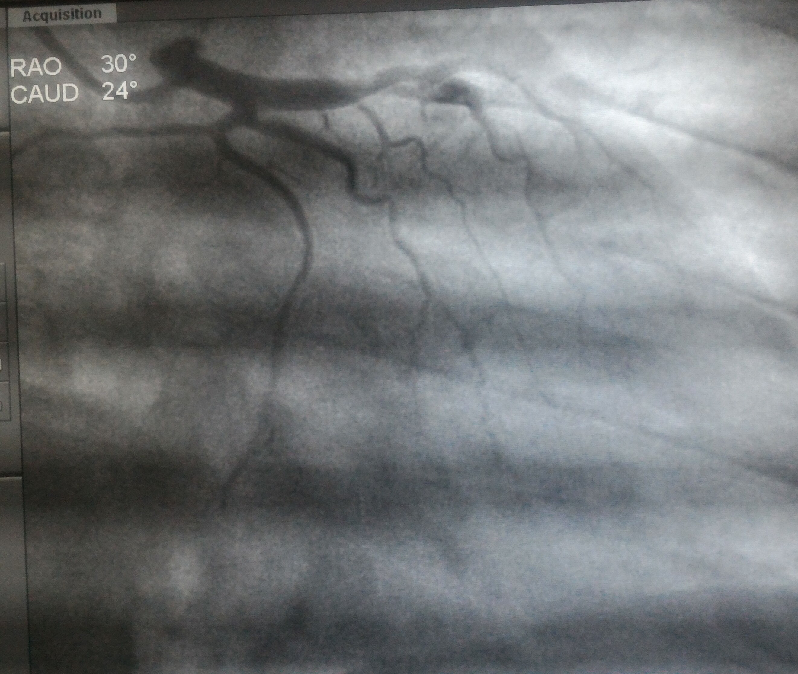
Left main simply continues as left main* after giving off a casual side branch from mid left main shaft .Yes , Its a innocuous looking LCX which would be non dominant as expected

LCX arises exactly mid way in left main , (Technically LAD begins at this point ) but , can you find any difference in the left main after giving off  LCX branch.

Can we say left main continues as LAD without a bifurcation ?

Or shall we say  left main gives off a premature early side branch ( true LCX)  non bifurcating  branch ?

It is an unusual anatomy and  as expected , this patient had a dominant  RCA .

What could be the clinical implication for such a premature  LCX ?

We can only guess . May be nothing !  Obviously ,these patients are immune to develop true bifurcation lesion. Does it in any way mean they have anatomically blessed coronaries !


Reference 

1.Finet G1, Gilard M, Perrenot B Fractal geometry of arterial coronary bifurcations: a quantitative coronary angiography and intravascular ultrasound analysis. , EuroIntervention. 2008 Jan;3(4):490-8.

Read Full Post »

Heart is not like a rigid structure built with  bricks . . . . so , its too architectural mindset to describe cardiac chambers to be made up of walls. Rather , Its a four chambered muscle mass moulded together in a complex 3D interface with distinct surfaces rather than walls. It’s also important to realise, since the heart is positioned (rather hanging )delicately in the middle mediastinum resting on the diaphragm , its subjected to one more dynamism due to respiratory motion blurring the definition of surfaces as well. (Vertical vs Horizontal)

4415_21_26-heart-human-posteriorly

Posterior surface is now referred to as infero-posterior

The posterior aspect of heart contains essentially the venous channels and the atrium (LA in particular)pulmonary veins and coronary sinus.  This happens right from 8 week heart open stage when venous end of lower straight heart tube folds up and posteriorly .

cardaic-looping-posterior-wall

It should be recalled only a small portion of lower aspect of posterior wall is alloted to left ventrilce.Instead the Infero diaphragmatic surface is formed by two-thirds  the LV and one-third Right ventricle.

anatomy-of-heart-posterior-wall-mi-lcx-rca-grays-grants-anatomy-netter

Image courtesy : From the great Netter

Nomenclature  issue 

The term posterior wall is now abandoned in most Echocardiography texts its replaced by inferior .The implication is more for Electrophysiologists with reference to accessory pathway localization

What is true posterior wall MI ?

As discussed before ,posterior surface of heart is different from posterior aspect of left and and right ventricle.

What does leads V7 V8 V9 record ?

It actually records electrical signals arising from posterior  aspect  of heart.  Left atrium,  pulmonary vein along with insulatory  effect of lungs dampens the potential . This makes the sensitivity of ST elevation  in posterior MI is low.

Blood supply of posterior surface

It’s highly variable.Both RCA and LCX arteries contribute with its  posterior left ventricular branches (PLV)

It can be inferred , LCX has more territorial rights than RCA in this unique zone of heart as the artery covers more posterior areas.

Read a related article

Back pain from anterior MI : Is it possible ?


Read Full Post »