Feeds:
Posts
Comments

Posts Tagged ‘posterior stemi’

Heart is not like a rigid structure built with  bricks . . . . so , its too architectural mindset to describe cardiac chambers to be made up of walls. Rather , Its a four chambered muscle mass moulded together in a complex 3D interface with distinct surfaces rather than walls. It’s also important to realise, since the heart is positioned (rather hanging )delicately in the middle mediastinum resting on the diaphragm , its subjected to one more dynamism due to respiratory motion blurring the definition of surfaces as well. (Vertical vs Horizontal)

4415_21_26-heart-human-posteriorly

Posterior surface is now referred to as infero-posterior

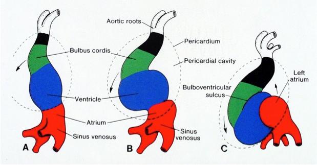
The posterior aspect of heart contains essentially the venous channels and the atrium (LA in particular)pulmonary veins and coronary sinus.  This happens right from 8 week heart open stage when venous end of lower straight heart tube folds up and posteriorly .

cardaic-looping-posterior-wall

It should be recalled only a small portion of lower aspect of posterior wall is alloted to left ventrilce.Instead the Infero diaphragmatic surface is formed by two-thirds  the LV and one-third Right ventricle.

anatomy-of-heart-posterior-wall-mi-lcx-rca-grays-grants-anatomy-netter

Image courtesy : From the great Netter

Nomenclature  issue 

The term posterior wall is now abandoned in most Echocardiography texts its replaced by inferior .The implication is more for Electrophysiologists with reference to accessory pathway localization

What is true posterior wall MI ?

As discussed before ,posterior surface of heart is different from posterior aspect of left and and right ventricle.

What does leads V7 V8 V9 record ?

It actually records electrical signals arising from posterior  aspect  of heart.  Left atrium,  pulmonary vein along with insulatory  effect of lungs dampens the potential . This makes the sensitivity of ST elevation  in posterior MI is low.

Blood supply of posterior surface

It’s highly variable.Both RCA and LCX arteries contribute with its  posterior left ventricular branches (PLV)

It can be inferred , LCX has more territorial rights than RCA in this unique zone of heart as the artery covers more posterior areas.

Read a related article

Back pain from anterior MI : Is it possible ?


Read Full Post »

Thrombolysis is specifically indicated when there is  ST elevation in ECG. ST elevation is a surrogate marker for  total coronary occlusion. It occurs due to current of injury flowing   towards* the lead  facing the  infarct territory . There is only one situation where you can safely and effectively administer thrombolysis in the presence of ST depression ie

Note : There is no accepted terminology  to label a MI as  ST depression MI . Here it is used to emphasise ST elevation is not the only indication for thrombolysis .In posterior MI there is infact ST elevation but it is failed to pick up by standard 12 lead ECG.

NSTEMI is a different entity altogether and  thromolysis is never indicated.

Isolated ST depression in V1 V2 V3 .It almost always indicate isolated posterior STEMI. This can be confirmed by posterior chest ECG leads V7-V10 .

*One will be surprised, to know  the mechanism of ST elevation in STEMI is still not fully elucidated .Technically speaking the net movement of current is away from electrode as  there is only a baseline  diastolic shift  which  gets neutralised in systole  mimicking an ST elevation .(Electro-optical illusion !)

How sensitive is these leads to detect isolated posterior STEMI  ?

Fairly sensitive. Both scapula and  para spinal muscles can be a  significant electrical  barrier that can prevent ST elevation from inscribed .In case of doubtful ST elevation in posterior leads , mit is always better to rely on the clinical presentation.Acute chest pain , consistent with ACS and a new onset ST depression >2mm V1 to v3 is a definite indication for thrombolysis .

Link between posterior MI and RV MI ?

They are closely linked entities .In fact posterior surface of heart is contributed significantly by RV.

What is the angiographic correlation of  isolated ST depression in V1 to V3 ?

It almost always localise the lesion to left circumflex artery . If it is dominant , it can involve lateral and RV territories.

Is isolated posterior MI  less dangerous ?

May be yes , but only after the patient reaches the hospital as electrical risk is same in every STEMI .

The area of infarct  is less , LV failure is less common. While conduction disorders and ischemic mitral regurgitation   can occur  significantly.

Also read ,  Why thrombolysis is contraindicated in UA/NSTEMI ? in this blog

Read Full Post »