Feeds:
Posts
Comments

Archive for the ‘Infrequently asked questions in cardiology (iFAQs)’ Category

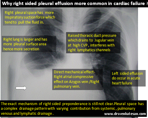
Here is a case report from Dr. Brugada’s group. What is your diagnosis?

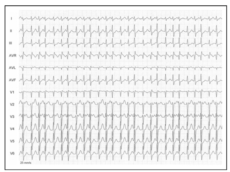
Source & Courtesy Sergio Richter, Joseph Brugada et all , 100(1), 154–156. doi:10.1016/j.amjcard.2007.02.067

Whoever diagnosed AF in the above ECG need not feel bad. The rhythm is not AF, though it mimics very closely. In cardiology, especially in electrophysiology, we can get surprises on a daily basis. (Read below)

Why the ventricular rate is irregular in AF?

Atrial fibrillation (AF)  may sound like a  simple clinical arrhythmia until we ask this delicate question. The traditional and fairly accepted answer is that, AV node with all its collective decremental property filters the incoming atrial impulses (Which varies 400-600/mt) in a random fashion and allows only about 1/3rd of impulses. So, technically it is  AV block of various degrees that makes the ventricular response irregular.

Any other explanation possible?

How about AV node playing out a silent game with Atria, deciding to block everything and start its own fast escape rhythm, rather than leaking out selectively atrial impulses. Some think this is fictional, some others feel it can be real too. When this happens it can be referred to as irregular junctional tachycardia or AF with varying AV blocks. It has been tough, to prove it is only the atrial impulses penetrating through the AV node complex and exiting on the ventricular side unscathed?

Understanding AV node is not easy 

AV node morphology and function still remain a mystery.( Katritsis DG.  Arrhythm Electrophysiol Rev. 2020)The AV node shows huge variation in its size, shape, orientation with LV long axis and AV plane in short axis. The approach to slow pathways with multiple inferior nodal extensions makes a dual (or even poly ) AV pathway in any human being real. How common is dualism or multilateralism within the AV node in the general population? (More than 30-40 % ?) . Let us also mind the traffic in this busy & complex AV flyover can change on a moment-to-moment basis based on neurohormonal and autonomic tone.

Any tachycardia can become irregular if the AV node wishes so !

Though rare ,multiple physiological splits in the AV node make it possible for a single atrial impulse can generate 2 or 3, even more, ventricular impulses. (1: 2 or 3  AV conduction) Since these pathways are dynamic they can make the ventricular response irregular as well (Unlike the regularly coupled Echo beats in classical AVNRT substrate ). Hence, any supraventricular tachycardia can masquerade as AF if AV nodal pathways decide to split and share the impulses this way. It is also interesting to note there has been a documented link between AVNRT and AF (.Ref 2) . Also, adenosine-induced AF is known (James E. Ip et al Circulation: Arrhythmia and Electrophysiology. 2013;6:e34–e37)

Final message

Irregular RR interval with absent/or invisible P waves is not always AF. It can be due to the aberrant behavior of the AV node.( anatomical or functional) It is termed Pseudo Atrial fibrillation as in the above case report. Why do we need to be aware of this entity? We need to be cautious, as any overzealous efforts to ablate the pulmonary veins in such patients will go in vain.

Reference

1.Sergio Richter; Antonio Berruezo; Lluis Mont; Tim Boussy; Andrea Sarkozy; Pedro Brugada; Josep Brugada (2007). Pseudo–Atrial Fibrillation, Rare Manifestation of Multiple Anterograde Atrioventricular Nodal Pathways. , 100(1), 154–156. doi:10.1016/j.amjcard.2007.02.067 

2.Schernthaner C, Danmayr F, Strohmer B. Coexistence of atrioventricular nodal reentrant tachycardia with other forms of arrhythmias. Med Princ Pract. 2014;23(6):543-50. doi: 10.1159/000365418. Epub 2014 Sep 3. PMID: 25196716; PMCID: PMC5586929.

Read Full Post »

Pericardial effusion is often detected in patients with Infective endocarditis. Incidence can be as high as 25% . Most often it is mild, can be moderate in few.

Mechanism

  1. Sympathetic effusion in response to endocardial infection. It’s never more than minimal. (Evidence ? it’s only an assumption)
  2. IE related cardiac failure (Raised systemic venous pressure to which pericardial veins drain)
  3. Local sepsis, Abcess formation tracks to pericardial space through transmural lymphatics
  4. Fungal , granulomatous , Tuberculous IE (Rare) Here IE and PE  share the same pathology
  5. Part of systemic sepsis activated Immune mechanism (Polyseroists)
  6. Renal Involvement of IE-Renal failure
  7. Postoperative pericardial effusion in Prosthetic valve IE (Common, often loculated)

Clinical Implication

  • If the pericardial effusion is more than mild, it often denotes worse outcome. This implies more extensive infection or a marker of extracardiac causes of effusion like renal dysfunction.
  • Effusion may predispose to local dissemination of infection and ends up as peri-annular abscess is whether it is a cause or effect of effusion remains to be understood.It is often exudate as one would expect, but transudative  effusions also occur and would indicate more benign course.
  • The sterility of pericardial fluid has not been proven. Culture studies are rarely done from effusions associated with IE.
  • Pericardial effusions appear more often seen in IE of right heart valves. They turn out to be  IV drug abusers.
  • Contained rupture of an abscess needs to be differentiated from effusion

Can we give steroids for PE associated with IE?

Steroids can rapidly plug the inflammatory pores in the from the pericardial surface.It may also prevent future constriction. Currently, routine steroid therapy is not advised in infective pathology . If the infection is confirmed and is being taken care of by antimicrobial therapy there could be a role for steroids with user discretion.

Final message

During the echocardiographic evaluation of IE, the presence of pericardial effusion should be specifically looked for. These patients should be flagged and will require monitoring as the prognosis of PE complicating IE is a concern unless proved benign.

Reference

Two studies one from Spain and other from Egypt looked into this issue specifically.


Read Full Post »

Background STEMI knowledge check : Evidence-based Ignorance

I think , It is unfortunate, In the management of STEMI , the two popular strategies of myocardial reperfusion is made to fight with each other as if they are perennial enemies for over two decades. Suddenly, someone with a rare coronary insight thought, why fight each other , they can have a friendly hug and work together. That brought the concept of pharmco -Invasive approach or strategy(PIA) backed up by STREAM, FAST-MI, and TRANSFER AMI studies.Yes, it appears to work well and devoid of all the early adverse events of pPCI. (Much to the dismay of ardent fans of Primary PCI )

*May I add one more shocker of a fact . Deep subset data mining from the above trials did show very early lysis may even act as a perfect stand-alone therapy negating the need for acutely one pharmaco Invasive PCI altogether.(Which was never published) Don’t get alarmed the concept is nothing but , the good old lysis , followed by leisure & elective Ischemia guided PCI in all uncomplicated STEMI.

Now coming to the FAQ in Cardiology Boards: Why is the time window for PIA is 3 to 24 hrs ?

The simple answer for an uncomplicated fellow is “published studies have shown benefit only in this time window. If you do PCI early (,<3h) after lysis paradoxically both bleeding and pro-thrombotic complication over the stented lesions are more common. The upper limit is 24 hrs , since by that time we lose all the potential for myocardial salvage”

End-

Larger version of the answer

(Advanced readers who are willing to get confused, may read further)

1. Lysis and immediate PCI doesn’t go well at least in trial world. (FINESSE study, by Ellis et all NEJM 2008) Though cardiologists tend to blame lysis (effect of) to Interfere with their hand skills, it can very well be the opposite. The PCI undo the true benefit of lysis. For cardiologists to accrue maximum benefit in the early time window, they need to be too fast, in the process, they accelerate and fuse adverse events of both modalities.

2. The time window 3 to 24h could simply be evidence-based empiricism. In the major STREAM trial, invasive limb happened between 6 and 16 hours only. We stretched both in the top and bottom in the time clock and made it 3 to 24 hours with other trial data.

3. One realistic reason could be this. It requires a minimum of three hours for a patient to reach a place of coronary Invasion after lysis. So one may argue its time allowance for transport .It comes in handy at times.

4 .If the patient reaches earlier, we need to delay the PCI intentionally to please the evidence based medicine. Mind you, every minute delay increases the chance of no reflow as the microvasculature goes for edematous and porous death.

5. Please note, the time window for pharmaco Invasive strategy will go for a tail spin if the initial lysis is failed. Here, we have to rush I guess. Mind you, In this situation, the evidence based blaming that early PCI increases the adverse events immediately following lysis goes topsy turvy . This is where , we should recall old studies of routine rescue PCI (without clinical criteria) rarely succeeded to correct failed thrombolysis (SWIFT trial)

6.Now, why not PCI after 24hrs? The game can be played reversed if you document ongoing Ischemia in IRA or Non IRA, one may do it . The problem arises when the flawed thought process of a cardiologist could legally justify all PCI beyond 24 h /class 3 Indication after STEMI.The argument goes like this. I think this patient has residual silent Ischemia in- spite of severe LV dysfunction (Suspicion is the justification, to which ,unfortunately no one can dispute) It only suggests open artery hypothesis is still trying to raise from the graveyard more than a decade after its near burial.

Final message

To all those energetic, evidence-based cardiac physicians, we all know coronary care is all about time. In fact, we need to be blessed much more than a sense of time. There is something called medically( or spontaneously )stabilized ACS.  Please realise , “timely and safe intervention” for your patients could simply mean either playing the time button slow/ fast / slow or fast forward / pause or simply shutdown the cath lab, reach home early and enjoy some music or movie in your favorite streaming player.

Reference

1.Ellis SG, Tendera M, De Belder MA, FINESSE Investigators Facilitated PCI in patients with ST-elevation myocardial infarction. N Engl J Med. 2008;358(21):2205–2217. [PubMed]

2. Armstrong PW, Gershlick AH, Goldstein STREAM Investigative Team Fibrinolysis or primary PCI in ST-segment elevation myocardial infarction. N Engl J Med. 2013;368(15):1379–1387. [PubMed]

3. Danchin N, Puymirat E, Steg PG, T, on behalf of the FAST-MI 2005 investigators Five-year survival in patients with ST-segment-elevation myocardial infarction according to modalities of reperfusion therapy: the French Registry on Acute ST-Elevation and Non-ST-Elevation Myocardial Infarction (FAST-MI) 2005 Circulation. 2014;129(16):1629–1636. [PubMed]

4. Cantor WJ, Fitchett D, Borgundvaag B, TRANSFER-AMI Trial Investigators Routine early angioplasty after fibrinolysis for acute myocardial infarction. N Engl J Med. 2009;360(26):2705–2718.. [PubMed]
5.. Bonnefoy E, Steg PG, Boutitie F, , CAPTIM Investigators Comparison of primary angioplasty and pre-hospital fibrinolysis in acute myocardial infarction (CAPTIM) trial: a 5-year follow-up. Eur Heart J. 2009;30(13):1598–1606. . [PubMed]

Read Full Post »

We know pleural effusion (hydrothorax) is disproportionately more common on right side in cardiac failure.Though its a well observed phenomenon, the mechanism of which  has not been clear to us. It could be due to multiple  anatomical , physiological factors.


*The are  right and left lymphatic (Thoracic) ducts that drain the corresponding lungs and pleural space . There can be overlap and contribute to the differential occurrence of pleural effusion


Reference 

A meticulous paper written some 75 years ago (1946) from Harvard medical school teach us some important points in this phenomenon.

There is still lot, to be understood about pleural effusion in cardiac failure. We need to know why some pleural effusions tend to occur independent of hydrostatic forces.  It is also noted long-standing transudative effusions can become true exudates. Role of local pleural capillary hypoxia resulting increasing permeability is underestimated.Hepatic congestion and trans-abdominal seepage of fluid is a distinct possibility.

One more area we are not clear is  the relationship  between the  genesis of  pericardial effusion in cardiac failure and concomitant pleural effusion. Post operatively , after univentricular repair (as in Fontan ), pleural effusions can be much problematic with high venous pressure interfering with  pleural drainage.

Impact on symptoms

Finally, even mild pleural effusion can increase the work of breathing and result in dyspnea which is out of proportion to cardiac dysfunction.While we expect the diurteics to clear the effusion of cardiac failure, it doesn’t happen always arguing for a non transudative mechanism in at least some of them.

Further reading

Discerned readers are advised to study the pleural space dynamics in detail.

Link to the original Article of Edgar Mcpeak and Levine 1946


Read Full Post »

Bernoulli principle states that , when a high pressure jet (Air, Water, blood etc ) moves over a conduit, the pressure exerted by the jet on its sides (Lateral wall) reduces . The velocity gain is equal to pressure drop .This is why we take velocity as a rough guide to pressure gradient and the sacred formula in doppler echocardiography 4V2 came in to vogue . (Incidentally, Bernoulli principle shares the same principle when aircrafts lifts from runway at its peak speed as the pressure above the wings drops to zero or negative and the plane lifts up.)

Please note , the pressure should drop both above and below the aircraft by Bernoulli principle .But, the engine and wings are arranged in such a way , the air speed below the aircraft is slower and hence the pressure is high below and low above and the lift occurs promptly at take of velocity. Imagine , how the valve leaflets in heart is subjected to lift and drag forces every time the blood gushes with high velocity flows.This is also the reason for the Pulsus bisferiens, SAM in HOCM, Coanda effect in supra valvular stenosis, and any post stenotic dilatation.

In Echocardiography the Bernoulli equation is modified.

In clinical doppler echocardiography, we have liberally simplified the original Bernoulli equation by ignoring the the proximal sub valvular velocity V1 . Further , two more components in the equation is also amputated for our convenience ! (Flow acceleration and the viscous friction) .This is the reason we tend to err many times especially in outflow tract gradients and prosthetic valve gradients .

Pressure recovery phenomenon.

This is another hemodynamic lacunae in clinical echocardiography. We know, thepeak velocity of blood is attained just distal to site of obstruction. As the distal velocity beyond the obstruction begins to fall, the pressure tends to recover corresponding to the loss of velocity. This happens to certain distance beyond the obstruction. Since continuous wave doppler measures the pressure in its entire axis of alignment , it is likely to pick more pressure samples from the recovered areas and net result is, it measures more than the true difference in gradient across the valve.The phenomenon is most relevant in assessment of Aortic stenosis and results in over estimation of severity of stenosis.

Importance of Aortic root dimension

Pressure recovery is more likely to occur with small Aortic root. A stiff  so be careful when interpreting echo gradients in small aorta. Relationship between size of aorta and pressure recovery is complex .(Niederberger of pressure recovery for the assessment of aortic stenosis by Doppler ultrasound. Role of aortic size, aortic valve area and direction of the stenotic jet in vitro. Circulation 1996; 94:1934–40)

How much can be the overestimation ?

It can be up to 30 % or even more.Especially in prosthetic Aortic valves.

How to recognise it and overcome it ?

  1. First of all, recognise such a hemodynamic phenomenon exists and the sacred 4v square can be a myth !
  2. Never go with gradient alone in diagnosing valve stenosis. Look for 2D features also.This is more vital when you suspect acute valve obstruction.
  3. Always add the proximal sub valvular velocity (V1 ) in your Bernoulli equation .It need to be subtracted.
  4. The effect of heart rate on pressure recovery has not been properly studied.(The impact of which could be vital and hence too many false prosthetic emergencies could be avoided, as cardiologists tend to rely mostly on gradient than anatomical diagnosis of valve obstruction like visualising thrombus or struck leaflet by TEE or fluro.

Does this phenomenon happen with cath gradient ?(Generally it’s more pronounced in doppler echo )

Yes, It does happen in cath lab also , as its related to physics of flow. It can be minimised if we can use two simultaneous catheters ,one in LV and the other Aortic catheter placed very close to the leaflets.

pressure recovery in aortic stenosis animation

Click below for an Animated version

pressure recovery phenomenon in aortic stenosis 005

Note the pressure recovers from P 2 to P3

Reference

Pressure recovery phenomenon in doppler echocardiography

pressure recovery phenomenon doppler echocardiography

Read Full Post »

One of my fellows gave a discharge summary  for a 62 year old patient with stable diabetic  CAD  who had Triple vessel disease with a final advice reading as CABG / PCI/or OMT .

There was a near fury over his angiogram report in the cath meet. How can be  eligible for all the three Intervention at the same time ?.(PCI -Percutaneous coroanry Inervention ,CABG-Coroanry artery by-pass graft, OMT-Optimal medical therapy )

The lesion in question was , Triple vessel disease(Non critical LAD) and significant LCX and again a non critical RCA .Syntax was less than 22 for sure , however the patient  had class 2 angina (now reducing ) .When asked to explain  , the fellow  argued since the patient  is symptomatic , has DM with TVD  he is eligible for CABG , since  LCX lesion was discrete and PCI was distinctly possible , of course as all three  lesions would be  eligible for OMT on any given day  ! he inferred .

How can  a cardiologist be so casual and non-commital in an important medical decision where a life of a heart is at stake.There was a unanimous condemnation about the report. As a consultant he has to be specific , one can’t leave the decision to  your patient’s whims  . . . rather it’s our scientific whims  that should prevail  !


MEDICAL VS PCI VS CABG OMT COURAGE BARI 2D FREEDOM FAME STUDY MASS 2 CASS OPTOMAL MEDICAL MANAGEMENT SYNTAX ACC AHA ESC GUIDELINES PTCA STS EUROSCORE NEJM

The curiosity continued and looked amusing for many. I was the only one supporting  his argument ! After all , he is being frank and understood the futility of  applying  evolving knowledge base in critical decision making. But, I  asked him to grade the choices .In my opinion  OMT should be the first choice if it can be administered , but reality tells me  true OMT is rare as a modality  at-least in  this  part of world . However every one should insist for it.

Apart from poor  compliance for OMT , pressure  mounts for a procedure from peers and non peers . I am  sure  many  patients  will end up with an  invasive modality sooner or later  backed by a  second or  third opinion  driven by that elusive googled intellect !

Final message

When clinical decision making is debatable with available knowledge (Especially with futile and evolving knowledge base !) , please include your patient into the debate and you may even consider giving him the veto power.If Hippocrates is alive today , I am sure he will argue for medical  knowledge and ignorance should be equally shared with their  patients.

Counter thoughts

Don’t give the choice to your patient  . . . that would mean you lack  clarity, wisdom and confidence !

No, I don’t agree , I know there are  some  patients who are  well informed , rational , more focused than even a professional  !


Read Full Post »

Junctional tachycardia(JT) is often a misunderstood arrhythmia. Technically,  any tachycardia arising from the AV junction could be termed as JT.Even AVNRT was considered as a form of Junctional tachycardia till recently.The crux of the issue is , true anatomical extent and borders of  so called AV junction is  yet to be clearly demarcated .The common perception that  AV node is a discrete  structure is  an anatomical illusion  , rather its collection of  condensed fibers with proximal  nodal approach and distal fanning .

Now , we have a  proper definition by the apex scientific bodies  ACC/AHA/HRS 2015)

definition of junctional tachycardia

Source :2015 ACC/AHA/HRS guideline for the management of adult patients with supraventricular tachycardia: Executive summary A Report of the American College of Cardiology/American Heart Association Task Force on Clinical Practice Guidelines and the Heart Rhythm Society April 2016Volume 13, Issue 4, Pages e92–e135

Please note :The key point is , JT by definition  should  be a focal  /automatic tachycardia either due to triggered activity or after depolarisation and the boundaries of  junctional tissue is liberally extended up to  His bundle.

Read  related post  :What does the term junctional tachycardia mean in current era?

Reference

http://www.heartrhythmjournal.com/article/S1547-5271%2815%2901188-1/pdf


Read Full Post »

Inferior STEMI is as  common as Anterior  STEMI .Unlike the anterior  STMI  which  auto localises  to LAD , inferior STEMI has to be fixed either RCA or LCX.

Following ECG features help localize Inferior STEMI  .

  • ST elevation in lead 3 > lead 2  suggest RCA (Not always true )
  • ST depression in lead V1,V2,V3 strongly suggest LCX. (More objectively the sum of  ST depression in V1, 2 , 3 divided by sum ST elevation in 2,3, AVF ,  if less than 1 indicate LCX.   Or simply ST depression  V3 > Lead 3 indicate LCX.)
  • ST depression in lead 1 indicate RCA
  • ST elevation in lead V6 strongly suggest LCX

Finally , and most importantly RV infarction as documented  by  ST elevation in V4R almost always localises the lesion in proximal RCA.

Role of Echo

If ECG  features  are not clear , a rapid bed side echo has a very good  localizing value. To fix RCA  look specifically for wall motion defect between “6 to  8”  O-clock position .It corresponds to  infero basal septum  that is invariably  supplied by RCA. For LCX involvement concentrate  on “3 to 6” o clock position.

stemi localisation by echo inferior rca lcx

Image source and courtesy http://www.aseuniversity.org

Which has better  outcome RCA or LCX STEMI ?

  • Though RV infarction  does not occur with  LCX , incidence  of MR is more with LCX and  can be truly troublesome. This probably negates the potential advantage of  “protected RV”  in  LCX  STEMI.
  • Since LV lateral free wall involvement  is extremely rare with RCA STEMI , it  has a lesser  impact on LV function while LCX STEMI can  give a double blow to LV   (MR and LV dysfunction)
  • On the down side ,coronary artery spasm and thrombus load are more with RCA .

Interventions in RCA is fairly straightforward ,while acute LCX PCI  has some  issues . Apart from technicalities of  intubating  the posteriorly  curving LCX ,realistically it involves fishing in troubled waters , as we need to cross the left main , likely physical contacts with LAD ostium , which is the sole supply chain for the injured and ischemic LV myocardium . Meanwhile ,  If RCA  is the culprit  , its a well cordoned crime scene where one can spend time liberally and fix the lesion.

Final message 

It is easier to localisethe culprit artery in inferior STEMI ,but its a tricky  to  predict outcome .Both can be troublesome .It depends on  dominance of the RCA/LCX ,proximal nature of lesion, the number and caliber of OMs, and PLVs and RV branch .However, it remains a fact  LCX STEMI has a  overall turbulent course.

Read Full Post »

Mitral regurgitation is expected to occur only in systole during  left ventricular contraction. In rare pathological states , if   LV pressure exceeds the mean LA pressure at any point in diastole , small  puffs of regurgitation into LA can occur.The genesis of this MR and its  hemodynamic  significance has generated much interest .

Causes

  1. Aortic regurgitation -Severe . (Occurs mainly in acute AR or chronic AR with decompensated LV )
  2. AV blocks (especially complete AV block )
  3. Any cardiomyopathy with severely elevated LV diastolic pressures

Mechanism

No single mechanism is  responsible.

  • Common hemodynamic denominator  is transient cross over of LV pressure over and above LA pressure curve .
  • This tends to happen often soon after the atria contracts  specifically so , if the atrial contraction is not followed by a QRS complex as in heart block .
  • The fact that its reported even in the presence of atrial fibrillation (As in some cardiomyopathy ) atrial mechanism is not exclusive.
  • In Aortic regurgitation the mechanism is  different (More of  volume dependent ,  Read below )

diastolic mitral regurgitation animationTiming of  diastolic MR

It occurs in later part of diastole as it takes a time lapse for raising LV diastolic pressure  to cross the LAP and generate a reversed ventricular gradient.

Will there be a clinical evidence for this MR ?

Its silent in most cases .Some patients with complete heart block may  generate  mid diastolic murmur . (Rytand AHJ 1946) .Retrospectively this could be due to diastolic MR

Is there a link between Austin flint murmur and diastolic MR ?

Many researchers believe the generation of diastolic murmur in severe AR is attributable to premature closure of mitral valve and the poorly compliant LV  is not able to accommodate the leaking blood and it tends to regurgitate into  LA  through partially closed mitral valve in diastole (Ochaya  S,  Am Heart J. 74 1967:161-169)

Echo features

  • Doppler flow signal in mitral inflow is diagnostic
  • Color M-mode is ideal to map  diastolic MR.

Cath correlation

Wong has demonstrated this phenomenon by direct hemodynamic  recording in 4 patients

Further research

While the field of diastology is growing , still we are not clear how significant this MR in clinical diastolic dysfunction and acute LV failure that results in flash  edema.

Reference

5.Diastolic atrioventricular valve closure and regurgitation following atrial contraction: their relation to timing of atrial contraction.Okamoto M1, Tsubokura T, Kajiyama GClin Cardiol. 1989 Mar;12(3):149-53.
7. An auricular diastolic murmur with heart block in elderly patients.Rytand  DA; Am Heart J. 32 1946:578-598.
8. Late diastolic mitral regurgitation secondary to aortic regurgitation its relationship to the Austin-Flint murmur.ochaya  S, Igarashi  M, Schaffer  AB; Am Heart J. 74 1967:161-169

Read Full Post »

Atrial fibrillation is the most  common arrhythmia we encounter in clinical cardiology .Ironically it is  uncommon during ACS and extremely rare in association with UA/NSTEMI. Surprisingly , an entity ” Ischemic AF” is not to be found in cardiology literature.

The incidence of AF in STEMI is less than 5%. Occurs more often due to factors other than primary ischemia of atrial musculature. Of-course , AF in association with Infero posterio MI and RVMI is an important trigger for AF.LCX disease is more often associated with AF as it gives up a consistent branch to left atrium.

Though it is tempting to implicate ischemia as a trigger for AF ,most often it occurs , in elderly ,associated COPD ,hypoxia preexisting atrial disease .Acute elevation of LVEDP and stretch of left atrium could be a more logical mechanism.

Hemodynamic impact

  • AF can bring down the blood pressure.
  • Worsen ischemia by increasing the MVO2
  • Could be very destabilising in RV infarction
  • Surprisingly it is well tolerated in many STEMI patients.

AF in STEMI- Is it an emergency  ?

It would appear so. But , if hemodyanmicaly stable one need not panic.Many times they are transient .Correcting  hypoxia, optimizing beta blocker would help.

Role of DC Shock  , Precautions before shocking  & Post shock events

  • DC shock is done only if there is hemodynamic instability  or ongoing ischemia .(Very difficult to rule out the later )
  • Mural LV clots can form even within 24 hours and DC shock embolic strokes may ensue .
  • Hence it is mandatory to do an echocardiogram prior to shocking.

Drug of choice

  • Betablocker
  • Class 1c -Flecanide.
  • Class 3 -Amiodarone./Ibutilide/

Role of Digoxin

There used to be a concern about usage of Digoxin in the setting of ACS as it pro-arrhythmic , but it remains useful in the management of AF .There is no other  anti-arrhymic drug available to control, the heart rate without depression of  the LV  function

Rate control vs rhythm control

Always aim for rhythm control in the setting  of ACS.Rate control is may not be a  logical concept in acute settings though Amiodarone does both.

Wide QRS Atrial fibrillation

As we know , AF in STEMI can conduct with aberrancy , and we have a traditional teaching all wide qrs tachycardia are VT in the setting of MI making our patients statistically vulnerable.

After all , both entities lack discernible p waves. At high rates it may be difficult  to identify irregularity  RR interval. However , one would shock such patients  and both AF and VT would respond .All is well that ends well.

Summary

AF during STEMI is a risky arrhythmia and needs urgent intervention , but one need  not be alarmed .There is a set of protocol . Only hemodynamically unstable AF require DC shock .Many times it is just transient.There has been instances of  physician panicky that has resulted in more adverse events .

Read Full Post »

Older Posts »