Feeds:
Posts
Comments

Archive for the ‘Cardiology-Arrhythmias’ Category

The term Ischemic heart disease (IHD) was once very popular, but many abandoned it as it became an academic cliche.  CAD & CAHD are the other terms that are equally popular and prevalent. Stable IHD was in vogue till recently, which was again replaced by “chronic coronary syndrome’ now. Honestly, I feel the original term IHD to be restored however outdated it may look. it encompasses the entire spectrum of clinical cardiac disorders.

Manifestation of Ischemia heart disease 

  1. Angina
  2. Infarction
  3. Cardiac failure
  4. Arrhythmias 
  5. Silent ischemia
  6. Sudden cardiac death

 The purpose of this post is to share some thoughts on the link between Ischemia and cardiac arrhythmia. 


What is the relation between  Ischemia and cardiac arrhythmia (especially VT)

A.Strong relation

B.Weak relation 

C.No relation 

D. Recurrent ischemia protects against arrhythmia.

The fact that. acute Ischemia triggers primary VT and VF and is the leading cause of electrical death is sufficient to fix the answer without any doubt. But, the  truth is, the link between Ischemia and cardiac arrhythmia is more complex  

If we could agree ischemia is a powerful trigger of ventricular arrhythmia, will every patient with chronic stable angina be at risk of  VT after walking a certain distance?  

So, where does cardiac arrhythmia fall in the Ischemic cascade of events? the fact that chronic ischemia on exertion rarely precipitates an arrhythmia conveys a strong hidden message. 

Coming back to, STEMI where the arrhythmic risk is powerful, still, if the same acute ischemia, presents as UA/NSTEMI, with severe compromise of resting blood flow, it doesn’t trigger a VT usually. This fact should baffle us and question why even acute ischemia carries low arrhythmic risk except when it happens with STEMI.

The potential mechanisms of lack of VT in UA* (No evidence /Class C evidence) 

  • In UA, ischemia is primarily subendocardial, and the neuronal innervation which is more in the epicardial plexus fails to get stimulated. This is in contrast to what happens in STEMI. Here It is transmural ischemia and the sub-epicardial is always involved.
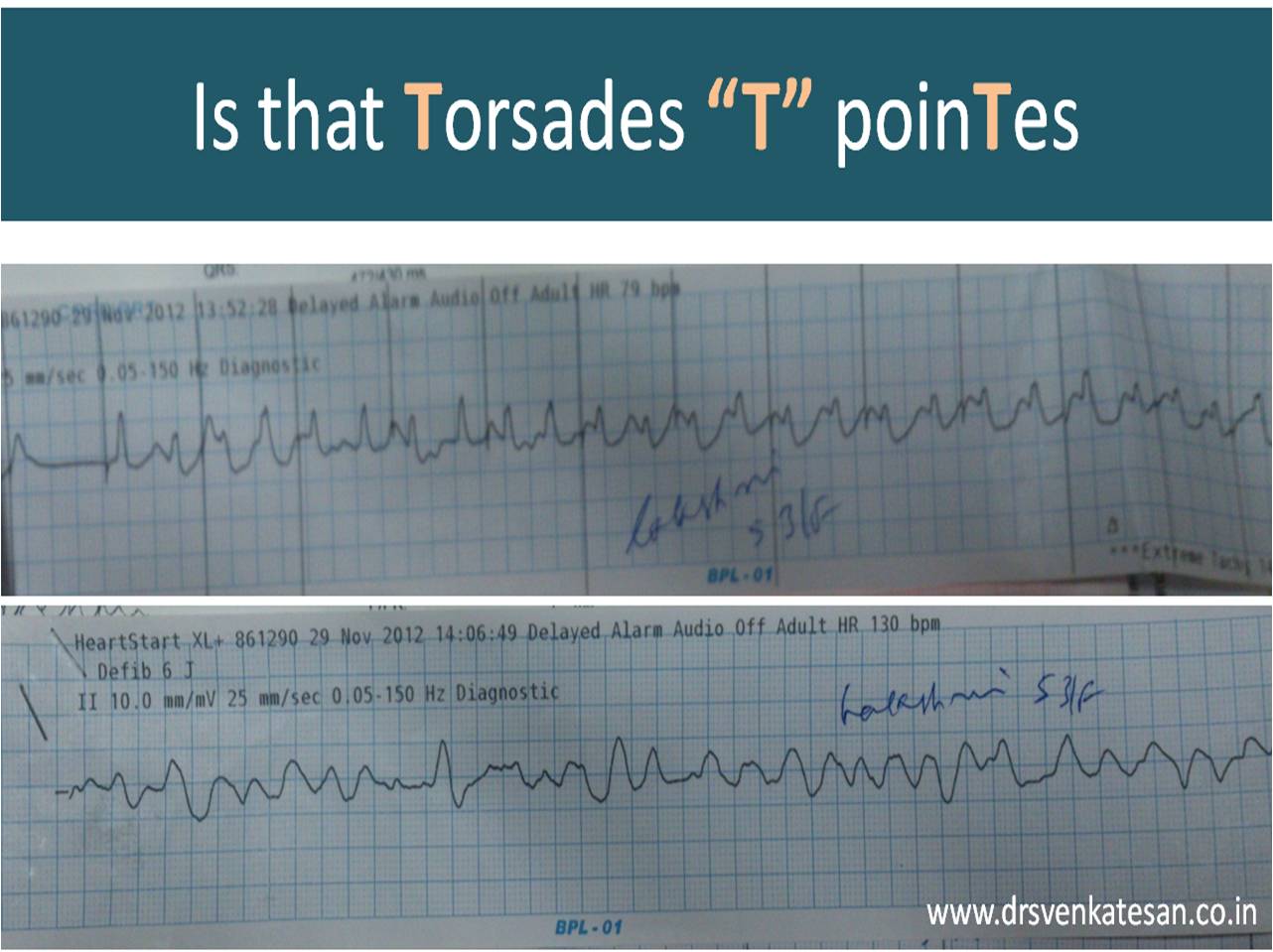
  • In this context, the high prevalence of VT in Prinzmeal angina where there is subepicardial injury is a point to be noted. In young NSTEMI/STEMI crossover entities like Wallens and De-winters, there is a high adrenergic drive, which triggers the VT rather than ischemia per-se.
  • Finally, even in STEMI, only a minority of 15-20 % of myocardium become arrhythmogenic and face fatal complications. What protects the rest of the 75 %? is genetics, epigenetics, or fate.? 

The practical implication of this question

  1. The VT in ischemic cardiomyopathy is related more, to the scar burden, strategically placed islands of dead and live tissues, and the overall severity of LV dysfunction. This makes the Ischemic VT, as a term,  could be a misnomer.  In fact, it is the viable ischemic tissue that is a powerful trigger.
  2. If baseline chronic ischemia is less likely to trigger any VT, revascularisation in chronic CAD (PCI/CABG) is unlikely to give relief from it as well.

Anti-arrhythmic adaptation in chronic Ischemia 

We know about ischemic preconditioning and angina relief. It may apply to arrhythmic preconditions as well. Recurrent ischemia, while we expect to elevate the arrhythmic risk, it is a curious and exciting possibility it might work as an “anti-arrhythmia vaccination” at the molecular level. 

Final message 

So, what is the true relation between ischemia and cardiac arrhythmia?  

It is not as strong as one would believe it to be( Except in the early hours of STEMI and a very small subset of NSTEMI.

Curiously, in certain lucky beings, recurrent baseline ischemia may protect against future arrhythmias.

Reference 

1.A V Ghuran, A J Camm, Ischaemic heart disease presenting as arrhythmias, British Medical Bulletin, Volume 59, Issue 1, October 2001, Pages 193–210, https://doi.org/10.1093/bmb/59.1.193

2.Janse MJ, Wit AL. Electrophysiological mechanisms of ventricular arrhythmias resulting from myocardial ischemia and infarction. Physiol Rev 1989:  69 ; 1049 –169

Few more questions worth pondering 

Why do certain VTs struggle to become sustained?

Will discuss this later (Will need to talk about the diameter of the primary Rotor, the Curvature of rotors and source-sink mismatch, etc. )

How often  Ischemia triggers AF?

I haven’t heard of Ischemic SVT, or ischemic AF much. Trying to accumulate more Info on this.

Read Full Post »

A 5-minute session: Answers are my own. Please cross-check.

1. Is Brugada syndrome clinical or ECG diagnosis?

Always clinical. Never get confused on this.

2. Spontaneous type 1 vs Induced Type 1 (from type 2) which carries more risk?

Both are risky since they are close cousins. But, spontaneous type 1 is the dreaded devil. 

3. Is Brugada primarily a defect of myocardial depolarization or repolarisation?

Not clear. Often in both. In fact a mismatch between them. (Don’t ask how Na+ Channel defect affects repolarisation !)

4. Is Brugada VT is monomorphic, polymorphic?

Both. What determines morphology is not clear though. (All de-nova monomorphic VT will degenerate to polymorphic en route to cardiac arrest)

5. Should  Fever induced Brugada pattern be investigated further?

Better, it is not to be reported in ECG. May not be important in the majority if there is no adverse family history. (If the patient is well educated and afflicted  by Dr.Google and cardiologists can’t escape from ordering sophisticated tests)     

6. What is the overlap between ERS and Brugada?

It is all about the Idiosyncrasy of the K+ channel phenotypes ( Transmural dispersion heterogeneity )  

7. Is a benign Brugada better than a malignant ERS?

Yes, it would seem so. (Inferior or Infero -lateral ERS prone for primary VF in case they develop ischemic / ? also non-ischemic stress)

8. How important is the link between Brugada and Long QT 3 syndrome?

A rare entity, but It is double jeopardy for VT risk. The entire action potential width is vulnerable right from phase 0 to 3 or 4 A case report Sandhu A Clin Case Rep. 2017;5(8):1315-1319.

9. Is Amiodarone really contraindicated in VT?

Not really. Though Amiodarone unmasks Brugada, it can still be used during episodes of VT in patients with manifest or unmanifest Brugada. Maybe in Long QT 3 overlap, it may perpetuate the VT.

10. How important is the structural myocardial defect in Brugada?

Not important in the majority. Though localized RVOT abnormalities are noted in some..RV abaltion can be succesful in odd case.

11. What happens to the ST segment in Brugada during exercise stress?

ST-segment may normalize in some. A stress test can help to risk stratify.  Subramanian M, J Cardiovasc Electrophysiol. 2017 Jun;28(6):677-683.0

12. Which drug is probably best for Brugada as of now?

Quininde , A fairly specific blocker of Ito current. However, it needs to be used diligently. Management of Brugada Syndrome: Belhassen B, Rahkovich M, Michowitz Y, Glick A, Viskin S Circ Arrhythm Electrophysiol. 2015 Dec; 8(6):1393-402.

13. Is ICD definitive therapy?

Obviously not. But, definitely life-saving in high-risk survivors. I guess definitive therapy is possible for future generations through the science of genetic reprogramming of Na+ channels. (Of course, our planet shouldn’t succumb to man-made climatic arrhythmia, by then ) 

14. Does widespread genetic testing & screening of families help in the management and reduce anxiety?

Cracking the genomic code of cardiac ion channels is the ultimate sophistication (Blueprint of fate ?) However, there is no guarantee this information is going to ease out the family members who harbor a genocopy with or without a phenocopy. 

15. Is Brugada getting undue attention in cardiology literature compared to many other common arrhythmias?

      You can answer this …………………………………….


Further reading

Li KHC, Lee S, Yin C, et al. Brugada syndrome: A comprehensive review of pathophysiological mechanisms and risk stratification strategies [published correction appears in Int J Cardiol Heart Vasc. 2020 Dec 19;32:100699]. Int J Cardiol Heart Vasc. 2020;26:100468. Published 2020 Jan 21. doi:10.1016/j.ijcha.2020.100468


Read Full Post »

How many times you have treated cardiac arrhythmia in both emergency & non-emergency situations?

Infinite times.

How many times did you really bother to know the mechanism of a given arrhythmia before ordering medication or shocking?

Hmm,.. let me think. (Except for AVNRT/ AVRT, and few VTs, very rarely I have worried about the mechanism  !)

Why is it so? because treatment takes priority and we are able to tame the arrhythmia even without knowing the real mechanism.

The following slide is a gross summary of the cardiac arrhythmia mechanism

Understanding cardiac arrhythmia is vitally important for a few reasons in a few settings.

  • In acute settings, we need to know automatic tachycardias will not respond to shocks. Reentry tachycardias will respond more promptly. (Of course, we may not know it till we shock ) Calcium blockers like verapamil might block triggered activity in MAT. Overdrive pacing is the answer for many automatic tachycardias and some refractory reentrant tachycardias (ATP protocols in ICD has taught us this ) 
  • In the chronic setting when you contemplate mapping, locating, and ablating arrhythmias, mechanisms are important. The task here is locating slow conduction paths and decoding the diastolic circuit around the scar  (If you plan ICD, knowledge about mechanism  becomes redundant again)

  • Finally, knowing the mechanism of arrhythmia is a fascination by itself to help understand the great subject called cardiac electrophysiology, where 100s of ion channels work nonstop drawing the action potential on a moment to moment basis sustaining our life.

A challenge

Can you localize a VT and find the mechanism in a patient who is Ischemic /hypoxic and acidotic? You can never do it. Please note, most polymorphic VTs can’t be localized. The mechanism is either automaticity, trigger activity, or even micro-reentry. You need to shock and look for the causes.(Link to How does the treatment of monomorphic VT differ from Polymorphic VT? )

Final message

Should we need to know about the mechanism of arrhythmia we treat?  Definitely yes, if you have that passion to know the truth, or else just order Amiodarone or shock and check out of CCU. (Of course, we have a very good option of calling EP consult the next day.)

 A review article on mechannism of cardiac arrhymias

Rev Esp Cardiol. 2012;65(2):174–185

Read Full Post »


A 50-year-old man was referred for dizziness, bradycardia and dysphagia .He was very clear in describing his symptoms and  landed up in Gastro- enterology  OPD , from there was referred to my clinic for cardiac work up . His ECG showed a sinus bradycardia HR of 48 /mt.

Screenshot_2017-07-05-19-09-12

Echocardiogram revealed a structurally normal heart as we expected , but was surprised to spot suspicious shadow in para-sternal long axis view , beneath left atrium.

A well demarcated large mass compressing left atrium.  Trans Thoracic Echocardiography  may not be looking at the heart alone ,(Its technically Thoracic Ultrasound though we may refer it as Echocardiogram   )

  • Aortic aneurysm ?
  • Mediastinal teratoma?
  • Bronchial adenoma ?
  • Esophageal mass ?

The Answer is none of the above

As I was wondering what it was, the staff nurse in charge threw a heavy folder with well worked up gastro Investigations.

That moment , diagnosis became obvious , without a need for further scrutiny to my medical acumen.

Note: The barium swallow of the Esophagus reveals the Intimate relationship between the food tube and the heart as it descends vertically downwards posteriorly  . Realise , how the proximity of these two structures could  confuse a physician when symptoms spill over on either way. (I would have expected a lateral view to show the compressive effect of Esophagus on the left atrium the radiologists felt its not important !)

Yes , it is Achalasia of the cardia , dilating the lower end of esophagus with fluid /mass effect  , compressing the posterior surface of Left atrium.He underwent a myomectomy surgery.

Why bradycardia  ?

There is well described esophago-vagal reflex reproducible by stressful swallow or balloon inflation in the lower end of esophagus at D7 level.(Ki Hoon Kang,Korean J Intern Med. 2005 Mar; 20(1): 68–71.)

Achalasia cardia is known to be associated with symptomatic bradycardia, dizziness, and rarely swallow syncope,though this patient didn’t have a classical syncope.The bradycardia is probably due to high vagotonia, (Hugging effect on posterior surface of heart known for rich innervation of vagus.) . Complete reversal  of bradycardia after esophago -gastric surgery is expected.

Implication for cardiologists

There has been instances of patients with esophageal syncope and reflex bradycardia getting permanent pacemaker therapy. I think , clinical or sub clinical esophageal disorders should be included in the work bradycardia before labelling them as intrinsic sinus node dysfunction .(Ref 1,4)

Final message 

The field of Cardiology  is often referred to as a super specialty atleast in India . I disagree with it strongly. Cardiologists are neither super(eme) nor special .We need to be reminded  its afterall a sub-specialty of Internal medicine and each specialist should undergo retro-training in medicine periodically .This patient is a typical example of a gastric problem entering the domain of cardiac Imaging.Strong foundations in symptom analysis and some degree of medical  curiosity will enable an occasional cardiologist to make a correct diagnosis belonging to a remote foreign specialty.

Reference 

1. Palmer ED. The abnormal upper gastrointestinal vagovagal reflexes that affect the heart. Am J Gastroenterol. 1976;66:513–522. [PubMed]

2.Armstrong PW, McMillan DG, Simon JB. Swallow syncope. Can Med Assoc J. 1985;132:1281–1284. [PMC free article] [PubMed]

3.Turan I, Ersoz GBor S..Swallow-induced syncope in a patient with achalasia
4.Dysphagia. 2005 Summer;20(3):238-40  4.Basker MR, Cooper DK. Oesophageal syncope. Ann R Coll Surg Engl. 2000;82:249–253.

Read Full Post »

Junctional tachycardia(JT) is often a misunderstood arrhythmia. Technically,  any tachycardia arising from the AV junction could be termed as JT.Even AVNRT was considered as a form of Junctional tachycardia till recently.The crux of the issue is , true anatomical extent and borders of  so called AV junction is  yet to be clearly demarcated .The common perception that  AV node is a discrete  structure is  an anatomical illusion  , rather its collection of  condensed fibers with proximal  nodal approach and distal fanning .

Now , we have a  proper definition by the apex scientific bodies  ACC/AHA/HRS 2015)

definition of junctional tachycardia

Source :2015 ACC/AHA/HRS guideline for the management of adult patients with supraventricular tachycardia: Executive summary A Report of the American College of Cardiology/American Heart Association Task Force on Clinical Practice Guidelines and the Heart Rhythm Society April 2016Volume 13, Issue 4, Pages e92–e135

Please note :The key point is , JT by definition  should  be a focal  /automatic tachycardia either due to triggered activity or after depolarisation and the boundaries of  junctional tissue is liberally extended up to  His bundle.

Read  related post  :What does the term junctional tachycardia mean in current era?

Reference

http://www.heartrhythmjournal.com/article/S1547-5271%2815%2901188-1/pdf


Read Full Post »

J point is a critical point in the  ECG  when the ventricles hand over the baton in  the  electrical relay race from depolarization to repolarization .This the time the sodium  channels extinguish itself  and the potassium current begins its activity  from Phase 0 to 1 .

If the  potassium channels  activate little early and snatch the baton prematurely from sodium , we get early repolarization pattern .When this happens , the J point of ECG show a conspicuous wave  called J wave , originally  denoting  Junctional wave between QRS/ST segment  (Now  perceived as  Jitter waves ?) The other implication of premature K+ activity is , lifting up of  ST segment , making it the most common cause of non ischemic ST elevation.

* J wave in hypothermia is referred to as Osborne wave and  may not be  not related to ERS(Ref.4)

J wave and J point early repolarisation syndrome

Image source.www.cardiology.org

The Ito current is responsible for the phase  1 of action potential (AP), where a rapid outward k + ion flux take place and draws the dome of AP . The dynamics of Ito is complex .It depends  upon the density of epicardial K + channels , which are  clustered in a heterogeneous manner .There seems to be a concentration gradient   along the epicardium and endocardium , making the wave appear prominent in some. This is especially true in healthy, athletic  male population  where we have some evidence for androgen  to  play a role on how  these channels will behave.Here comes the overlap between Brugada  syndrome and ERS as well.

The subset of patients with J wave pattern were recently shown to have increased risk of primary VF due to phase 2 reentry ,  when they develop ACS. (Rather J wave pattern was more common in patients who had primary VF following STEMI(Ref 1).This resulted in a spate of worrying articles .Now we know , the  fear is  largely unfounded ,the risk is far less.

Current thinking is,  persons who have asymptomatic ERS pattern with prominent J waves should not be investigated electro-physiologically . (Please remember , every human  heart can be induced  to VF in EP lab  if appropriately  stimulated ! )

In fact , I used to tell the  young men  who  harbor  prominent J wave , as a marker of healthy heart  rather. Let us not  fear them with a remote risk  that could be as  negligible as risk of  intercontinental flight crashing into the ocean  !

References

1.Haissaguerre M, Derval N, Sacher F, et al. Sudden cardiac arrest associated with early repolarization. N Engl J Med. 2008;358:2016–2023.

2.Idiopathic Ventricular Fibrillation “Le Syndrome d’Haïssaguerre” and the Fear of J Waves , Sami Viskin, J Am Coll Cardiol. 2009;53(7):620-622. 11

Read Full Post »

Cannon waves occur when Atria contracts against a closing tricuspid valve of  right ventricle .( There  would be a equivalent left atrial cannon which  goes into pulmonary vein as well  , it is discussed elsewhere !)

Cannon waves  happen only when P waves fall within QT interval in ECG as QT represents the electro-mechanical systole of  ventricles.  (Since P wave represents atrial systole , it is simple to understand when it falls within QT both atria and ventricular contractions collide to produce a cannon wave into the neck or pulmonary veins.)

The following two images of cannon waves  taken from the legend  Dr Paul woods own tracing  .

irregular cannon waves in jvp  complete heart block

regular cannon waves in jvp  svt avnrt  11 va conduction  002

Regular cannon waves

Occur during SVT  with 1:1 VA conduction.*

1 : 1  VA conduction  can be considered as  absence of  AV dissociation  (Rather  disciplined  VA association with every beat ) This is essential to create a hemodynamic  milieu for regular cannon waves.

* In AVNRT , VA conduction in strict  sense  is a misnomer  .It is simply a retrograde conduction thorough  the AV node .

Irregular cannon waves 

  1. Complete heart block .
  2. Multiple random VPDs
  3. Some patients with VT.*(Who are those patients ?  Those with AV dissociation when retrograde “P” wave falls  within QT interval cannon occurs. As expected this occurs in random fashion  which makes  the cannon fire irregular.

Can we get regular cannon in VT ?

Yes , but rare . As explained earlier this can happen only if AV  association occur on a retrograde fashion.

Further reading in this site

What-is-a-cannon-sound  , how is it related to cannon wave ?

Read Full Post »

Wandering pacemaker is benign  cardiac arrhythmia . The only danger  is , it can create false alarm .This patient was referred   as  AV dissociation

wandering pacemaker

Read a related article  from this site .  ( A restless pacemaker goes for a walk down the  lane )

wandering pacemaker

wandering pacemaker

Read Full Post »

Pacemaker current is   strangely  referred  by physiologists  as  funny current (I f ) . I am yet to find the exact reason .  This is the current  that  sustain  our life right from the day 22  of  embryonic life when the  cardiac jelly beats for the first time.   SA node  solemnly  follow our  entire life  before  making  a  bid-adieu !

pacemaker  potential sa node 5


pacemaker current if funny current poential 002

pacemaker current if funny current poential 003

What is contribution of  If  current in the overall Pace-making  activity ?
This  has not been quantified . The fact that ,  Ivabradine induced  If  current  blockade does not result in serious bradycardia indicate  , SA node has alternate reserve currents as well . ( SA node  is a such a mystery  structure , it would never be a  surprise , if we  find many more  “not so funny”  currents !)

Read Full Post »

It is believed  T wave alternans  is a marker of impending ventricular fibrillation. Though it is not applicable in every clinical setting it is indeed true if we observe T wave alternans in an acute ischemic setting .Here is a patient with  ACS and inferior MI who developed T wave alternans after temporary pacing.

T waves alternans torades de pointes 3

t wave alternans

T waves alternans torades de pointes

went in  for a chaotic  T wave rhtythm  and ended up in VF that  required s shock.T wave  alternans is other wise known as repolarisation alternans .

Twist dance of Heart

Torsedes is twist around it’s axis.   Any   ECG wave  can twist in it’s axis .If T wave alternans  becomes gross it will twist 180 degrees   .Once this happens the heart can go for  fibrillation any moment !

Final message

Extreme form of T wave alternans would result in  complete twisting of repolarization vector which is a  harbinger of ventricular fibrillation

I wish  this can be referred to as Torsades  “T” pointes instead of  Torseades “de” pointes


Read Full Post »

Older Posts »