Feeds:
Posts
Comments

Archive for November, 2019

Whenever a patient is getting discharged after a PCI, the treating cardiologist often faces this situation.

So, you fixed the block in my coronary artery doctor. Thank you so much. Now, I can have a peaceful life, free from  future heart problems. “Am I right doctor”?

I wish I can answer “Yes”  to your query but I can’t for the following reasons.

I have fixed only a lesion that caused maximum obstruction. Atherosclerosis is a diffuse disease and you have minor plaques scattered across your coronary artery. These can grow at its own will. So you carry a definite risk remote from the current problem. (Don’t get frightened, read further, you have definite solutions to reduce this risk.)

How common is the progression of native vessel disease?

It varies from 10 to 40%. Mind you, the exact incidence directly depends upon the compliance of medical management, risk factor reduction, and adaptation to a new life healthy lifestyle. In effect, you (the patients) decide the incidence.

One surprise phenomenon (though unproven) might happen. Since the tightest lesion is jailed with a scaffold the minor lesion is preselected to an accelerated process of atherosclerosis if medical treatment is not properly followed.

Dr.Zellweger from the university hospital, Basel, did an extraordinary study with 400 patients, meticulous 5 years follow up with SPECT and found remote lesions accounted for 40% of future events (Basel Stent Kosten-Effektivitäts Trial [BASKET]) The other study by Glazer and concurred with this. These studies reiterate the importance of taking care of the entire coronary artery instead of focused piecemeal care by scaffolds.

Does a proximal DES protect a  distal lesion in the same artery by the drug effect?

It is a good thing to happen at least on paper. A proximal LAD with the latest generation Everolimus coated stent is expected to keep the distal LAD drugged for few months at leas.( with anti-mitotic activity) Thus preventing the progression of distal lesions.

No, I can’t believe this.In this era of momentary touch on sidewalls of artery by drug-eluting balloon (DEB) shown to do wonders, anything is feasible. Chacko’s (Ref 2 : JACC CV Interventions 2009)observation has a possible answer for this. It showed BMS vs DES didn’t make any difference in remote lesion progression.

Final message 

These studies reaffirm one vital truth. Stents are temporary solutions to a permanent, systemic disease of the vascular system .Stents are indeed a major revolution in CAD, “if and only if” it’s used in a highly selected CAD population. Global attempts to project cath labs as a tool to control human atherosclerosis is a typical example of flawed science. The only effective way to tackle this menace is to faithfully follow overall healthy living,  assisted by drugs.

This is the Editorial in response to Zellweger’s article


Reference

1.Glaser RSelzer FFaxon DP,Clinical progression of incidental, asymptomatic lesions discovered during culprit vessel coronary intervention. 2005 Jan 18;111(2):143-9 2004 Dec 27
3.

Postamble

One of my patients asked some time ago. If stents are the definite remedy for severe arterial narrowing, why not stent all my lesions (even the minor ones ) prophylactically doctor, so that it will not become tight at a later date?

That’s a good query. Your doubt is genuine , appear logical as well. But, unfortunately, it will be the most dangerous thing to do*. Metals are never friendly with the coronary arterial wall. We should use it extremely judiciously and only with tight flow-limiting lesions. These metals require annual (rather permanent) maintenance. Its taken care by multiple antiplatelet drugs. If for some reason your maintenance is erratic or the drugs fail to act you are at more risk of a future event.

(* This is what has  happened (happening) in the past, that demanded urgent publication of appropriate usage criteria)

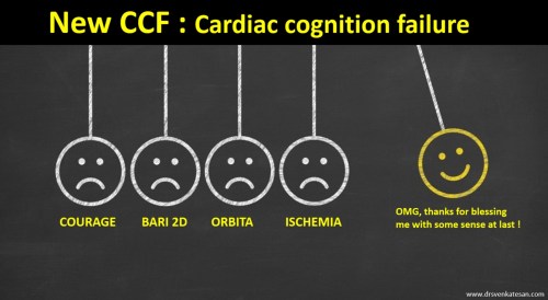
Now, the current belief among the “fair thinking cardiology community” is dramatically changing. It’s leaning towards non-stent management even with significant flow-limiting obstructions in otherwise stable patients(SIHD). This belief is accruing more and more evidence base (The COURAGE 15 year follow up / ORBITA/ISCHEMIA)   All these studies confirm the emerging doctrine and bring back some semblance of sense into the cardiology community.

Read Full Post »

*ISCHEMIA trial breaks not in NEJM or Lancet but in Washington Post and Wall street Journal

After three decades into cardiology profession, one thing is very clear. We work so hard to create pseudo-knowledge and struggle with it for so long and feel awkward and guilty to come out of the mess. But we have to  . . .  in the overall interest of mankind, isn’t?

We aptly call the whole process as continuing medical education, but in the melee, often we ditch some of the precious gems as obsolete. (This tempts me to suggest discontinuing false education is also an option for medical knowledge seekers !)

Confucius has something to say about this issue , which appears more relevant to the medical profession in current times.

Postamble 

We don’t know what’s in store for 2020

Read Full Post »

I have never found it difficult to retrogradely cross a dangerous epicardial collateral in complex CTO. Delivering a twin stent in a partial culotte strategy for a bifurc lesion has never tested my talents. Stenting a left main across the LAD, jailing the LCX with OCT support is my favorite time pass. Crushing a calcium infested diffuse long lesion with diamond-tipped ablator appear as breezy as shopping in a mall.

But this one is really challenging 

What is that?

Understanding these four studies (Ref 1-4 ). They dogmatically say medical management confers definitive protection in chronic coronary syndromes. It stretches our limits of Imaginary Intelligence! How can a near tight coronary obstruction sitting right across your eyes, be left untouched? The latest one seems to suggest we can even ignore FFR positive lesions.

COURAGE BARI 2D ORBITA ISCHEMIA DRSVENKATESAN SHD CHRONIC STABLE HEART DISEASE PTCA PCI ACC ESC SCAI 2GUIDLEINES

It requires 4 negative forces . . . to bring one big positive Impact!

I don’t understand who is funding these negative trials and glorify it, and trying to defame the talents in me. All these studies have a huge lacuna. They conveniently exclude high-risk cases and allowed liberal cross over to PCI later on. Even the just-released ISCHEMIA trial had 38 % patient with no angina. (But why they received PCI ?) How to Interpret these trials and extract the true conclusion? .One consolation is, I know these negative trials have a very short memory and expiry date. Very soon I shall be liberated from the clutches of this negativism.

Even as I scribble this, my inner conscious is telling a completely different story. I agree we do Indulge a lot in stable Ischemic Heart disease. (SIHD).  I am yet to be clear what exactly we mean by SIHD. How is that near 90 % mid LAD guy ran 12 met exercise with negligible perfusion defect and still FFR was .7 ?

OMG, save me from this academic conundrum and help to acquire true wisdom.

Reference

1.Weintraub WS, Spertus JA, Kolm P, Maron DJ, Zhang Z, Jurkovitz C, et al. For the COURAGE Trial Research Group. Effect of PCI on quality of life in patients with stable coronary disease. N Engl J Med. 2008;359(7):677–687. [PubMed[]

2.BARI 2D Study Group. Frye RL, August P, Brooks MM, Hardison RM, Kelsey SF, MacGregor JM, et al. A randomized trial of therapies for type 2 diabetes and coronary artery disease. N Engl J Med. 2009;360(24):2503–2515. [PMC free article] [PubMed[]

3.Al-Lamee R, Thompson D, Dehbi HM, Sen S, Tang K, Davies J, et al. ORBITA Investigators Percutaneous coronary intervention in stable angina (ORBITA): a double-blind, randomised controlled trial. Lancet. 2018;391(10115):31–40. [PubMed[]

4. International Study of Comparative Health Effectiveness With Medical and Invasive Approaches – ISCHEMIA


Read Full Post »

Differential cyanosis classically occurs in PDA with reversal of shunt when raised PA pressures /PVR is able to supersede the systemic Aortic pressure and drive the blood from LPA to descending Aorta bringing down the lower limb saturation.

Of course,  this can be undone by the presence of any other intra-cardiac shunts or aberrant left subclavian that arising from the desaturated descending aorta.

Other causes of reversed differential cyanosis 

Where the upper body is cyanosed (desaturated) and the lower half is not. There is a conventional list of conditions.

  1. Transposition of the great arteries (TGA) with patent ductus arteriosis (PDA) and elevated pulmonary vascular resistance
  2. TGA with PDA and pre-ductal aortic interruption or coarctation
  3. Supracardiac TAPVC* + PDA
  4. Anomalous right subclavian artery connected to hypertensive ductus through RPA

(*This occurs due to streaming effect ) Highly saturated superior vena cava (SVC) blood into the right ventricle, reach MPA / through a PDA, and to the descending aorta, with streaming of more desaturated blood from the inferior vena cava (IVC) into the LA through PFO (Ref Yap S H Pediatr Cardiol. 2009 )

Now let us add one more cause for  reversed differential cyanosis in the Modern Era

It is seen with ECMO in VA connection (Often reported in babies ) . The Aorta has high oxygen content entering from the femoral cannula going up into the Aortic arch., while deoxygenated blood from LV (because of failing lungs) reach antegradely to the Aorta. Ideally, the ECMO is expected to supply the entire aortic arch and hence oxygenation is uniform all over the body. It rarely happens as some amount of flow will come from LV unless its in asystole. However, If the severely dysfunctional heart tends to recover & lung oxygenation is very poor as well, the LV stroke volume competes with highly oxygenated blood coming from below ( femoral inflow ) into the Aorta , creating a watershed zone . This makes the deoxygenated blood perfusing upper half of the body and hyper oxygen saturation lower half. This is been referred to as North-south syndrome or (Harlequin syndrome the famous Italian comical character)

How to manage North-South syndrome?

  • Try to Improve the oxygen perfusion with high-frequency ventilation(This is logical first step , to improve the native lung function)
  • ECMO flow rate may be increased and overdrive the LV ejection .(This can be counter-productive as we are hitting a recovering ventricle)
  • Converting to VV ECMO if the hemodynamics allows. This is possible as North-south syndrome is a sign of recovering cardia function VV ECMO will convert it into a primary lung support

Reference

ECMO review article

Read Full Post »

LV dysfunction is one of the most commonly used terminology by cardiac professionals.It can be systolic, diastolic or global, regional etc. But, before dysfunction sets in, the heart fights. The Left ventricle can behave in many different ways when confronted with stress. It increases the force of contraction, elevates it’s Intra cavitary filling pressure and still accomplishes its task of pumping adequately. Further, It can build fresh muscle (LVH). It can double up with more heartbeats. (All these factors are referred to as cardiac reserve mechanisms)

These reserve mechanisms can be activated in the short or long term. In the long term, autonomic activation with neuroendocrine factors joins the compensation process.  These will work for some time till the circulatory system settles down to new homeostasis. However, they become counterproductive and becomes decompensated, ultimately heart failure sets in(Unless Intervened)


Is LV dilatation a mechanism of cardiac reserve ?

No one calls LV dilatation as a reserve or compensatory mechanism. (I wonder, why not ?) I think like RV ,  LV too has some potential to reversibly dilate . The quantum of which we are unable to estimate.This happens usually in response to chronic  volume stress* like regurgitant valves or high output states. Though cardiomegaly and a huge heart convey a sinister outcome, many hearts shrink if the primary issue is corrected.(Typically in Anemia, Beri Berri. We also know LV may transiently dilate in response to some toxic /pregnancy-related cardiomyopathy.

* Mind you LV poorly tolerates acute volume stress as in Acute AR/MR

The critical gap in our understanding is about this question.

When does LV dilate physiologically and when pathological persistent LV dilation sets in (The absolute state of irreversibly lost cardiac elasticity.) We also know dilated LV will consume more oxygen due to enhanced wall stress (Laplace law) and hence its possible LV dilatation begets further dilatation. Optimal timing of mitral and aortic valve replacement in patients with AR and MR directly depend on this knowledge.

Final message

We need clarity in the following queries

  • Is LV dilatation (with normal EF ) a sign of LV dysfunction?
  • If so at what level of dilatation?
  • Since LV dilatation  occurs in diastole can we fit this entity “Isolated LV dilatation” in the already confused spectrum of diastolic dysfunction?

Let us wait for the knowledge to evolve. Young cardiologists could take up this area for research.

Read Full Post »

I asked some of my experienced colleagues, how much time they inflate the balloon to deliver a stent? Most answers were spontaneous and unanimous “It’s hardly 10 seconds,  few said maybe up to 15s.

Can prolonged balloon inflation time reduce the need for post dilatation and prevent mal-apposition?

We know high-pressure Inflation( up to 20 atmospheres ) was a big revelation in the science of PTCA more than two decades ago. (Antonio Colombo JACC 1995  ) He proposed and proved high-pressure inflation eliminated the need for routine anticoagulation following stenting as approximation was better. He also pioneered the concept of dual antiplatelet therapy (DAPT) in the PCI arena.

Similarly, prolonged balloon Inflation  (30 to 60sec) could be another trendsetting tip to prevent malposition. It delivers more sustained pressure. Its believed the imparted centrifugal force and the inbuilt radial forces add up to the stent vessel wall Interface and prevent mal-apposition.

Is there a downside to high-pressure Inflation?

There must be few.  Potential new Ischemic events and arrhythmia. In calcium laden plaques( spur) risk of perforation may be enhanced.

Final message 

I don’t know why this concept never took off. Many of us still fear to inflate the stent balloon no longer than 10 to 20 seconds? Adhoc post dilatation with short NC balloon appears mandatory in areas of mal-apposition. Meanwhile, we also understand sustained (30-60s) high-pressure initial inflation could deliver the stent in a more synchronized and smooth fashion with a perfect metal/vessel wall interface. Further , prolonged balloon inflation times could make a routine (By the way who does routine ?)  IVUS/OCT redundant.

What do the experts say?  What does science say?  There is one meta-analysis that clearly says the advantage of long inflation time. This issue becomes much more relevant as it could avoid post dilatation which all of us know can be tricky. In fact, every balloon dilatation should be technically counted as another PTCA procedure and adds up to net total risk.

Reference

1.M. Saad, M. Bavineni, B. F. Uretsky, and S. Vallurupalli, “Improved stent expansion with prolonged compared with short balloon inflation: a meta-analysis,” Catheterization and Cardiovascular Interventions, vol. 92, pp. 873–880, 2018. View at Google Scholar 

2.https://www.researchgate.net/publication/317175130_Shorter_duration_of_balloon_inflation_time_results_in_greater_malapposition_during_PCI_with_DES_in_patients_with_stable_coronary_artery_disease_a_randomised_control_trial_of_the_second_STent_OPtimisat

Read Full Post »