Feeds:
Posts
Comments

Archive for May, 2012

Hypoxia is most important feature of acute pulmonary embolism.

It occurs due to variety of mechanisms

  1. Ventilation perfusion mismatch is the major  mechanism  ( Normal ventilation /Reduced perfusion)
  2. Atelectasis of lung  ( Left to right shunt)
  3. Loss of lung volume due to pulmonary infarct  contribute later
  4. Low mixed venous Oxygen saturation  (Tissue hypoxia -more extraction )
  5. One more important cause is right to left shunting  across PFO  due to sudden elevation of right atrial mean pressure reflected from RVEDP .

Can  acute pulmonary embolism be diagnosed  with out Hypoxia ?

Surprisingly many standard text books mention hypoxia is a soft sign . In fact , Braunwald’s  text book of cardiology  do mention about it .

Significant acute pulmonary embolism can not occur without affecting o2 saturation .

However , it is possible sub acute  pulmonary embolism could occur with normal oxygen saturation.

Final message

Hypoxia is indeed a hard sign  for most events  of major pulmonary embolism . It can even be termed as an essential criteria .A hypoxic , tachypenic patient in  sinus tachycardia with echo evidence of  new onset RA or RV dilatation is almost 100 % specific for acute pulmonary embolism . ( This becomes 200 % if he or she has DVT as well !)

Read Full Post »

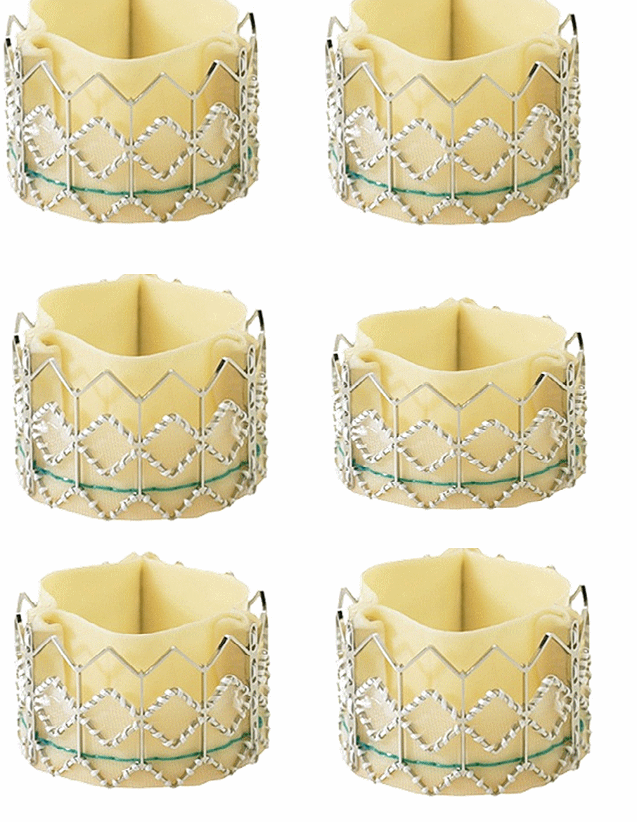
Cardiologists are  closing in ,  trying to capture the final frontiers. The  trans-cutaneous Aortic valve Implantation now has  a two year follow up. (NEJM March 2012  Issue) . The results are encouraging .

While two companies are fighting for the supremacy in TAVI ,   the real  threat is for the cardiac surgeons. Currently Edward  Sapiens  has an edge over Medtronic core valve as it  has a provision to redeploy or fine-tune the  final geo- position.

Reference

PARTNER 1

PARTNER 2

Medtronic core valve

Open access  article  by Martin Leon

http://www.rmmj.org.il/userimages/22/1/PublishFiles/25Article.pdf

Read Full Post »

We owe a lot to our past genius minds for our current understanding of  cardiology.Youngsters   should  know how the filed of cardiology  evolved .Few  great  brains  taught us how to think   hemodynamically  in the setting of  STEMI.

The Diamond and Forrester classification is  an  undisputed achievement of  modern cardiac  hemodynamics.They gently converted the  clinical classification of  Killip into more scientific  hemodynamic  one .Both these classification continue to fascinate  us even in the era of instant PCI for STEMI .

And youngsters  should read this again and again and critically evaluate their patients  within this system.The two key parameters he used was PCWP of  18mmhg /And cardiac Index 2.2liters . He also suggested a simplified version where  intra- arterial monitoring is not feasible.  The   cardiac Index could be replaced by systemic blood pressure  lung congestion   represents PCWP >18mmhg .

The DF classification would become

An important inference from DF classification !

The class 3  of   DF   grading  has no pulmonary congestion  but persistent hypotension . What does it mean ?

It is a stunning proof of a great concept.  As the patient moves (Worsens)  from  DF  two  to   DF three  , the lung congestion tends  to regress . This sub-set  actually  means   development of  bi-ventricular failure or isolated RV failure  . This is an ominous sign and indicate a bad prognosis . ( One may call it a paradox  , according to conventional thinking   “The more the lung crackles  , dismal  is the outcome”   DF  grading clearly proves this is  not  always true ,  as long as  the systemic pressure is maintained  crackles can be managed effectively  . In  DF 3  the right ventricle  as a pump is  becoming so weak it is not able to congest the lungs  at the same  process leads to  systemic hypotension.

James Forrester

http://www.cedars-sinai.edu/Bios—Physician/A-G/James-Forrester-MD.aspx

Forrester is also a pioneer in how we evaluate chest pain in the emergency rooms and cardiology OPDs .  His thoughts on utilization of Besean theorem revolutionized   the interpretation of exercise stress testing.

* Killip is a genius of different caliber would be discussed later .

Reference

Forrester, J, Diamond, G, Chatterjie, K, et al Medical therapy of acute myocardial infarction by application of hemodynamic subsets (first of two parts). N Engl J Med 1976;295,1356-1362


Read Full Post »

Magnesium   is a powerful anti-arrhythmic drug . It has a well  established role in controlling VT when administered  Intravenously   especially in polymorphic VT .

Mechanism of action

  • It acts at the cell membrane.
  • It has a unique action of blocking calcium channels  that reduces the number of oscillations of  both  early and late  after potentials

Link for more  on mechanism  of action

https://drsvenkatesan.wordpress.com/2010/01/13/how-does-magnesium-acts-as-an-antiarrhythmic-drug/

How often cardiologists administer oral magnesium for long-term control of VT ?

As for as I know ,  no one uses it ! but dietary  supplements are used for general well  being .

Why ? Is it because

  1. Magnesium does not get absorbed in the gut
  2. Magnesium levels are un- predictable in plasma if administered orally

Answer : No one has really tried  it as a  chronic therapy in VT  yet  !

Final Message

Tablet Magnesium can give a tough fight to Amiodarone and Flecanaide in refractory VT at a fraction of the cost !

Who has the audacity  to  compare Magnesium  with Amiodarone head on ?

Reference

Magnesium as health supplement . 

Magnesium is available  in tablet form as  Malate , Stearate, Taurate and Aspartate  along with calcium and Zinc etc .

Read Full Post »

The entity of stress cardiomyopathy ,  other wise referred to as  Takotsubo  cardiomyopathy is a popular clinical entity in recent decades.The heart and mind are closely linked entities even though they are  situated apart physically . Extensive neural and hormonal control  mechanisms  exist.

In extreme stress ,the hyper- sympathetic  drive triggers a rush of adrenaline ,  which some how makes the  left ventricle  to bulge out !

The clinical features  are varied .

  • It can exactly mimic an acute coronary syndrome .
  • ECG may  show ST elevation and mimic an anterior STEMI
  • Echo shows a wall motion abnormality  classically  described  as the apex alone dilates /Bulges or elongates
  • LV  may acquire a shape of a  banana. (See below )

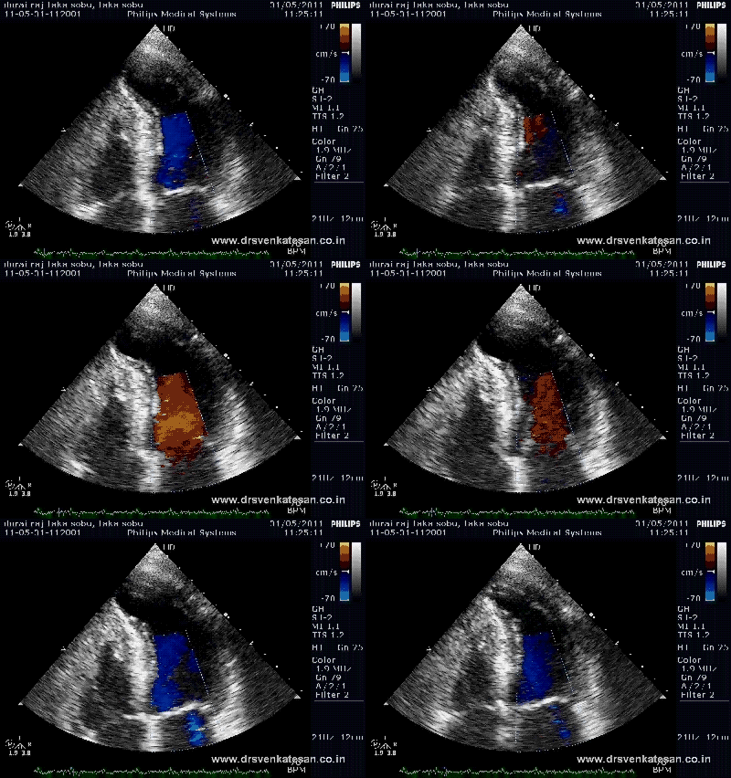
A 45 year old man came to the ER with severe chest pain , dyspnea and minimal ST elevation in anterior leads. He  was a smoker and was experiencing  recent major office stress  . Echo showed an elongated LV apex with some thinning .We made a diagnosis of stress cardiomyopathy .( It was disputed by my professor as the LV  apex was contracting well   ! but we  learnt later there are many varieties of Takatsubo )

Echo showed an elongated LV apex with some thinning . Note the LV apex goes  out of plane  with RV apex.

Color  Doppler revealed Trivial Mitral regurgitation

Follow up

He underwent coronary angiogram.  Had  no significant lesions ,   in 48 hours time the wall motion defect disappeared and was discharged with beta blockers.

Incidence

Up to 2 % of ACS could be related to Takatsubo . More common in women especially post menopausal  , with stressful/emotional background like loss of loved ones.

Synonyms

Apical ballooning , Broken heart syndrome ,  Stress cardiomyopathy.

Mechanism

Not clear . Microvascular spasm , excessive catecholamines  ,  are thought to be major culprits.

Echocardiography

Hyperkinetic base and akinetic or dyskinetic LV apex .

Lots of variations are reported .

Shimizu described 4 types

Courtesy : Shimizu et al J Cardiol. 2006 Jan;47(1):31-7.

  1. Apical akinesia and basal hyperkinesia,
  2. Reverse  Takotsubo  (Basal akinesia and apical hyperkinesia)
  3. Mid-ventricular ballooning   with  basal and apical hyperkinesia
  4. Localised  to any one segment

*The Banana type which  is described here (Elongation  of LV apex > Widening )

Histopathology

Focal myocytolysis are described. (Broken heart)   Monocytic infiltrations are common.These are  believed  to be transient .

How to differentiate it between a STEMI ?

  • Enzymes are only mildly elevated.
  • Wall motion defect do not confine to a specific arterial territory.
  • Most importantly coronary angiogram do not reveal any significant obstructions.

Prognosis and outcome

  • Generally good
  • The initial presentation may be turbulent in few with cardiac failure or arrhythmia .Other wise these patients do well

Treatment

  • Mainly supportive
  • Major principle is to avoid inotropic agents as they  are already  heavily expose to it
  • Beta blockers  could be the mainstay therapy .

Final messge

Think about  Takatsubo  whenever an acute coronary syndrome presents atypically . Not surprisingly few of them land in the cath lab !

Reference

http://www.cardiologyrounds.org/crus/cardus1206.pdf

Read Full Post »

Irregular  wide qrs tachycardia is a fairly common clinical entity in any cardiac emergency room. The moment you ask about  such tachycardia ,  9/10  fellows will  come out with a  prompt answer   ” AF with WPW syndrome” even before you complete the question !  It is not that common  as we perceive .The problem is with  our traditional teaching methods and the attraction of human brains to  rare and exotic disorders.

traditionally   SVT with aberrancy  is   diagnosed  mainly  in the setting of regular tachycardia .

We often  forget  “AF with aberrancy”  is equally common  , and  it presents   with a  irregular  wide qrs tachycardia . 

I  wonder whether  this phenomenon  can be termed as  orthodromic aberrancy .This can directly compete  in the differential diagnosis  of  antidromic AF  with  WPW !

It should also be mentioned antidromic  AF can run into very high rates  as accessory pathways do not check the incoming signals while orthodromic aberrancy the ventricular rates can not exceed 220 or so at least theoretically . (This simple clue can clinch the issue in favor of  WPW )

There is no proper  published data available for the true  incidence of AF with orthodromic aberrancy in general population

In fact , there are  many  electrical  environments for AF  to  become a  wide qrs AF

1. AF  with  Antidromic conduction through accessory WPW pathway.

2. AF with Orthodromic aberrancy ( Non WPW – Similar to  any SVT with aberrancy )

3. AF with pre existing LBBB

4. AF  with Amiodarone effect. (Especially with DCM and cumulative load of Amiodarone )

5. AF with electrolytic /  especially excess  intra-cellualr  potassium

6. Finally , even  Atrial based pacing (DDD)  can cause wide qrs irregular tachycardia when  mode switching  fails .Here the  ventricles  may track the  atrial irregularity  and respond with a  wide qrs  bizarre tachycardia .

Final message

There are many causes for  wide qrs tachycardias  in  Atrial fibrillation . WPW with anti-dromic conduction is just  one of them .We need to approach the issue with an open mind .Please  be reminded , once contemplated  WPW syndrome  can be a powerful thought blocker  !

Note : *We are not including   polymorphic ventricular tachycardia here .It is an  important subset of  wide qrs irregular  tachycardia.

** VT can co-exist with AF .This is not   surprising  as  many of the diffuse cardiomyopathies  involve  both atria and ventricle  with extensive scarring and fibrosis  a perfect trigger for  both atrial and ventricular arrhythmias .

Read Full Post »

Interventions in Eisenmenger  syndrome  or severe PAH in  left to right shunt continues to be a major diagnostic issue.The challenge lies   not only in  assessing whether the progression of PAH can be prevented by  blocking the left  to right shunt , but also  to assess  it’s impact  on  survival.

The factors  involved are

  1. Pulmonary artery pressures
  2. Pulmonary blood flow
  3. Pulmonary vascular  resistance
  4. RV function
  5. Co-morbid /general condition of the patient

While cardiologists worry more about LV , surgeons have different issue .  In left to right  shunts with PAH  RV function bothers them more , as the high pulmonary artery pressure may never allow the surgeons to come off the pump , once the decompression provided by ASD/ VSD  is removed

How relevant is Ohm’s Law in complex shunt with leaky valves and bidirectional shunting ?

The fundamental hemodynamic equation  is derived from  Ohm’s law .How relevant  is  Ohm’s law in Eisenmenger  is not clear.  For decades we have been using complicated calculations with many presumed  and assumed parameters.  The calculation of effective pulmonary blood flow in bidirectional shunt may be most complex equation in clinical  cardiology. One can only imagine how one error could amplifies the other.

The hemodynamic equivalent of  Ohm’s law states

R = Pressure / Flow .The current thinking is  If the PVR is between 6-8 it is operable .

Is it really that simple ?

We know pressures  can be measured with a fair degree of accuracy . Flow  and resistance are  subjected to change in a  moment  to moment basis  .They are  determined by a gamut of  neural and humoral factors.

Ironically , we are not yet clear , whether flow determines  the pressure or pressure determine  the flow .

The right heart blood flow can get complicated by not only bi-directional shunt but also  by pulmonary  and tricuspid regurgitation ,

There is a huge perception problem here .  We are tuned  to think ,  reversibilty of PAH is  same as operability  of shunt lesion . Definitively not !  This is the reason why there is  a vast difference in  ultimate outcome  with  little correlation with PVR !

In  Eisenmenger   physiology  , critical decisions  regarding surgery  are made outside  the cath lab 

  • Good clinical  acumen,
  • A meticulous echocardiography
  • Hard parameters  like  pulmonary  artery diastolic pressure and pulse pressure
  • Above all a  harmonious  Cardiologist – Cardiac surgeon team is vital to plan  this  complex surgery

So, now it would seem  cath studies  are  primarily done for  academic pursuit ,  and  it  rarely helps  in genuine decision-making process.

The following table  synthesized in our hospital (Mainly with  clinical data ) can be a useful tool.

Reference: Learnt in the bedside from poor children of India

We had a situation like this   . A patient was  in class 3 or 4  and calculated PVR was less than 6 Wood units what will you do ?

Never give importance to numbers .  These  patients  will 99% of times won’t survive a shunt closure surgery.

Future development

With  the availability of modern drugs like Nitric oxide, prostocyclins, Sildenafil  analogues  medical management has a potential to improve upon surgical results. Unfortunately large studies are not possible in these population . In the surgical front, fenestrated  VSD closures peri-operative intensive nitric oxide   show some promise.

 Final message

I think  we are about to say a  final   good-bye* to oxymetry  ( or even cath study )  in  the  work up of  PAH  due to shunts.

*Still, pressures of  right heart chambers and pulmonary artery  is vital .Echo can not be expected to provide accurate measure of PA pressure .(Even though there some echo studies  available to calculate  qp/qs and PVR non invasive)

Reference

Pulmonary artery pulse pressure : A simple parameter to assess reversibility  of PAH

Read Full Post »
We know prompt reperfusion of infarct related artery( IRA) by any means  constitute the specific management of  STEMI .However, It needs  to be emphasized ,  treatment process of STEMI  is not over after  primary  PCI or thrombolysis .Early hours after a PCI or thrombolysis  is vital as well .The ill-fated coronary arteries are as  vulnerable as before.  In the setting of multi-vessel CAD  (Which usually is the case) the unpredictability is still more.

Image courtesy New york times , January 5 , 2009

When a patient complaints of chest pain  24 hours after a STEMI . Think about any of the possibilities and act accordingly.

  1. Infarct related pain ( Dull aching pain from residual neural signals from infarct zone,  till type C  un-medullated  nerve endings  die of hypoxia )
  2. Post infarct angina –From IRA zone (Residual ischemia)
  3. Post infarct angina-From Non IRA zone(New Remote ischemia)
  4. Re-Infarction
  5. Infarct expansion/ Extension /mechanical stretch
  6. Pericarditis
  7. Intra coronary dissection adjoining  a plaque (Plaque fissures  are same as dissections if they extend into media ! But plaque fissures are painless since they lack nerve endings  )
  8. Myocardial tear /Rupture (Generates  severe pain , usually transmit to back , patient often become violent and poorly respond  even to narcotics)
  9. Post resuscitation/DC shock / chest wall contusion . ( I know at least one patient  who was rushed to cath lab for a  suspected  acute stent thrombosis  ,  it was indeed   a rib fracture during an  earlier resuscitation at ER  on his arrival !)
  10. Finally ,when the  pain is refractory and atypical   non cardiac chest pain which might have been pre existing to be considered as remote possibility .

Read Full Post »

Sons  are too glad to  inherit wealth from their father .  But destiny  maintains  a fine  balance . It makes sure  they do  inherit adverse  biological events as well .

A 68 year old man who had a TIA and was completely evaluated . Except for a mild elevation of systolic bl0od pressure and   dyslipidemia (Hgh TGL)  other  parameters were normal. Carotid vertebral  Doppler study  were normal even though the  Intimal-medial  thickness was  borderline.  His  CRP was normal . His neurologists warned him about possibility of  recurrent  TIA or cardiac events and prescribed  statins /Amlodipine .

Even as every one was worried about their  father  his eldest  son aged 44 developed a full fledged stroke just a month later !

What is the inference and final message ?

The vascular risk is a continuum .The risk  is transmitted vertically to the family members.  After all , the father and son share at least 30 % of vascular endothelium by means of structural and genetic blue print.

 “Father’s  Aorta  could continue as   son’s carotid artery !  (What   a  crazy  statement ! )

So ,  whenever you have an elderly man with a vascular  event ,  screen  entire family and preferably start  vascular prophylaxis. The problem with vascular inheritance is  ,  the children  may be conferred  more  or less  risk . The exact   quantum can not be predicted.

Final message

Beware , children  can  inherit  diseases form their  parents  even before  the parent manifest the  full expression of the index disease.It  was  an  example of  instantaneous inheritance here  .

The irony is complete  as the father develops   warning shots (TIA)  and the son suffers permanent damage (Stroke )

We can’t  expect genes to behave in rational way .  More   importantly   genes do get modified with environment in a significant fashion. What is preventing two  biological system created by same  genes one goes for full-blown vascular event other escapes  with a minor event .  One simple  explanation is  , while vascular aging is physiological  , the younger vascular system faces much more stress and strain due to altered  living  conditions.

Read Full Post »

Jugular vein is a natural non invasive right heart catheter inserted permanently in the right atrium . It faithfully reflects the right heart hemo-dynamics  during  every heart beat.

The information you gather is dependent upon the time you spend and mind you you apply on this biological catheter.Wenke back did so nicely he was able to identify progressive a and c interval and a drop of c wave  before even the ECG machine was invented.

The following table  illustrates  the difference

Read Full Post »

Older Posts »