Feeds:
Posts
Comments

Archive for March, 2013

Have you ever wondered a given chest x-ray is taken in systole or diastole ?  We should . . .  isn’t ?  Statistically chances of a  X ray to  fall in  diastole is 10-20 % more than systole as the later phase is longer . The peculiarity of cardiac anatomy is that ,  the  profile of the heart alters so little between systole and diastole  .Still the blood is pumped  efficiently into both pulmonary and systemic circulation . The left ventricle shortens by 35%  and ejects 65 % of blood . Similarly RV shortens but with  lesser quantum.

ct ratio in systole and diastole influnce of cardiac cycle on ct ratio

In a simple and elegant study  by Stephen Gammill  in 1970  published in Radiology journal,

he concluded the following about the CT ratio between systole and diastole.

  • 52 %   showed changes of 0.3 cm
  • 41 per cent showed alterations of 0.4 to 0.9 cm,
  • Only  7 per cent  showed a significant  variation of 1.0 to 1.7 cm in transverse cardiac diameter.

(I wonder why any follow up studies on this vital issue is scarce !)

xray chest 002

In spite both ventricles contracting during systole the radiological transverse cardiac diameter is relatively undisturbed ! 

Importance of  Rotary , Twist ,Torsional  and Longitudinal motion

The fact that CT ratio does not alter significantly in most ,  imply the heart has some other  kinetic motion which does not compromise the transverse diameter during systole. They are the rotary , and twist  motion .The relative constancy  of  CT ratio  is a good evidence  for existence for such alternate motions .We have since  confirmed  this  by sophisticated echocardiographic techniques .

Another evidence for rotary motion  recognised in the bed side when the apical impulse hits you in the fingers even as the ventricle is supposed to go away from chest wall during systole . This is  the torsional  movement of  LV  apex  and adjacent inter ventricular septum .

Reference

http://radiology.rsna.org/content/94/1/115.abstract

Coming soon

Inspiratory and expiratory  x ray chest and  the effect on cardiac contours .

Read Full Post »

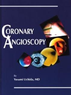
In the early 1980s , when cardiac physicians were confronting how to tackle intra coronary thrombus , one man from Japan  was  looking directly at the ground zero with fiber-optic coronary angioscope .He  provided live images  of coronary plaques and thrombus (long before the IVUS and OCT era) because of technical difficulties it did not get into  clinical utility  but gave us vital information like plaque morphology and behavior.

  • The concept of red and white thrombus
  • The yellow lipid enriched vulnerable plaques
  • Post lytic  clot surface
  • The fibrin strands within the clot etc.

coronary angioscopy Yasumi Uchida

The angioscopes have now given way to IVUS and OCT which provide indirect vision of the coronary arteries .Uchida has written a book tilted coronary angioscaopy which is a must read for all clinical cardiologists.

I think Japanese are  leading in this aspect of cardiac Imaging .Yasunori Ueda is another person who has  done lot of work on angioscopy . here is an  Image from his paper. Exciting  stuff  is isn’t !

coronary angioscopy  Yasunori Ueda www.invasivecardiology

Image source : Yasunori Ueda http://www.invasivecardiology.com

  Reference 

http://circ.ahajournals.org/content/104/24/e143.full

http://www.invasivecardiology.com/article/5571?page=3

Read Full Post »

Is Transient Ischemic attacks (TIAs)  belong to the  exclusive domain of cerebral circulation ?  Can it occur in the coroanry arteries ?  If so what situations ?

This is a presentation in one of the cardiological society of India annual scientific sessions . A pdf download is  provided

transient ischemic attacks attack of heart coronary tia

Download  a PDF presentation

Read Full Post »

Primary PCI is presumed to be the ultimate  , undisputed reperfusion  strategy  in STEMI .  Still , time and again one study or  other strips down  this   “Numero Uno”  status of pPCI  .  If it is really supreme ,  such awkward  situation shouldn’t arise  too often . More importantly , the  major reason for  dubious real world record of  pPCI  goes beyond  the time and logistic factors (which is considered the only issue  for pPCI by most interventionist ! ) There is something more to it that is invisible ! (Is it the no reflow ?)

The nearly flawless study from Belgium ( STREAM Just released in ACC 2013/Sanfransisco ) , pre-hopsital or early fibrinolysis has proven to be superior in the prevention major end points at 30 days .

  1. Death
  2. Re-infarction
  3. CHF

STREAM STUDY NEJM PRIMARY PCI VS FIBRINOLYSIS

The major surprise was pre-hospital  fibrinolysis  showed less  incidence of cardiogenic shock . ( pPCI

group had more of this ( 4.4 VS 5.9 %  in STREAM )

Now . . .  shall I make a provocative statement ?

while pPCI may be treatment of choice for cardiogenic shock . . . but it may  also confer a risk of cardiogenic shock in otherwise low risk MI !

Caution  and  conclusion

STREAM population applies strictly to 1 to 3 hour time window . It does not apply to either before or after that ! Simply put,we do not have  guts to compare fibrinolysis and pPCI  in patients who arrive  within one hour into a facility where 24 hour cath lab facility is available .  We call it unethical to do a study like that !  I personally feel it is really unethical  if we do not do a study in this time frame . The reasoning is  simple and very personal .In a  large  Government  hospital   where  we do not have primary PCI program  our net mortality for STEMI never exceeded 7-8 %  over a period of 10 years  , Which  is almost at par with global data on pPCI. (Our door to needle time is an unbelivebale  8-12 minutes ! that  too only streptokinase !)

Adding Further controversy

pPCI  is indeed a superior reperfusion strategy . No one can dispute that .But its superiority  is not  realised  in  every patient  who gets it.  The benefits are accrued if and only if it is  used most judiciously . In Low risk , small regional  , branch vessel STEMI ,  pPCI has never been  shown superior . It is well recognised ,  upto 15 % of STEMI is likely to spontaneously abort or experience very good spontaneous recannalisation . By rushing these  patients very early into cath lab pPCI   meddles with the natural anti fibrinolytic mechanisms . It is this population who  invite all the procedural hazards. .

Is this the reason STREAM had  more  cardiogenic shocks in pPCI limb ?

I think STREAM has  strengthened the case in favor of fibrinolysis in this  ever ending debate .

I would  seriously believe  pPCI is hanging it’s superiority over fibrinolysis with a wafer thin mortality advantage . pPCI may  not be recommended in a routine fashion to all STEMI  population even if they arrive within 6 hours and able to perform the plasty fast .  Science is   . . .  after all . . .  continuing  confrontations with our  assumptions !

Counter point

STREAM is not an exclusive study comparing fibrinolysis and PCI . It is a  study comparing   Pharmaco Invasive approach vs  pure invasive approach . 80 %  of patients in the  fibrinolytic limb ultimately received PCI and  stenting . It simply doesnot make sense to conclude fibrinolysis is superior to PCI . Most of the beneficial  effects on 30 day outcome may reflect the timely PCI  in the lytic group.

//

Read Full Post »

Answer :

In cardiogenic shock it is A . In all others it is probably  C.

While D may be  considered as  an  essential target criteria  for completing the  rescue PCI

Read also

Why-we-often-follow-a-reckless-time-window-for-rescue-angioplasty ?

Read Full Post »

VPDs are such a common cardiac arrhythmia . We also know most are benign .Still modern science demands to rule out structural heart disease in any patient with multiple VPDs.

When ventricles get irritated it reacts with VPDs . ( The irritants  can be anatomical , physiological or primary electrical)

Echo can detect only anatomical irritants .We are recognising  more such focus for VPDs . Hence idiopathic VPDs  may simply reflect our ignorance !  A focused  echocardiogram is  required .

The following conditions are often observed in patients  with recurrent VPDs

  1. Posterior Mitral annular calcification (Especially in women ) –Annular VPDs
  2. Aortic valve degeneration /Bicuspid aortic valve with calcification – Cuspal VPDs
  3. Mitral valve prolapse in young -Stretch induced  Pap muscle VPDs
  4. Minimal  pericardial effusions with adherent epicarditis
  5. LV false tendons-Stretch VPDs
  6. RVOT lipid focus -Subclinical ARVD
  7. LVH and Hypertension –Fibrotic VPDs    
  8. Asymmetric septal hypertrophy
  9. Scars in MI/ DCMs
  10. infiltrations in RCMs (Any Interstitial heart disease )

(Conditions 7 and 8 are  common disorders myocardium  just included to  complete the list )

**Please note ,above mentioned entites are anatomical irritants .There is a whole lot of physiological  irritants

that can induce VPDs .  ( Hypoxia, Excess catecholamines ,  K + fluxes ,  acidotic milieu etc ) .

*** Another group is primary electrical diseases inherited channel disease can induce VPDs

Also read

A crash course on ventricular ectopics


Read Full Post »

Answer

The tie is between “B” and “D ”

We know in hypertensive hearts LV primarily fails in diastole . Lungs get congested due to raised LVEDP .Here is a catch . . .  if diastole is  terribly dysfunctional  how can be systole be near  normal  ? (After all  . . . systole is not a  distant cousin of diastole !)

How is  that  high blood pressure maintained in spite of LV failure* ?

Is it due to  well-preserved  EF and cardiac Index ?  or Is it due to extreme levels of peripheral sympathetic activity mediated by catecholamine surge triggered by LVF.

We have attempted to measure  LVEF in patients with flash pulmonary edema and acute severe hypertension .It was a real messy echocardiography . We could not conclude much but one thing is  clear in acute hypertensive  LVF   the LV was vigorously contracting in , probably making the option D  more correct .

* The other way of  reasoning is    . . .  it is because  of high blood pressure the LVF  has occurred . LV contractility has no contribution in maintaining the high BP ( Not in line with  the age  old concept of LV contractility  a major determinant of systolic blood pressure !)

(Having said that  . . . we also see patients with severe LV dysfunction with  severely  stunned , ventricles in association with hypertension and LVF . In fact many of the reversible DCMs are due to sudden surge in blood pressure )

Other mechansims of LVF and lung congestion is

  • Extreme tachycardia and shortening of diastole
  • Mitral regurgitation
  • Assocaited  CAD unmasked by sudden raaise  in heart rate .

Postamble

If  this article has confused  you a little  , It has achieved  one of it’s  objective .  !  I expect more  from   young cardiology fellows to address the issue !

Reference

This NEJM article   authored by Sanjay  Gandhi  has almost answered the hemodynamics of acute LVF and HT .

mechanism of acute lvf in hypertension flash pulmonary edema lvedp in ht nejm 2005 sanjay gandhi

Read Full Post »

Can VF be a non sustained  arrhythmia ?   This question was raised and a single case report was presented

in the annual scientific sessions of  Cardiological society of India Meet in  year 2008 in  Chennai.

I am just reposting it from my archives .

Slide1 Slide2 Slide3 Slide4 Slide5 Slide6 Slide7 Slide8 Slide9 Slide10 Slide11 Slide12 Slide13

Slide14

Read Full Post »

Myocardial salvage is like  coronary fire fighting.When fire is fought  very early after the accident  , benefits are accrued  more . Text book primary angioplasty is . . .  fire engine arriving at the scene when the house is on fire .

Rescue angioplasty is asking for more force ,  when the initial fire fighting  was inefficient to control the fire. So , it is obvious the rescue efforts should be fast and brisk.In fact the pace should me more than the primary (The the second engine should  reach the ground zero  faster than the first !  – Read as  door -balloon time ! )

But what happens in real world ? We would tell  time window for primary angioplasty even in sleep ! but will struggle to come   with clear cut  answer for the  same in  rescue angioplasty  even in a  fully awake state !

CaptureWiz

It is  an overwhelming fact , we have  not taken enough efforts to define strict time limit  for rescue .( Even though guidelines say it should not be beyond  24  hours , common sense will tell us rescue PCI should not go beyond 12-15 hour window ! .One more definition for rescue PCI could be within 3 hours after diagnosing failed thrombolysis. In real world  it is a race against time in a different perspective .In many centers  rescue angioplasty “enjoys time less windows “

I was funny witness in a big private  hospital  when a  colleague  of mine  has posted  a case for  “elective rescue angioplasty” and was  waiting in the side cabin  for his turn !

Coming back to the title question

Why we often follow  a reckless time window for rescue Angioplasty ?

The reason is simple

Time is not only  muscle . . . time  is  money too !

Read Full Post »

An elderly man  with past H/o CAD  was admitted with ischemic LVF and hypotension .Blood pressure was  90/60 mmhg  and pulse rate was 140 . Urine urine output  in the immediate past hour was 50 ml . Saturation was 95 % .He had fairly extensive  crackles in both lung fields.

A bed side echo showed  moderate LV dysfunction , with wall motion defect in LCX territory and  mild  MR .A dignosis of post  MI -ischemic LVF was made .

He  was  put on intensive anti failure protocol. I asked my  fellow to administer IV NTG  and left the ward .

On my next visit after few hours  . . . the patient was in much bad shape  , and when I enquired , I learnt  NTG was never administered . I was curious to know why the fellow  dis- obeyed my instruction .

He felt sorry  .  .  . But he earnestly told me  , he  could not comprehend the principle of administering NTG in a  patient  with shock ! . I was happy  to  note his  genuine concern  for   the patient  !  But  . . . I had to take a brief  lecture to convince the importance of NTG in some forms of shock !

What is the cause for hypotension in ischemic LVF ?

Lungs are flooded due to  very high LVEDP . Blood  not only struggles to  enter the LV  but also finds difficult to   leave the LV ,  former due to defective relaxation later due to poor pumping.

The extremely high pre-load actually stuns the left ventricle in diastole . (Primarily diastolic stunning  )  . Here is a hemodynamic paradox . Excess pre-load  occurs in  terms of pressure , but  in terms of volume there is miniscule amount  blood  that  traverses LV  .

This is pre-load mismatch  at play .Empty ventricles with high wall stress  and that is reflected in aortic afterword as well .

We have to some how reduce the  very high levels of LVEDP . IV NTG can  dramatically  reduce the pre load  ( and reduce the LVEDP .) The other major  benefit is ,  NTG   can reduce the MVO2 by improving sub endocardium coronary perfusion and de-stress the heart.

Once  LVEDP  is  lowered  , the ventricle will tend to recover and gain at least some  original elasticity ( Frank starling forces) . Of course it will be defective due to ongoing ischemia . Even slight fall of LVEDP (say from 25 to 18 mmhg  can have  significant benefits as the LV function curve labors on the steep shoulder region !) .

This is one situation where NTG can increase the blood pressure once the hemodynamics is favorably altered.

*Yes  . . . heavy doses  of  Frusemide injection can do the same job but it largely depends the kindey’s cooperation to flush out fluids  .In a shock like situation one can trust the kidney perfusion  !

Additional benefits of NTG

Mitral  regurgitation  is a serious destabilizer of LV function .NTG can reduce the regurgitant fraction in acute MR effectively .

Caution

NTG may worsen the hypotension of RV infarction . Make it very sure , you are not dealing with this unique  pre-load dependent circulation.

What happened to this patient ?

He  did show  improvement with IV NTG . Of course it was not dramatic as I have projected in this article .Still it was really helped him .He required simultaneous dobutamine infusion as well .The BP did not fall  further and lung congestion was relieved  .He went on to recover fully by 48 hours and was posted for elective cath study .

Final message

                                             We tend to  worry  more about falling blood pressure  when administering  NTG. . .It is a wonderfully effective drug especially in the setting of ischemia and cardiac failure  even if  the blood pressure is low !

Acute cardiac hemodynamics  is  complex phenomenon .No one has mastered it .Paradoxes are common . Hypotension in the back ground of  acute pulmonary edema  especially due to ischemic LVF  can be corrected by NTG . Of course physicians  need  some  courage to administer NTG in patients  with a systolic pressure of  80-90mmhg.

This should ideally be done with intra arterial line in place and a simultaneous inotropic line (Doubtamine /Nor-epinephrine ) back up in case of worsening perfusion pressure .

Read Full Post »

Older Posts »