Feeds:
Posts
Comments

Archive for March, 2024

The well known pro-coagulant state of pregnancy is an evolutionary protective process to make blood clot quicker, to save fetal loss in early pregnancy and mitigate postpartum bleeding. Still, in many women, this natural adaptive process confers an enhanced thrombotic risk. The molecular mechanisms for this pro-coagulant state are, there is increased factor VII, fibrinogen, reduced protein S. It is interesting to note, while plasminogen levels are elevated, D-dimer is also increased, indicating an ongoing fight between pro & anticoagulant forces, converting the physiological maternal- placental bed a mini harmless DIC equivalent zone.

There are several important systemic, placental, (Fetal) and cardiac indications for anticoagulants and antiplatelet agents in pregnancy. The list is increasing in a steady fashion. (Most IVF pregnancies seem to need it for some unknown reason)

  • Risk of VTE
  • Recurrent pregnancy loss(Placental micro-circulation clogging)
  • SLE/APLA syndrome /Scleroderma VTE related PAH
  • Mitral valve disease with AF. Cardiomyopathy
  • Finally most importantly prosthetic heart valves & other Intra cardiac devices.

We have few options

  • Warfarin (Molecular weight 300 Daltons) is used in dose of 2- 10mg
  • Un-fractioned regular Heparin , (40000 Daltons) -Not practical for long term. Used at peripartum phase , just before labor to take control over possible PPH.
  • LMWH (Molecular weight 5000 Daltons)
  • NOACs are not an option as of now
  • Aspirin alone might give partial or near complete protection in some of the above mentioned indication.

General rules

  • Warfarin is safe for mother, Heparin is safe for fetus .(both Un-fractioned heparin & LMWH )
  • Just because heparin is safe, we cant choose t, it must be equally efficacious too. (Till date no study on LMWH has come to show its efficacy any where closer to Warfarin efficacy, (forget about beating it) in protecting mechanical valve events)
  • The concept of bridging till 12 weeks is not mandatory in all
  • Switching to regular heparin at term is applicable for both
  • Lactation both Warfarin and heparin are safe.(But LMWH is more likely than regulars heparin to appear in breast milk because of low molecular wt. (Ref https://www.medsafe.govt.nz/Medicines/medicine-information.asp)

Is warfarin really unsafe ?

Any anti-coagulant usage in pregnancy is like playing with fire .They have narrow safety window. Further, we must have have a quick antidote in case of dose excess. Warfarin, a powerful VKA, is the time tested key drug despite the well known teratogenic effect. Now we have an alternative LMWH ,which has gained considerable popularity.

The risk of Teratogenicity in warfarin is absolute or is it dose dependent ?

Yes it is dose dependent. (Warfarin causes two phases of side effects one is embryopathy, it also affects later half of pregnancy ie fetopathy with neurological bleeding etc

The Italian connect

Answer to this question came from oldest Romanian city built by the Greeks, Naples, Italy . Dr.Vitale , from the department of Cardiac Surgery, Monaldi Hospital, did this landmark study, way back in 1999 , and convincingly proved , the dreaded embryological side effects are dose dependent. It was done with a meager 58 pregnant women . The conclusions of the study changed the way we used to worry about this drug. It said, warfarin is safe at low doses even in the first trimester , if used <5mg, in terms of embryo and fetal issues. Isn’t it curious that a dreaded drug was made pregnancy friendly by simple study from smart surgeon . It is a real surprise that the conclusion of this study is still can’t be disputed by another big one. Almost all current guidelines use this 25-year-old study to form the core algorithm of current anticoagulant protocol in pregnancy.

Warfarin vs LMWH debate

Teratogen or No-teratogen, coumadin still rules supreme in most high risk situations, especially in women with mechanical valves, (Despite the ease with which this molecule crosses the placental filter , because of low molecular weight -300)

Heparin one of miraculous drug of last century , remains a life saving anticoagulant for various medical conditions. However, its refined version LMWH, though made it more palatable & user friendly, it un-apologetically took the sting out of regular heparin, made it less efficacious (more glamorous though) LMWH usage is in CAD widespread , it has suspect value* in true ongoing ischemia in any active ACS situation. It is strange anti X-a is never monitored in CAD protocol , while in pregnancy we insist on intensive monitoring i. What does it imply ? Monitoring is primarily done to ensure adequacy of anticoagulant activity , rather than risk of bleeding .

In my 30 years I am yet o see a patient have fatal bleed to poorly monitored Enoxaparin. This is the reason the mid trimester LMWH heparin bridge to collapse in many pregnancy anti-coagulant protocols. Now ,we can understand why the veteran VKAs continues to be a flag bearing drug in pregnancy .Of course, INR-guided OAC therapy, though can be tricky, most of us are used to that, unlike the frightful anti X-a troughs and peaks.

*I am sure most Interventional cardiologists will hesitate to disagree with this observation.

2022 update on LMWH : More trouble for LMWH in pregnancy. There considerable concern , that twice a day sub-cutaneous injection may not maintain target anti -X a trough .6-.8U/ml and peak 1 to 1.2 U/ml and currently many centers advice LMWH three times a day ( Bai C, Wu . Medicine (Baltimore). 2022 Dec 30;101(52):e32550.)

Final message

So far, the traditional dictum has been, Warfarin is safe for mother & heparin is safe for fetus. One has to decide accordingly with patient ,spouse & family. I think, its time to tweak this rule, little bit. Warfarin is safe for both mother & fetus* in most patients till 36 weeks in low doses , while LMWH may be more safe , but lags far behind in efficacy, especially in high risk indication. (*Including first trimester but with a lesser proof though)

Postamble

Even in these era of shared decision making, it’s our duty to impress upon our patients (or even enforce) to choose warfarin over LMWH in appropriate times. Don’t simply leave this critical decision to patients.

Reference

1.Vitale N, De Feo M, De Santo LS, Pollice A, Tedesco N, Cotrufo M. Dose-dependent fetal complications of warfarin in pregnant women with mechanical heart valves. J Am Coll Cardiol. 1999 May;33(6):1637-41. doi: 10.1016/s0735-1097(99)00044-3. PMID: 10334435.

2.Elkayam U. Anticoagulation Therapy for Pregnant Women With Mechanical Prosthetic Heart Valves: How to Improve Safety? J Am Coll Cardiol. 2017 Jun 6;69(22):2692-2695. doi: 10.1016/j.jacc.2017.04.034. PMID: 28571632.

3.Goland S, Schwartzenberg S, Fan J, Kozak N, Khatri N, Elkayam U. Monitoring of anti-Xa in pregnant patients with mechanical prosthetic valves receiving low-molecular-weight heparin: peak or trough levels? J Cardiovasc Pharmacol Ther. 2014 Sep;19(5):451-6. doi: 10.1177/1074248414524302. Epub 2014 Mar 6. PMID: 24607762.

4.Yurdakök M. Fetal and neonatal effects of anticoagulants used in pregnancy: a review. Turk J Pediatr. 2012 May-Jun;54(3):207-15. PMID: 23094528.

Annexure-I (For those who won’t believe this 5mg warfarin narrative, can continue to travel over the turbulent LMWH bridge ) as per the famous UCLA protocol LMWH + Aspirin protocol (From Ref 2)

Further thoughts

I think we need another study ,5, 8 and 10 Warfarin vs LMWH with a prosthetic valve and analyze the fetal bleeding risk in mechanical valves. It may not be a surprise if the cut off of 5mg could move further up.

I don’t know, whether it is a good trend, to note more and more biological vales are implanted at an young age to avoid OAC .These valves have short life span demanding redo surgeries within 10-15 years which may not be not a righteous approach.

(*Mechanical valves can last 25-30 years or more)
Read Full Post »

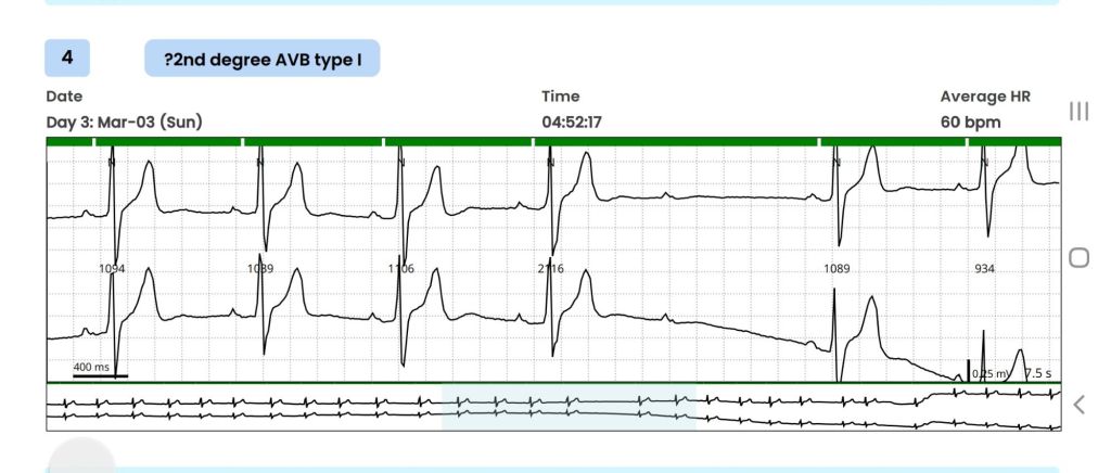
A 32-year-old high-profile businessman was advised Holter monitoring for a few ectopic beats during routine screening ECG. The 72-hour extended Holter monitoring picked up a single short pause with a blocked P wave and reported as doubtful Mobitz type 2 AV block.

The cardiologist in-charge, told the patient that findings are significant, and he would need further investigation. He was referred to their associate center for an EP study. After hearing about the procedure ,the patient was freighted about inserting multiple catheters inside his heart.

This was the time he consulted me with Holter report. It was indeed a missed QRS after a well inscribed p wave , recorded at 4.57 AM, It is a 2nd degree AV block, may be Mobitz type 2, . What if ? It could still be be blocked atrial ectopic. (Pseudo AV block) Both preceding and following PR intervals seemed to be non varying . The following QRS was narrow. I don’t know, whether a single blocked P could by any way a concealed Wenke -Bach. I didn’t have calipers to measure the PR accurately though. The baseline heart rate was around a vago-genic 60/mt, that was comforting . He had his echocardiogram done already and was normal.

What does the guidelines say ?

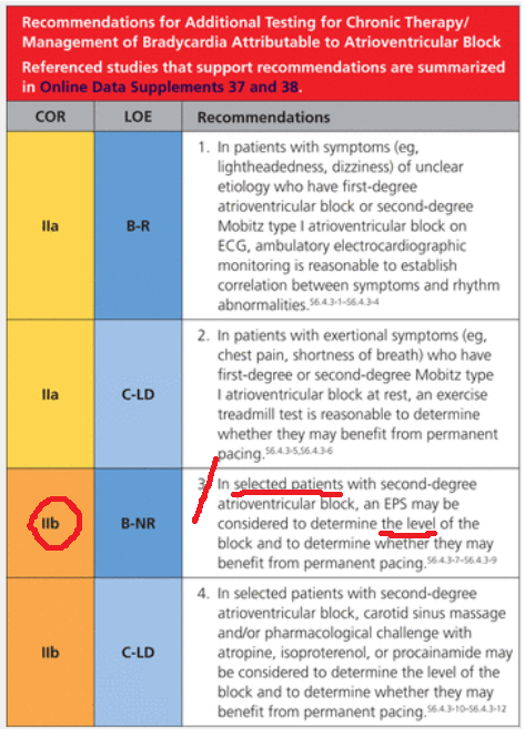
Guidelines are short of evidence , it was as vague as my thought process . It suggested EP study in selected patents with asymptomatic second degree AV block . My fellows tell, it is just 2B indication (To-be frank, 2-B indications should be called as a junk recommendation ) which would mean if you wish you can do a “potential harm”

I asked the patient two questions.

1.Does he have any symptoms like dizziness or syncope ?

Absolutely nil.

2.What is his functional capacity?

Excellent.

That’s great. Within a minute or so , I could confidently confirm, the non-seriousness of the Holter tracing. I asked him to forget everything, and sent him home, with reassurance, taking on myself a miniscule risk of missing a true AV block and its consequences. He thanked me profusely with so much gratitude. Every thing was hunky-dory , then , this thing happened. When he was above to leave the office, he came back. “Doctor, I forgot to tell, my father died suddenly at the age of 48 apparently by a heart attack” .I must admit, I was taken aback the moment he told this.

What an important past history, I failed to elicit earlier. As he left my room, I called my secretary to give a Suo-moto appointment to him 2 weeks later with a plan of TMT and possible CT -angiogram. Till late in the evening, this patient’s Holter recording ran in my mind. What was that reason for original VPDs that invited a Holter test and the subsequent documentation of Innocent appearing AV block ? Are they interconnected or inherited ? or Is it really Ischemic ones, that took his dad’s life?

The concern amplified, when I recalled about a review in EURO-PACE journal , that showed mutations of almost every structural sarcolemma proteins like Desmin and Desmoplakin can present with isolated electrical defects with or without LV dysfunction.(Brandão M, Desmoplakin Cardiomyopathy: Comprehensive Review of an Increasingly Recognized Entity. J Clin Med. 2023 )

Leaning on EP’s shoulder

That was enough for me to make a compelling call to my EP colleague, for a quick chat about this unique patient. We discussed for 15 minutes, right from Padua University paper to all the Brugada variants.(Ref 3) In the end, the basic doubts remained as before. However, the patient was advised for an EP study primarily to know the HV interval and the possibility of diffuse distal disease. The possible need for a MRI study to rule out silent arrhythmogenic intramural granulomas was also discussed. My EP friend poked me with more academic toxemia. He said a screening test called cardiac-arrhythmic genome analysis is available in certain European centers. Ref: Isbister, J.C., Semsarian, C. The role of the molecular autopsy in sudden cardiac death in young individuals. Nat Rev Cardiol 21, 215–216 (2024).

I said enough is enough , and requested for hanging up the chat.

Final message

AV blocks, even Mobitz type 2, can occur at normal times of heightened vagal tone.(Massie Block-Ref 1) But, if there is something unusual in the clinical history, be ready to investigate until the arrhythmia, or at least the anxiety disappears.

Reference

1.Massie B, Scheinman MM, Peters R, Desai J, Hirschfeld D, O’Young J. Clinical and electrophysiologic findings in patients with paroxysmal slowing of the sinus rate and apparent Mobitz type II atrioventricular block. Circulation. 1978 Aug;58(2):305-14. doi: 10.1161/01.cir.58.2.305. PMID: 668079.

2.ROTONDI, F., MARINO, L., LANZILLO, T., MANGANELLI, F., & ZEPPILLI, P. (2011). Prolonged Ventricular Pauses in an Asymptomatic Athlete with “Apparent Mobitz Type II Second-Degree Atrioventricular Block.” Pacing and Clinical Electrophysiology, 35(7), e210–e213.

3.Graziano F, Zorzi A, Cipriani A, De Lazzari M, Bauce B, Rigato I, Brunetti G, Pilichou K, Basso C, Perazzolo Marra M, Corrado D. The 2020 “Padua Criteria” for Diagnosis and Phenotype Characterization of Arrhythmogenic Cardiomyopathy in Clinical Practice. J Clin Med. 2022 Jan 5;11(1):279. doi: 10.3390/jcm11010279. PMID: 35012021; PMCID: PMC8746198.

Read Full Post »

CTOs are opened primarily for four reasons

  • Angina which is refractory to drugs
  • Stress test positivity with or without angina
  • Anxiety of having a blocked coronary artery in a self educated patient
  • Cardiologist’s clandestine pride & pursuit*

* Personal experience included

Some evidence based observation

Most of the studies as on today do not give survival advantage of opening a CTO.(DECISION-CTO,EURO-CTO,EXPLORE,IMPACTOR)

Opening a CTO, for reasons other than angina (i.e. for relief of dyspnea or improving functional capacity) is largely conjectural and based on randomly accrued data backed by poor interpretation. The role of collateral circulation in CTO that can compensate even during exercise is well known at patient level data. This has become a difficult area of research because it involves spending more time with the patient, and hence not studied much. We are in the era of artificial intelligence ,virtual patients and statistical extrapolations that can steer the Kaplan Meyer curves in the desired direction.

Pure academicians shall follow the current guidelines. Surprise… surprise !, There is some good news. The normally aggressive American guidelines exercise much caution with a 2B punch. Still , even today it is weird to see hours of academic time is consumed in CTO Interventions in any interventional cardiology meets. (May be , they could get a breakthrough benefit , which I couldn’t appreciate)

.

CTO-PCI follow up

The incidence of MACE including ACS varies between 12-28% depending on LV function.(Ref 2) How about Conferring 12% risk of ACS in a person who has normal LV by doing CTO-PCI ? Still it continue to be a smart move for many of us ? This is exactly the reason experts are struggling to come to term with truths behind negativity of most published CTO trials.

Image from Ref 2  Egypt Heart J 72, 28 (2020

Now, answer to the title question. What is the future risk of ACS in opening CTO related artery ?

Asymptomatic CTOs, with fair excercise capacity, should probably never be opened for the simple reason, a closed artery is naturally protected, against a future ACS at least in its territory

Final message

Currently, in the science of cardiac revascularization there is only evidence and it’s Interpretations, little patient level facts.

Reference

1. Hamzaraj K,Patient Selection and Clinical Indication for Chronic Total Occlusion Revascularization-A Workflow Focusing on Non-Invasive Cardiac Imaging. Life (Basel). 2022 Dec 20;13(1):4.)

2.El Awady, W.S., Samy, M., Al-Daydamony, M.M. et al. Periprocedural and clinical outcomes of percutaneous coronary intervention of chronic total occlusions in patients with low- and mid-range ejection fractions. Egypt Heart J 72, 28 (2020). https://doi.org/10.1186/s43044-020-00065-1

Post-amble

Living with a single coronary artery, is potentially a frightening scenario for the patient* which has to supply its own area and also, need to donate the occluded coronary artery . What will happen if a single donor (RCA/LCX) gets closed? One more remote risk in CTO is, acute collateral shutdown causing STEMI/NSTEMI. These statistically minuscule risks are well exploited by coronary caretakers. Meanwhile, there is little talk about the chances of CTO getting closed by itself after an apparently successful PCI. The consequences of anatomic and hemodynamic collapse of hitherto well flowing collaterals , after a CTO PCI will require a separate discussion.

*It is wiser to recall , left coronary artery is also single before bifurcating. Surviving an entire life span with a single10-20 mm tunnel called left main, rarely elicit the same fear in us.

Read Full Post »

It is a 120-year challenge. Can anyone replace Rontgen’s X-ray discovered in 1895 for medical imaging? The Nobel winning Invention redefined the way we looked at our body and management of diseases for over a century. However, the fact remained it is an invasive and injuring investigation. What is the alternative for the X-radiation ?

CT scan was a great invention, but it turned out to be a gigantic 360-degree clone of X-ray machine. Today’s cath lab, however sophisticated , is like spending hours together inside a hot Chernobyl coffee shop. MRI was a true game changer. With zero radiation, MRI came close in the fight with innocuous proton imaging. But for live cardiac interventions, MRI was not practical. Meanwhile, over the years, ultrasound moved up from the pelvis, abdomen, right into coronary arteries and heart. Intravascular ultrasound-based interventions are being done in coronary artery, in a few cases to avoid contrast in patients with CKD. (We call it zero-contrast IVUS-guided PCI). But, it is cumbersome and has some technical issues. Transesophageal echo (TEE) & Intracardiac echocardiography (ICE) do help us immensely in certain interventions.

Now is the era of Optics

If a torch light can illuminate and give us vision in absolute darkness ,how about acquiring a deep vision with scattered light ie photons. (Jnana-Chakshush ,third eye of Hindu God Shiva ?) The concept of Optical coherence imaging came (OCT) came in .It has limited use in deep vision of coronary wall anatomy and histology. As of now it has no role to play in catheter guidance.

Here comes the real Innovation . Fibro-Optic real shape( FORS) technology , which reconstructs image from optical data, and beam live fluro- like images, in 3 dimension. May be, we may soon, say good bye to electrons, protons, and welcome these harmless photons.

This video clip shows real time Intervention using FORS in Aortic endovascular stenting

One may Imagine FORS to Electro-physiologist’s electro- anatomic mapping made with a the GPS like pad attached beneath the cath table and reconstructing anatomical Images from the dynamic signals points generated from the catheter tip.

Final message

Now, we are looking at various different modalities to image without radiation injury to the patients, and more importantly the cardiologists .

  • Intra cardiac Echocardiography (ICE)
  • Proton imaging (MRI)
  • Electrical navigation (CARTO)
  • Fibro-Optic real shape (FORS)

FORS , is the new arrival. Let us hope it stands the test of time.

.

Read Full Post »