Feeds:
Posts
Comments

Posts Tagged ‘heart rhythm society’

The huge popularity of conduction system pacing (CSP) , especially the Left bundle branch Area Pacing (LBBAP) has been hailed as a paradigm shift in cardiac pacing. By utilising the native His-Purkinje network, CSP promises a physiological alternative to the dys-synchronous RV pacing. However, beneath this procedural enthusiasm lies a critical, clinical assumption .It is the disregard of distal LBB integrity. In our haste to bypass proximal blocks, the electrophysiology community has mastered the art of ignoring distal conduction system disease a practice that relies more on hope than long-term, evidence-based science.

The physiological basis of LBBAP  is based on capturing the main left bundle branch trunk or its proximal fascicles to downstream  distal healthy  Purkinje network1. In patients presenting with complete heart block , this approach looks fine , if the underlying pathology is strictly nodal or intra-Hisian. However, in progressive, degenerative conduction system disorders such as Lev’s or Lenègre’s disease fibrosis is rarely localized. It is a multi-level, diffuse process that tracks distally into the fascicles and terminal Purkinje ramifications2.

How, can we casually overlook the small but surely dangerous risk of progressive distal LBB disease in complete heart block, which can lead to unpredictable catastrophic events ? This electrical oversight finds a striking, parallel in historical pacing concepts. In patients with sinus node dysfunction (SND), the electrophysiology community long ago established that a standalone atrial lead (AAI pacing) is a clinical hazard3. The minor yet real risk of AV nodal disease progression (approximating 0.6% to 4.5% per year) almost universally mandates the placement of a backup ventricular lead, safely transitioning the strategy to dual-chamber (DDD) pacing4.

The current electrophysiology consensus downplays this distal disease , by invoking the physics of the virtual electrode. The argument is even if the localized nerve fibers directly contacting the lead tip degenerate over the subsequent decade, the high-programmed voltage outputs will maintain permanent myocardial capture, ensuring patient safety from asystole5. While this assumption satisfies the basic requirements of bradycardia support, it ignores a core fact.

Aprar from thew physiological uncertainty , there is also a  significant risk of procedural failure. Real-world registries reveal that approximately 5% to 15% of patients undergo acute on-table crossovers due to  septal calcification, anatomical variations, or an immediate functional failure to engage a viable conduction pathway7.

Conduction system pacing is undoubtedly a great addition to our EP armamentarium, but it must be practiced with  humility in the face of unproven long-term  putcome and procedural complexities. A clinical pragmatism is warranted. Conventional dual chamber (DDD) is backed by more than four decades of  randomized clinical evidence.9. Until we clarify the natural history of the chronically paced, diseased left bundle,  DDD pacing should remain  the standard of care.

References

  1. Huang W, Su L, Wu S, et al. A novel pacing strategy with low and stable threshold-left bundle branch pacing. Europace. 2019;21(3):471-477.
  2. Lev M. Anatomic, pathologic, and electrocardiographic correlations in the atrioventricular and distal conduction system. Prog Cardiovasc Dis. 1964;7(4):317-340.
    Andersen HR, Nielsen JC, Thomsen PE, et al. Long-term follow-up of patients paced in atrioventricular synchronous vs. single-chamber atrial mode for sick sinus syndrome. Lancet. 1997;350(9086):1210-1216.
  3. Brandt J, Anderson H, Fåhraeus T, et al. Natural history of AV conduction in patients with sick sinus syndrome as related to bilateral bundle branch block. Pacing Clin Electrophysiol. 1992;15(11 Pt 2):1914-1918.
  4. Vijayaraman P, Sharma PS, Cano Ó, et al. Comparison of Left Bundle Branch Area Pacing and His Bundle Pacing in Bradycardia Indications. JACC Clin Electrophysiol. 2021;7(11):1433-1442.
  5. Jastrzębski M, Kiełbasa G, Cano Ó, et al. Left bundle branch area pacing outcomes: the MELOS registry. Eur Heart J. 2022;43(40):4161-4173.
  6. Heckman LI, Luermans JG, Vos LM, et al. Left Bundle Branch Area Pacing: A Comprehensive Review of Technical Aspects, Clinical Outcomes, and Future Directions. J Clin Med. 2023;12(11):3611.
  7. Wijesuriya N, Niederer S, de Verteuil R, et al. Extraction of conduction system pacing leads: a systematic review and international survey. Europace. 2024;26(2):euae032.
  8. Wilkoff BL, Cook JR, Epstein AE, et al. Dual-chamber pacing or ventricular pacing in patients with an implantable defibrillator: the DAVID trial. JAMA. 2002;288(24):3115-3123.
  9. Khurshid S, Epstein AE, Verdino RJ, et al. Incidence and predictors of pacing-induced cardiomyopathy. Heart Rhythm. 2014;11(9):1618-1625.

Postamble

This opinion letter is being sent to EURO PACE journal : Expecting a fast tracked rejection letter within a week or two .(Based on my past experience) They view these type of articles as destructive criticism I was told by one editor. Some one please clarify me, the difference between a constructive and a destructive critic, please)


.

Read Full Post »

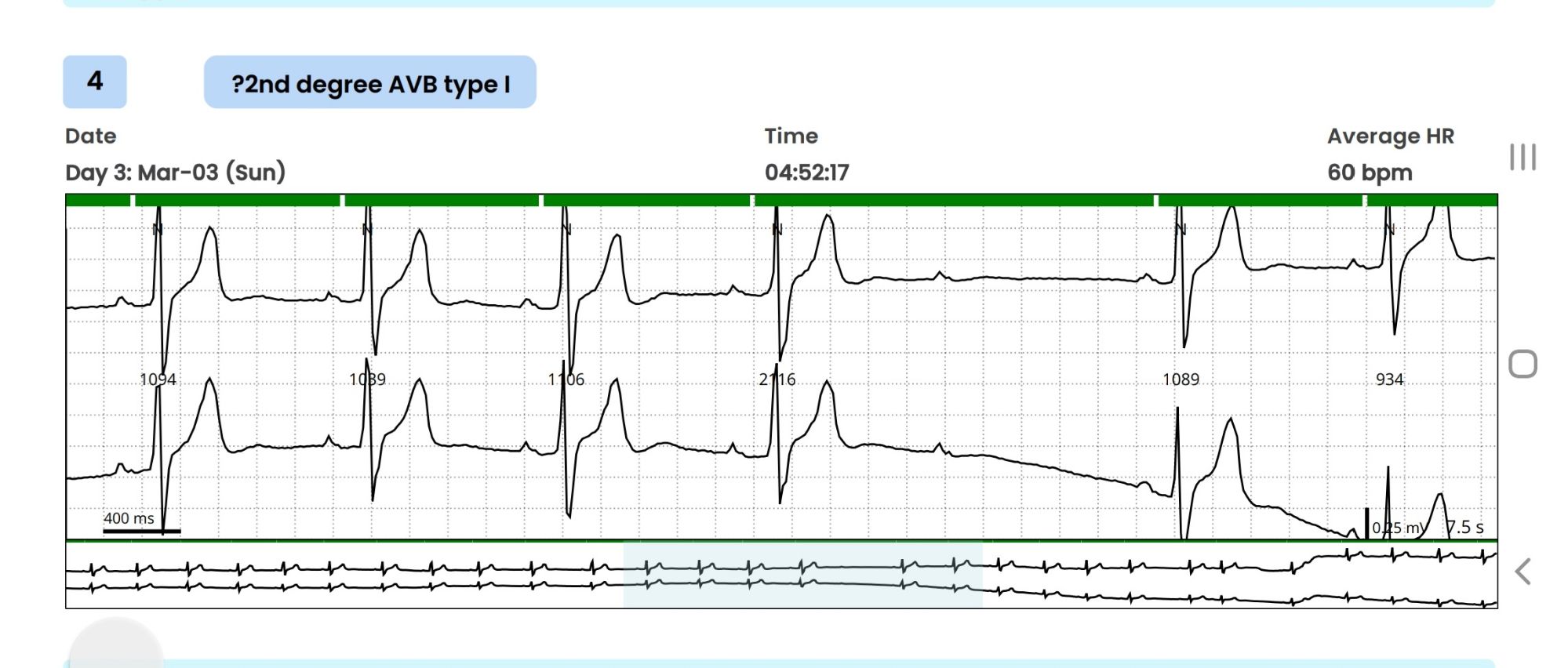
A 32-year-old high-profile businessman was advised Holter monitoring for a few ectopic beats during routine screening ECG. The 72-hour extended Holter monitoring picked up a single short pause with a blocked P wave and reported as doubtful Mobitz type 2 AV block.

The cardiologist in-charge, told the patient that findings are significant, and he would need further investigation. He was referred to their associate center for an EP study. After hearing about the procedure ,the patient was freighted about inserting multiple catheters inside his heart.

This was the time he consulted me with Holter report. It was indeed a missed QRS after a well inscribed p wave , recorded at 4.57 AM, It is a 2nd degree AV block, may be Mobitz type 2, . What if ? It could still be be blocked atrial ectopic. (Pseudo AV block) Both preceding and following PR intervals seemed to be non varying . The following QRS was narrow. I don’t know, whether a single blocked P could by any way a concealed Wenke -Bach. I didn’t have calipers to measure the PR accurately though. The baseline heart rate was around a vago-genic 60/mt, that was comforting . He had his echocardiogram done already and was normal.

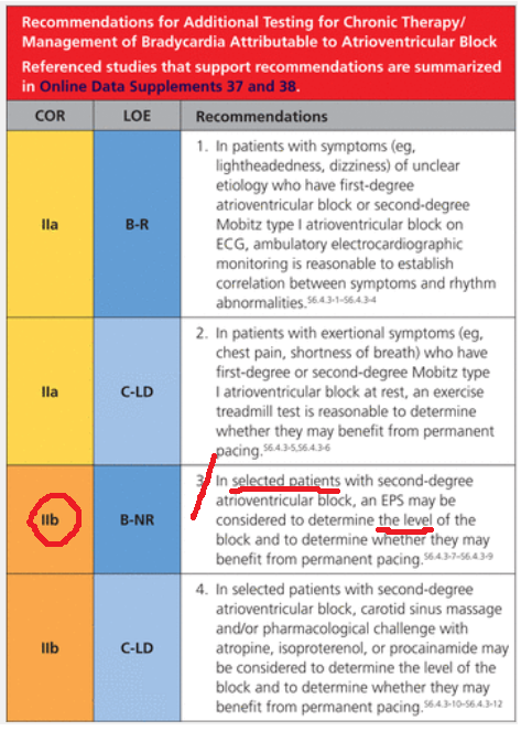
What does the guidelines say ?

Guidelines are short of evidence , it was as vague as my thought process . It suggested EP study in selected patents with asymptomatic second degree AV block . My fellows tell, it is just 2B indication (To-be frank, 2-B indications should be called as a junk recommendation ) which would mean if you wish you can do a “potential harm”

I asked the patient two questions.

1.Does he have any symptoms like dizziness or syncope ?

Absolutely nil.

2.What is his functional capacity?

Excellent.

That’s great. Within a minute or so , I could confidently confirm, the non-seriousness of the Holter tracing. I asked him to forget everything, and sent him home, with reassurance, taking on myself a miniscule risk of missing a true AV block and its consequences. He thanked me profusely with so much gratitude. Every thing was hunky-dory , then , this thing happened. When he was above to leave the office, he came back. “Doctor, I forgot to tell, my father died suddenly at the age of 48 apparently by a heart attack” .I must admit, I was taken aback the moment he told this.

What an important past history, I failed to elicit earlier. As he left my room, I called my secretary to give a Suo-moto appointment to him 2 weeks later with a plan of TMT and possible CT -angiogram. Till late in the evening, this patient’s Holter recording ran in my mind. What was that reason for original VPDs that invited a Holter test and the subsequent documentation of Innocent appearing AV block ? Are they interconnected or inherited ? or Is it really Ischemic ones, that took his dad’s life?

The concern amplified, when I recalled about a review in EURO-PACE journal , that showed mutations of almost every structural sarcolemma proteins like Desmin and Desmoplakin can present with isolated electrical defects with or without LV dysfunction.(Brandão M, Desmoplakin Cardiomyopathy: Comprehensive Review of an Increasingly Recognized Entity. J Clin Med. 2023 )

Leaning on EP’s shoulder

That was enough for me to make a compelling call to my EP colleague, for a quick chat about this unique patient. We discussed for 15 minutes, right from Padua University paper to all the Brugada variants.(Ref 3) In the end, the basic doubts remained as before. However, the patient was advised for an EP study primarily to know the HV interval and the possibility of diffuse distal disease. The possible need for a MRI study to rule out silent arrhythmogenic intramural granulomas was also discussed. My EP friend poked me with more academic toxemia. He said a screening test called cardiac-arrhythmic genome analysis is available in certain European centers. Ref: Isbister, J.C., Semsarian, C. The role of the molecular autopsy in sudden cardiac death in young individuals. Nat Rev Cardiol 21, 215–216 (2024).

I said enough is enough , and requested for hanging up the chat.

Final message

AV blocks, even Mobitz type 2, can occur at normal times of heightened vagal tone.(Massie Block-Ref 1) But, if there is something unusual in the clinical history, be ready to investigate until the arrhythmia, or at least the anxiety disappears.

Reference

1.Massie B, Scheinman MM, Peters R, Desai J, Hirschfeld D, O’Young J. Clinical and electrophysiologic findings in patients with paroxysmal slowing of the sinus rate and apparent Mobitz type II atrioventricular block. Circulation. 1978 Aug;58(2):305-14. doi: 10.1161/01.cir.58.2.305. PMID: 668079.

2.ROTONDI, F., MARINO, L., LANZILLO, T., MANGANELLI, F., & ZEPPILLI, P. (2011). Prolonged Ventricular Pauses in an Asymptomatic Athlete with “Apparent Mobitz Type II Second-Degree Atrioventricular Block.” Pacing and Clinical Electrophysiology, 35(7), e210–e213.

3.Graziano F, Zorzi A, Cipriani A, De Lazzari M, Bauce B, Rigato I, Brunetti G, Pilichou K, Basso C, Perazzolo Marra M, Corrado D. The 2020 “Padua Criteria” for Diagnosis and Phenotype Characterization of Arrhythmogenic Cardiomyopathy in Clinical Practice. J Clin Med. 2022 Jan 5;11(1):279. doi: 10.3390/jcm11010279. PMID: 35012021; PMCID: PMC8746198.

Read Full Post »

  • It is only rarely a journal of International caliber is published from India . IJEP is one such journal.
  • Cutting edge articles on Electrophysiological science  break here !
  • This is an online journal . No print issues . Enjoy, it is free !

Here is the  Link

Just sample an article  : A great review about cardiac arrhythmias in congenital heart diseases , Must read by  all cardiologists    http://www.ipej.org/0906/khairy.htm

Read Full Post »