Feeds:
Posts
Comments

Archive for September, 2026

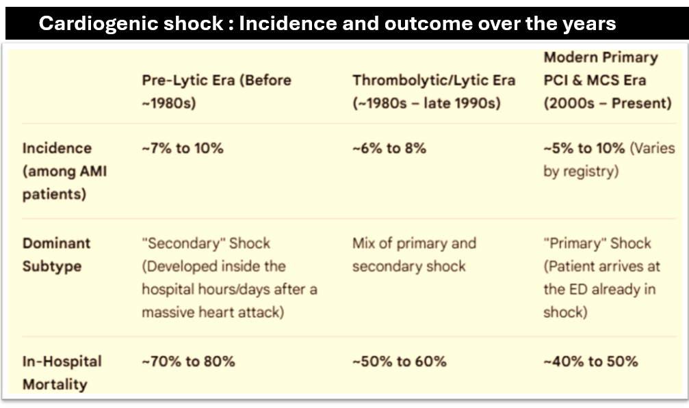
Cardiogenic shock occurs in 5% to 10% of all STEMI cases. The prefix “cardio” bothers us for all the wrong reasons. Though the shock component is initially triggered by primary cardiac pump failure, it soon spirals downward into a tissue level shock threatening the body’s 30 trillion cells. As cardiologists, we repeatedly fail to comprehend this. Instead, we aggressively attempt to revascularise the myocardium using multi-vessel PCI, IABP, ECMO, and LVADs. Yet, nothing seems to work.  When tissues are irreversibly damaged  a non-stop  lactate ooze  begins to ring a final pre death biochemical SOS call.

What is the Time Window for Tissue Perfusion?

For myocardial cell death, we have a defined therapeutic window of 12 hours, with under 6 hours being ideal. In cardiogenic shock, however, we face an unknown time window. During this uncertain period, cells within vital organs like the kidneys, liver, and lungs lose their structural integrity and face irreversible injury.

As far as I know, there is no defined timeline for intervention in cardiogenic shock. The inflection point between reversible and irreversible shock varies individually because it is a complex, systemic spiral. But, one thing we must be clear. The clock does not start at the moment of infarct-related artery (IRA) occlusion. Rather, it starts when cardiac index drops below critical threshold required to sustain cellular respiration. Note the emphasis on flow. Beyond a certain point, blood pressures have nothing to do with actual flow. This explains why systolic and mean arterial pressures do not correlate well with the severity of shock. We must never forget the concept of nomotensive shock .

Current Criteria to Diagnose Cardiogenic Shock

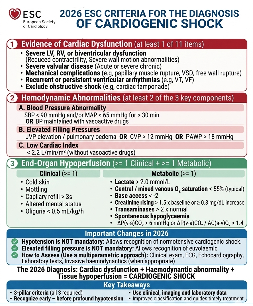
We have known this for a long time. To establish a diagnosis of cardiogenic shock, a patient must meet a triad of criteria:

Note the dramatic (& ironic)changes in the current guidelines : Neither the blood presure not the filling pressure matters in the diagnosis of cardiogenic shock

Myocardial No-Reflow vs. Tissue level No-Reflow

This concept has some evidence back up. Even if you somehow manage to revive the heart In CS,  it does not guarantee full systemic tissue recovery. This failure of organ perfusion at the  systemic level behaves exactly like the no-reflow phenomenon we witness at the myocardial microvascular bed.

So, what shall we do to tame this dreaded condition ?

We really don’t know. Preventing the onset seems to be the most effective Intervention.

How to prevent?

Have a close look at the table to gain some insights. Focus the third and last column. Ask this question. Why the incidence remains high even in the modern Interventional era,with little improvements in outcomes ?

It is prudent, we make every attempt to revive the heart, paradoxically, we must realise healing can still take place without myocardial excesses*Some times, attempting a cure , will interfere with prevention of  the event, we aim to cure.

Final Message

The cardiac component in cardiogenic shock becomes less relevant when vascular and tissue-level shock sets in, making PCIs or MCS devices almost redundant. I think , in the overall interest of the patients , It is advisable for cardiologists to transfer them early to an intensivist or anesthetist, as they are better equipped to manage shock syndromes including the cardiogenic ones.


*Postamble: The missing link in the SHOCK trial

When we look back at the landmark 1999 SHOCK Trial, we find an quixotic truth to ponder over.Nearly 34% of the patients randomized to the medical arm survived to one year without any emergency revascularisation.If cardiogenic shock were purely a mechanical problem that survival rate should have been zero. This study throws two facts, many of us  will be reluctant to accept.

  1. Cellular Resilience: The human cells do possess ancient survival mechanisms. When the coronary  flow crtically drops, some individuals have the microvascular mechanism to tolerate severe ischemia and “hibernate” until the native heart recovers.
  2. The harm of hyper-Intervention could  be real . Every extra wire, balloon, and large bore mechanical device we insert brings its own dose of  strqin, vascular trauma, and reperfusion injury. The reason for the  lucky  30% who survived in SHOCK trial, may simply because they were spared from the toxicity of excess Intervention.


Read Full Post »

Nothing happens without energy cost both in physics and biology. How much is the metabolic cost of an extra systole ? While an occasional VPD is benign and has a negligible effect, a high burden of VPDs can consume extra energy and drain myocardial metabolic reserves.

Three ways it can happen

  • Inefficiency: VPDs cause premature contractions before AV valves fill and hence there is a mechanically wasted work However the electrical cost is small.
  • Dyssynchrony: dys or desynchrony due to the VPDs also hikes myocardial oxygen consumption (MVO₂) for identical workloads.
  • Post-Extrasystolic Potentiation: We know, the beat following a VPD shows increased force due to calcium accumulation. This can elevates contractility and wall tension, increasing energy demands.

Molecular basis of energy depletion

At the cellular level, the variable timing of VPDs disrupts calcium homeostasis in phase 2 of action potential within the myocytes. The sarcoplasmic reticulum must continuously pump calcium ions against concentration gradients via SERCA2 channels, a process requiring ATP hydrolysis. Additionally, the chronic workload mismatch induces mitochondrial oxidative stress, which damages the electron transport chain, impairs ATP synthesis resulting in energy consumption mis mamtch.

What is the cut off number ?

A high VPD burden* (10% to 20% all heart beats in day ) is considered a chronic strain, leading to cardiomyopathy and heart failure.First step is a Holter monitor assessment. Yet, we are not clear about the exact number For example : Does bigeminy counted then 50 % of all beats )

*VPDS and elEctrical SCD is different topic , and is not discussed here.

Does suppression help ?

We think so. If and only if we can document porgressive LV dysfunction.

Final message

VPDs are usually benign unless linked to LV dysfunction, which complicates prognosis. The electrical energy cost of VPDs is negligible . But its impact on the mechanical behaviour on myocardial contractility is the issue. It’s crucial to determine if they are primarily electrical or secondary to associated LV dysfunction. Treatment is based on individual assessment.

Reference

Link to the article

Postamble

Difference between APD and VPDs in terms of energy consumption ?

Need to be discussed further.(We know frequent focal APDs or ATs, MAT can result in AF or atrial cardiomyopathy)

Read Full Post »

Final message

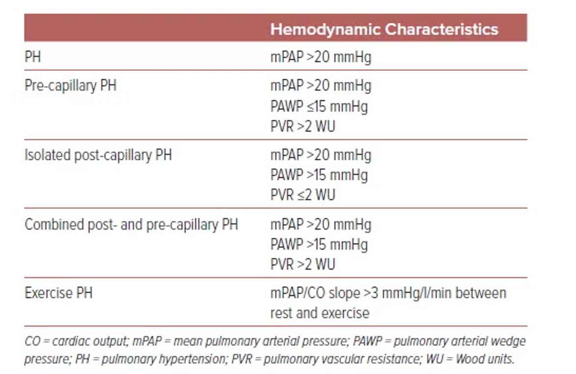
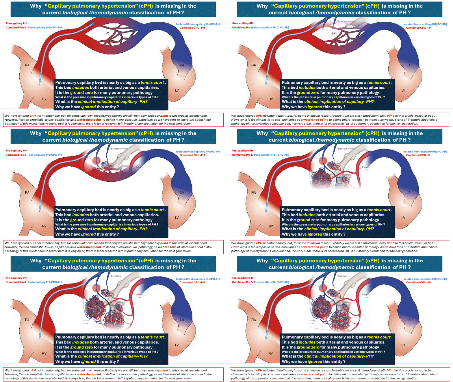
While the term “pre & post capillary pulmonary hypertension” is useful for hemodynamic understanding , it creates a false impression biologically. Pre-capillary PH is a pan-vasculopathy where the capillary bed is routinely remodeled, congested, or even entirely destroyed. Please mind, this happens in almost in all the 4 groups of PH , at least in the advanced stages.

Read Full Post »

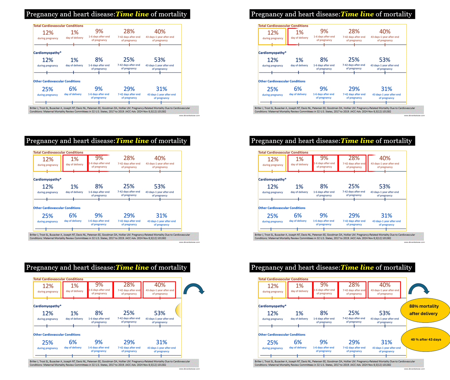
Heart disease in pregnancy is the third important cause for maternal mortality in most part of the world. (Apart from Bleeding, Infection and obstetrical complications) We know pregnancy is hemodynamic stress test for any mother and obviously it is a challenge in a compromised heart. Most heart disease encountered are mild and the mother has natural capability to walk through the pregnancy. It is the high risk subset that pose a real challenge. It is surprising ,even in the high risk, most women touch the finish line. The MMR review committee from USA has come out with clear data about the time line of mortality in various heart disease*.In this comprehensive analysis , the fact that 88% of mortality occur after delivery is too striking and is to be considered a core concept around which cardiac care of pregnant women should be addressed.

The figure shows the percentage of pregnancy-related deaths with an MMRC-identified underlying cause of death of cardiovascular conditions at 5 time periods from pregnancy to 1-year postpartum.(Ref 1) Note : The percentages are displayed for total cardiovascular conditions, and then for cardiomyopathy and other cardiovascular conditions individually. Percentages might not sum to 100 because of rounding. 

Why this data is important ?

Though the peak hemodynamic stress happens around 24-28 weeks, it is the post partum period after the baby is out is very critical. In fact, nearly 40 to 50 % event happen up to one year. Some of them doesn’t even come under the definition of maternal mortality.

Is there any thing we can do during pregnancy , that can mitigate the post partum events?

Also, this delayed events give us a window of opportunity to act on these adversity.Postpartum care for patients with heart disease, including PIH, should be provided in a specialized unit at major hospitals that manage a high volume of pregnancies.

Reference

1.Briller J, Trost SL, Busacker A, Joseph NT, Davis NL, Petersen EE, Goodman DA, Hollier LM. Pregnancy-Related Mortality Due to Cardiovascular Conditions: Maternal Mortality Review Committees in 32 U.S. States, 2017 to 2019. JACC Adv. 2024 Nov 8;3(12):101382. doi: 10.1016/j.jacadv.2024.101382. PMID: 39583867; PMCID: PMC11585746.

Postamble

*We are not yet clear , whether PIH(Gestational HT) related cardio- vascular complications to be included in the heart disease basket. Logically and scientifically it should.

Read Full Post »

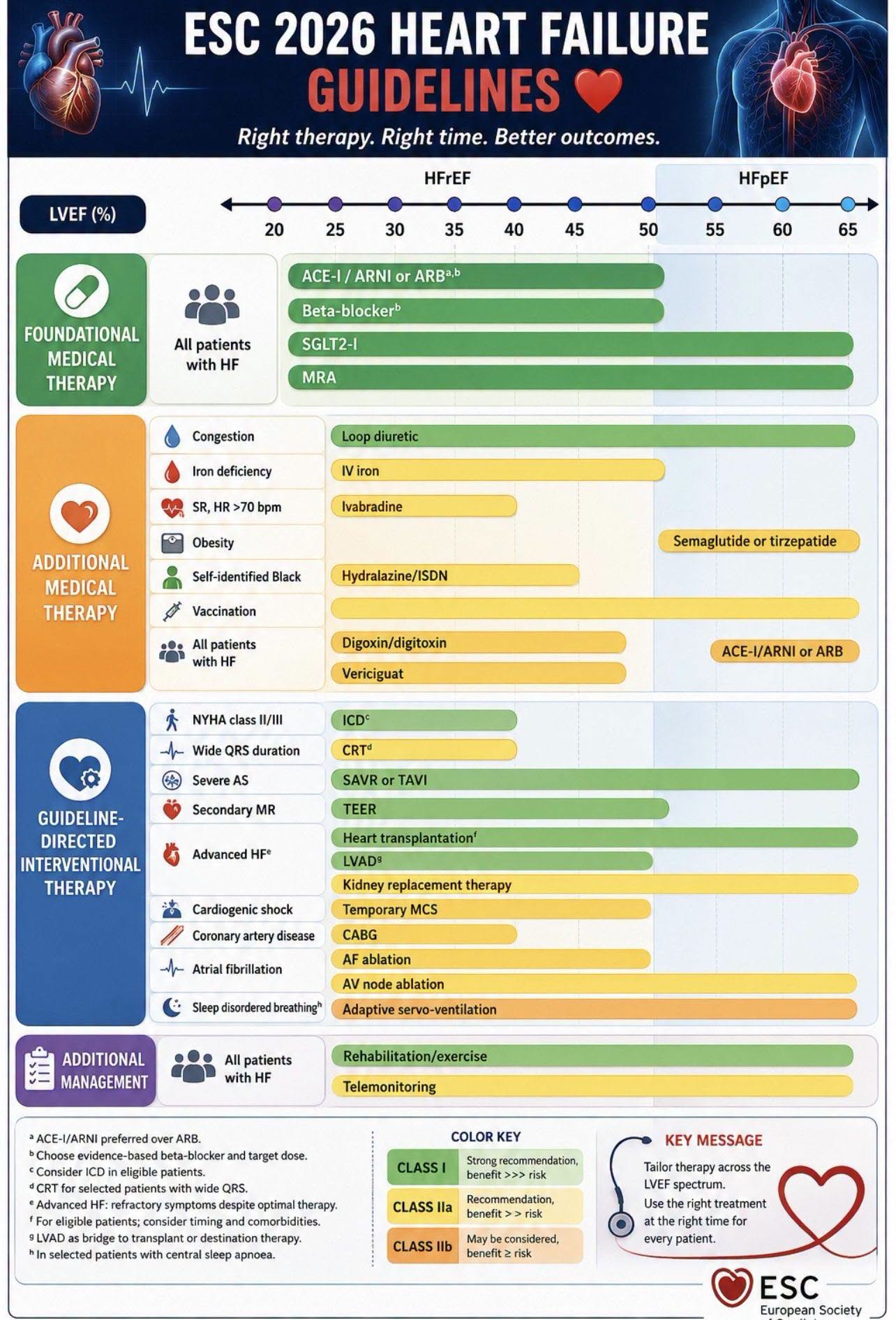
It is fashionable to frequently change the terminology, definitions, and classification of cardiac disorders. Now, the ESC is renaming therapeutic strategies in heart failure. Terms like OMT (Optimal Medical Therapy) and GDMT (Guideline-Directed Medical Therapy) have been taken off the table. A new term called FMT, (Foundational) AMT (Additional) and GDIT (Guideline directed Interventional therapy)

It is Intriguing to see more green color in the device arena and add on therapy is still yellowish.Note :  The diuretics, despite bring  the only green in the add on category, still considered  unfit, to be part of the foundation.

What could be the reason for this?

“Optimal” is entirely subjective, and guidelines are always fluid ( Including the current one!) A few things are particularly glaring in this update. For instance, the term “interventional therapy ” has been introduced, as if laying a pharmacological foundation is not an intervention by itself.

Why Digoxin is not in the foundation ?

Digoxin the wonder drug used for centuries was previously looked down upon. It was kicked out in earlier guidelines, but has now been brought back and placed under the category of “additional drugs”. As a cardiologist who has continued using this drug and rejecting those old guidelines, I was called unprofessional and unscientific. Now, I realize: who was really being unscientific all this time?

*Ask a simple question, can any physician manage heart failure without diuretic ?  How many lives a loop diruerrics are saving every day  across the globe  &  how many lives ARNI’s are unbale to save  ? Do we have a one to one study ? We have a  serious problem in asking right research questions.

I am sure, the committee will  ultimately realize their  mistake and will be compelled to move  digoxin and diuretics  to the foundational therapy.Meanwhile , we should appreciate them  for including rehabilitation and exercise as a green intervention, even though it was pushed to the bottom of the list.

Final message
Frequent tinkering of nomenclature, classifications, and definitions in cardiology has become an academic time pass. It is not going to alter the actual clinical outcomes of heart failure. We need a lot more application of mind to remove the clutter from genuinely beneficial drugs and modalities.

Read Full Post »