Feeds:
Posts
Comments

Archive for August, 2026

Localising the infarct-related artery (IRA) In NSTEMI , when there is multi-vessel coronary artery disease (CAD) is a classic clinical dilemma. Because NSTEMI lacks the clear, localising markers as in STEMI, identifying the “culprit” among multiple chronic blockages requires a more careful approach. It requires combined analysis of electrocardiographic clues , Echocardiographic WMA, angiographic analysis, and finally as a last resort intracoronary Imaging. Note : In UA,without NSTEMI it can be called as ARA-Angina related artery.
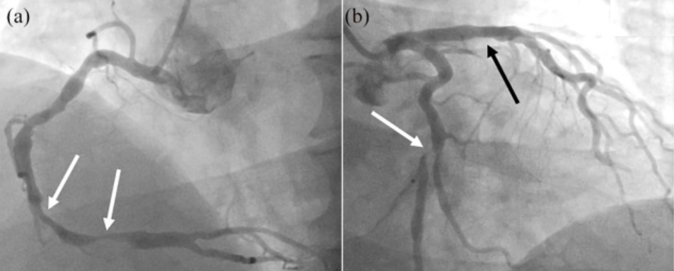
Image source : Baumann et al. Ther Adv Chronic Dis. 2020 Jul 1;11:2040622320938527

A meticulous review of the invasive coronary angiogram (ICA) will identify the culprit lesion in the majority of cases. Look for these specific features

  • Plaque Morphology: Eccentric , hazy lesions, irregular borders, or filling defects indicative of an intraluminal thrombus
  • Lesion Ulceration: Look for ulcerated edges, aneurysmal segments, or evidence of a dissection flap. Any vessel showing TIMI-1 or TIMI-2 flow, or prolonged contrast stasis during washout suggest microvascular plugging downstream from a fresh rupture.

ECG -ECHO correlation of WMA is the key

  • Wall motion defect correlation: Matching the distribution territory of the suspected culprit vessel LADcs LCX or RCA can be most useful . Still extensive collaterals can prevents us, to make a rule of thumb
  • Typical patterns like Wellens can help localise the IRA. Transient ST-depressions or deep T-wave inversions
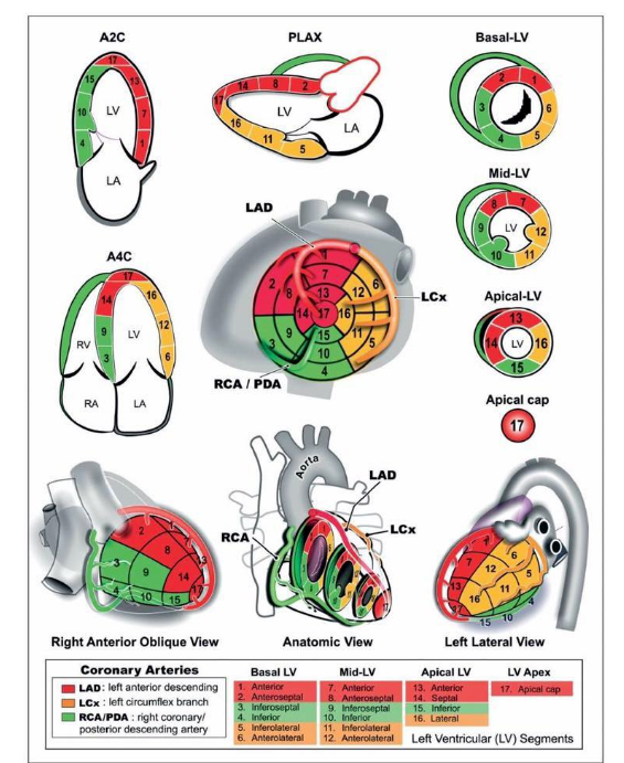
The most important illustration in coronary artery disease

Note* WMA can be deceptive as well, due to old MI, remote ischemia , or either due to collateral support or lack of it

Intra coronary Imaging

When angiography shows multiple severe lesions with no obvious acute features, coronary imaging like OCT/IVUS can be helpful. It visualises plaque rupture, thin-capped fibroatheromas (TCFA), red/white thrombi, and superficial plaque erosions. IVUS can help identify ruptured fibrous caps, and intramural haematomas.However, the temptation to do imaging should be resisited as performing this in ACS adds its own risk.
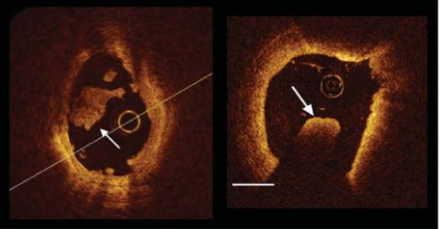
Thromotic lesion in ACS revealed by OCT . Image source Francesco Prati et al European Heart Journal 31(4):401-415

What about FFR & iFR ?

Physiological tools like FFR / iFR / iFR are less useful to identify culprit lesion rather it tell us about the flow across the lesion.

Final messsage

Identifying IRA in NSTEMI/UA is more challenging than in STEMI, relying on basic ECG and ECHO tests. It is good to realise, this effort may be redundant if we are considering only medical management.(Which is often the case in many low risk patients)

Reference
1.Baumann AAW, Mishra A, Worthley MI, Nelson AJ, Psaltis PJ. Management of multivessel coronary artery disease in patients with non-ST-elevation myocardial infarction: a complex path to precision medicine. Ther Adv Chronic Dis. 2020 Jul 1;11:2040622320938527. doi: 10.1177/2040622320938527. PMID: 32655848; PMCID: PMC7331770.

Read Full Post »

The huge popularity of conduction system pacing (CSP) , especially the Left bundle branch Area Pacing (LBBAP) has been hailed as a paradigm shift in cardiac pacing. By utilising the native His-Purkinje network, CSP promises a physiological alternative to the dys-synchronous RV pacing. However, beneath this procedural enthusiasm lies a critical, clinical assumption .It is the disregard of distal LBB integrity. In our haste to bypass proximal blocks, the electrophysiology community has mastered the art of ignoring distal conduction system disease a practice that relies more on hope than long-term, evidence-based science.

The physiological basis of LBBAP  is based on capturing the main left bundle branch trunk or its proximal fascicles to downstream  distal healthy  Purkinje network1. In patients presenting with complete heart block , this approach looks fine , if the underlying pathology is strictly nodal or intra-Hisian. However, in progressive, degenerative conduction system disorders such as Lev’s or Lenègre’s disease fibrosis is rarely localized. It is a multi-level, diffuse process that tracks distally into the fascicles and terminal Purkinje ramifications2.

How, can we casually overlook the small but surely dangerous risk of progressive distal LBB disease in complete heart block, which can lead to unpredictable catastrophic events ? This electrical oversight finds a striking, parallel in historical pacing concepts. In patients with sinus node dysfunction (SND), the electrophysiology community long ago established that a standalone atrial lead (AAI pacing) is a clinical hazard3. The minor yet real risk of AV nodal disease progression (approximating 0.6% to 4.5% per year) almost universally mandates the placement of a backup ventricular lead, safely transitioning the strategy to dual-chamber (DDD) pacing4.

The current electrophysiology consensus downplays this distal disease , by invoking the physics of the virtual electrode. The argument is even if the localized nerve fibers directly contacting the lead tip degenerate over the subsequent decade, the high-programmed voltage outputs will maintain permanent myocardial capture, ensuring patient safety from asystole5. While this assumption satisfies the basic requirements of bradycardia support, it ignores a core fact.

Aprar from thew physiological uncertainty , there is also a  significant risk of procedural failure. Real-world registries reveal that approximately 5% to 15% of patients undergo acute on-table crossovers due to  septal calcification, anatomical variations, or an immediate functional failure to engage a viable conduction pathway7.

Conduction system pacing is undoubtedly a great addition to our EP armamentarium, but it must be practiced with  humility in the face of unproven long-term  putcome and procedural complexities. A clinical pragmatism is warranted. Conventional dual chamber (DDD) is backed by more than four decades of  randomized clinical evidence.9. Until we clarify the natural history of the chronically paced, diseased left bundle,  DDD pacing should remain  the standard of care.

References

  1. Huang W, Su L, Wu S, et al. A novel pacing strategy with low and stable threshold-left bundle branch pacing. Europace. 2019;21(3):471-477.
  2. Lev M. Anatomic, pathologic, and electrocardiographic correlations in the atrioventricular and distal conduction system. Prog Cardiovasc Dis. 1964;7(4):317-340.
    Andersen HR, Nielsen JC, Thomsen PE, et al. Long-term follow-up of patients paced in atrioventricular synchronous vs. single-chamber atrial mode for sick sinus syndrome. Lancet. 1997;350(9086):1210-1216.
  3. Brandt J, Anderson H, Fåhraeus T, et al. Natural history of AV conduction in patients with sick sinus syndrome as related to bilateral bundle branch block. Pacing Clin Electrophysiol. 1992;15(11 Pt 2):1914-1918.
  4. Vijayaraman P, Sharma PS, Cano Ó, et al. Comparison of Left Bundle Branch Area Pacing and His Bundle Pacing in Bradycardia Indications. JACC Clin Electrophysiol. 2021;7(11):1433-1442.
  5. Jastrzębski M, Kiełbasa G, Cano Ó, et al. Left bundle branch area pacing outcomes: the MELOS registry. Eur Heart J. 2022;43(40):4161-4173.
  6. Heckman LI, Luermans JG, Vos LM, et al. Left Bundle Branch Area Pacing: A Comprehensive Review of Technical Aspects, Clinical Outcomes, and Future Directions. J Clin Med. 2023;12(11):3611.
  7. Wijesuriya N, Niederer S, de Verteuil R, et al. Extraction of conduction system pacing leads: a systematic review and international survey. Europace. 2024;26(2):euae032.
  8. Wilkoff BL, Cook JR, Epstein AE, et al. Dual-chamber pacing or ventricular pacing in patients with an implantable defibrillator: the DAVID trial. JAMA. 2002;288(24):3115-3123.
  9. Khurshid S, Epstein AE, Verdino RJ, et al. Incidence and predictors of pacing-induced cardiomyopathy. Heart Rhythm. 2014;11(9):1618-1625.

Postamble

This opinion letter is being sent to EURO PACE journal : Expecting a fast tracked rejection letter within a week or two .(Based on my past experience) They view these type of articles as destructive criticism I was told by one editor. Some one please clarify me, the difference between a constructive and a destructive critic, please)


.


Read Full Post »

Patient No 1 :

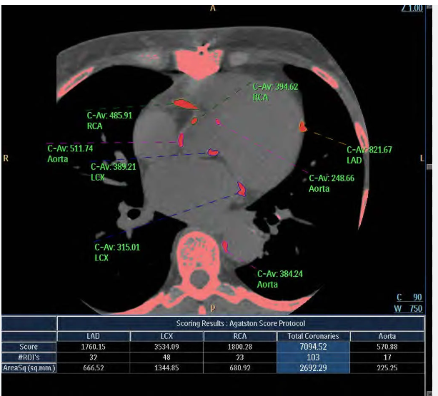
“Doctor, you had  put  me on high dose statins since my CAC score was high. Now, my CAC is increased further. What is this ? should I not get anxious ?”

“No.Not at all . It is a good sign and it indicates your plaques are getting stabilised”

How is that possible ?

It is a therapeutic paradox. We start statins based on a CAC score in a low-risk population only to find the CAC score increase further in many people. In multiple analyses, we have seen the CAC score even increase up to 30 % . Feel happy about it, as we presume it is a sign of plaque stabilization.

That’s interesting Doctor. Now,.. Doctor, my dad already has a good CAC score of 300 , without statins he should be lucky , is it not ?

No you are wrong. “Natural CAC score is a marker of plaque burden, and we can never consider it as an index of stability, while the statin-induced high CAC score is absolute bliss &  index of pure stability

Patient no 2 : My CAC is not increasing with statins , what to do ? Is it sign of statin failure ?

May be yes. I am not sure . It may indicate a poor response to statin and  inability to convert the plaques to its logical destination ie hardening and micro calcification.

Final message

Most coronary calcification whether God or statin made , imply that plaques are stable biologically. (Except the nodular /eruptive ones or those in  the shoulder region of the plaque, which can make a plaque physically stressed . Cardiologists  hate calcium essentially because, it is a hinderance to deploy a stent)

We rarely realise , how often we formulate important concepts in cardiology based on very superficial or incomplete knowledge

Reference

1.Dykun I, Lehmann N, Kälsch H, Möhlenkamp S, Moebus S, Budde T, Seibel R, Grönemeyer D, Jöckel KH, Erbel R, Mahabadi AA. Statin Medication Enhances Progression of Coronary Artery Calcification: The Heinz Nixdorf Recall Study. J Am Coll Cardiol. 2016 Nov 8;68(19):2123-2125. doi: 10.1016/j.jacc.2016.08.040. PMID: 27810054.

2.Henein M, Granåsen G, Wiklund U, Schmermund A, Guerci A, Erbel R, Raggi P. High dose and long-term statin therapy accelerate coronary artery calcification. Int J Cardiol. 2015 Apr 1;184:581-586. doi: 10.1016/j.ijcard.2015.02.072. Epub 2015 Feb 24. PMID: 25769003.


Read Full Post »

Eight point summary

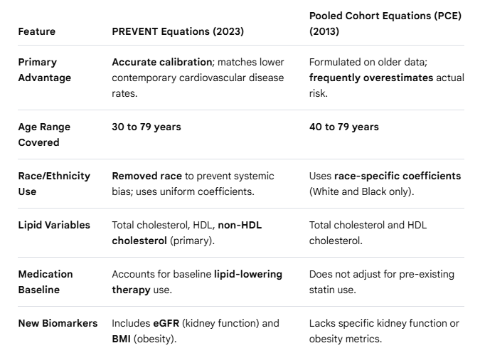
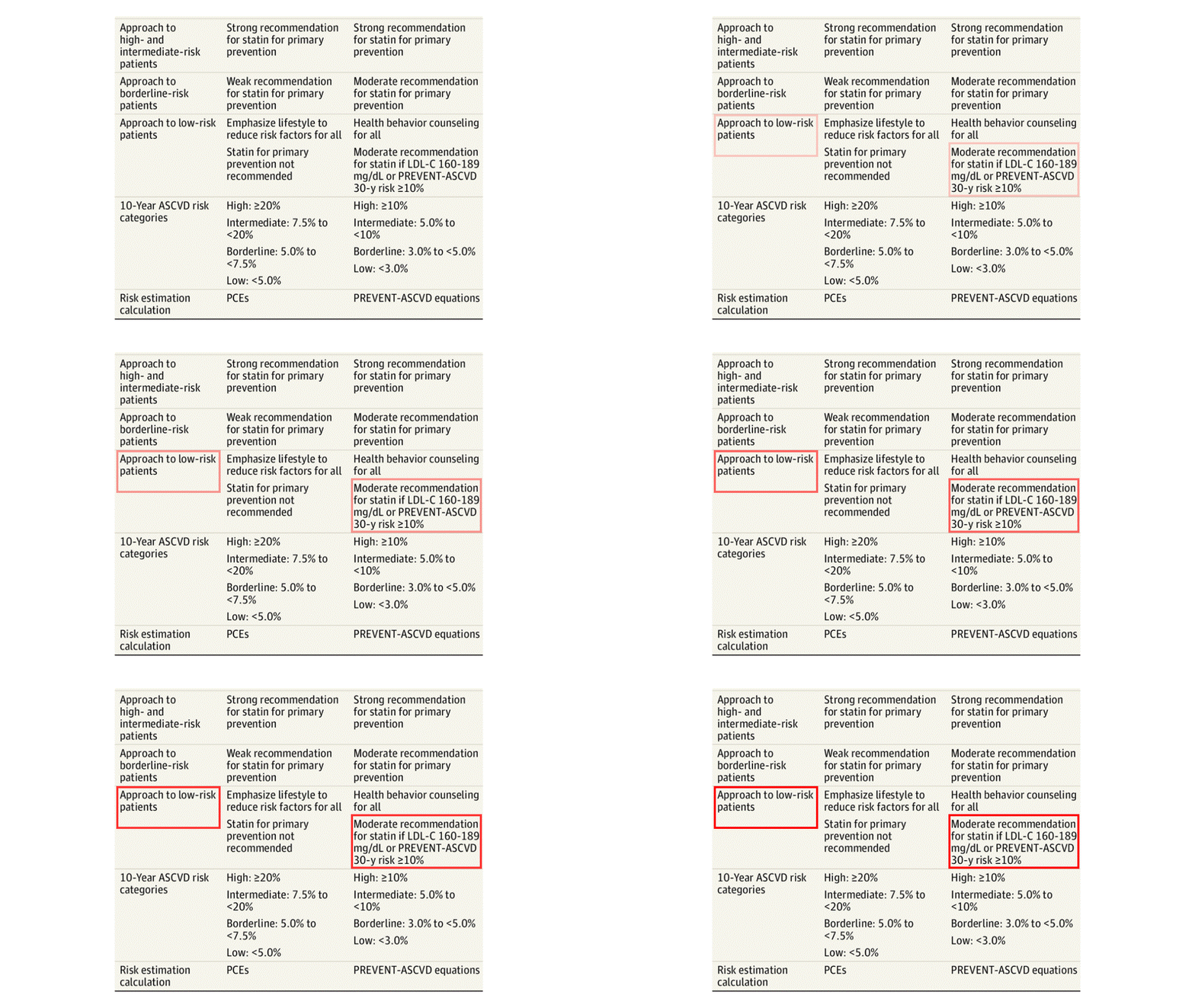
1.PREVENT-ASCVD Equations: Replaces the older Pooled Cohort Equations with more accurate, contemporary equations for adults aged 30–79, establishing new risk thresholds to guide lipid-lowering therapy.


2.Reintroduction of Lipoprotein Goals: Returns to using absolute target goals for low-density lipoprotein cholesterol (LDL-C) and non-high-density lipoprotein cholesterol (non-HDL-C) to effectively monitor and guide treatment intensity.


3.Preferred LDL-C Estimation: Mandates the use of either the Martin/Hopkins or Sampson/NIH equations over the traditional Friedewald calculation to ensure superior accuracy across varying triglyceride levels.


4.Universal Lipoprotein(a) Screening: Recommends measuring Lp(a) concentration at least once in all adults to refine cardiovascular risk assessment, recognizing levels ≥125 nmol/L as a key risk-enhancing factor.


5.Targeted ApoB Measurement: Recommends measuring Apolipoprotein B to guide further treatment intensification once standard lipid goals are met, particularly in patients with diabetes, CKM syndrome, or elevated triglycerides.


6.CAC Score : Upgrades Coronary Artery Calcium (CAC) scoring to a Class 1 recommendation to guide decisions to withhold, postpone, or initiate therapy when primary prevention decisions remain uncertain.


7.Severe dyslipidemia : Standardizes the addition of non-statin therapies like ezetimibe, PCSK9 monoclonal antibodies, or bempedoic acid to maximally tolerated statins to reach aggressive absolute lipid goals.


8.Dietary Supplements Discouraged: Recommends against using over-the-counter dietary supplements (such as fish oil, cinnamon, garlic, or turmeric) to lower lipids due to inconsistent data and lack of proven clinical benefit.

Three questions

1.What are key difference between PREVENT vs PCE equations?


2.Which can be ranked as the the most important change in the new guidelines ?

LDL reduction with statins as primary prevention in low risk individuals , is a major change

3.Which can be termed as the most questionable recommendation ?

CT calcium scoring for identifying candidates for primary prevention.A CAC score Zero only is safe . The new guidelines say even if it is 1 , a moderate-intensity statin is reasonable to achieve a ≥30% to 49% reduction in LDL-C

Reference

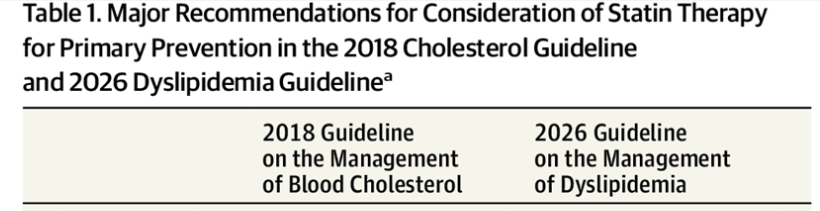
Blumenthal RS, Morris PB, Gaudino 2026 ACC/AHA/AACVPR/ABC/ACPM/ADA/AGS/APhA/ASPC/NLA/PCNA Guideline on the Management of Dyslipidemia: A Report of the American College of Cardiology/American Heart Association Joint Committee on Clinical Practice Guidelines. J Am Coll Cardiol. 2026 May 19;87(19):2624-2757. doi: 10.1016/j.jacc.2025.11.016. Epub 2026 Mar 13. PMID: 41824590.

Read Full Post »

Timing of AVR and MVR in AR and MR has always elicited huge discussion in cardiology literature. Mind you, if the patient is symptomatic and there is LV dysfunction, it is absolutely easy to make a decision.Here, the question of timing doesn’t arise at all. (Of course, the symptoms and LV dysfunction should be confirmed to arise because of valve disease, not because of any other systemic disease, or LV dysfunction might be due to associated CAD. *I know a women who got her leakinging mitral valve replaced because she developed symptoms of dyspnea due to anemia .

Timing becomes a big issue only in patients with no symptoms at all. Some cardiologists put them on a treadmill. It is controversial, as anyone will develop symptoms. But , somehow, we should make it sure, patients are truly asymptomatic.

For long, we relying on some gospel echo parameters. I don’t how good they are. But , no one really knows,how and when the onset of LV dysfunction would be , and how it will progress. But, we are bound by the guidelines , as on today. There is some critical difference between the echo parameters , based on we intervene.

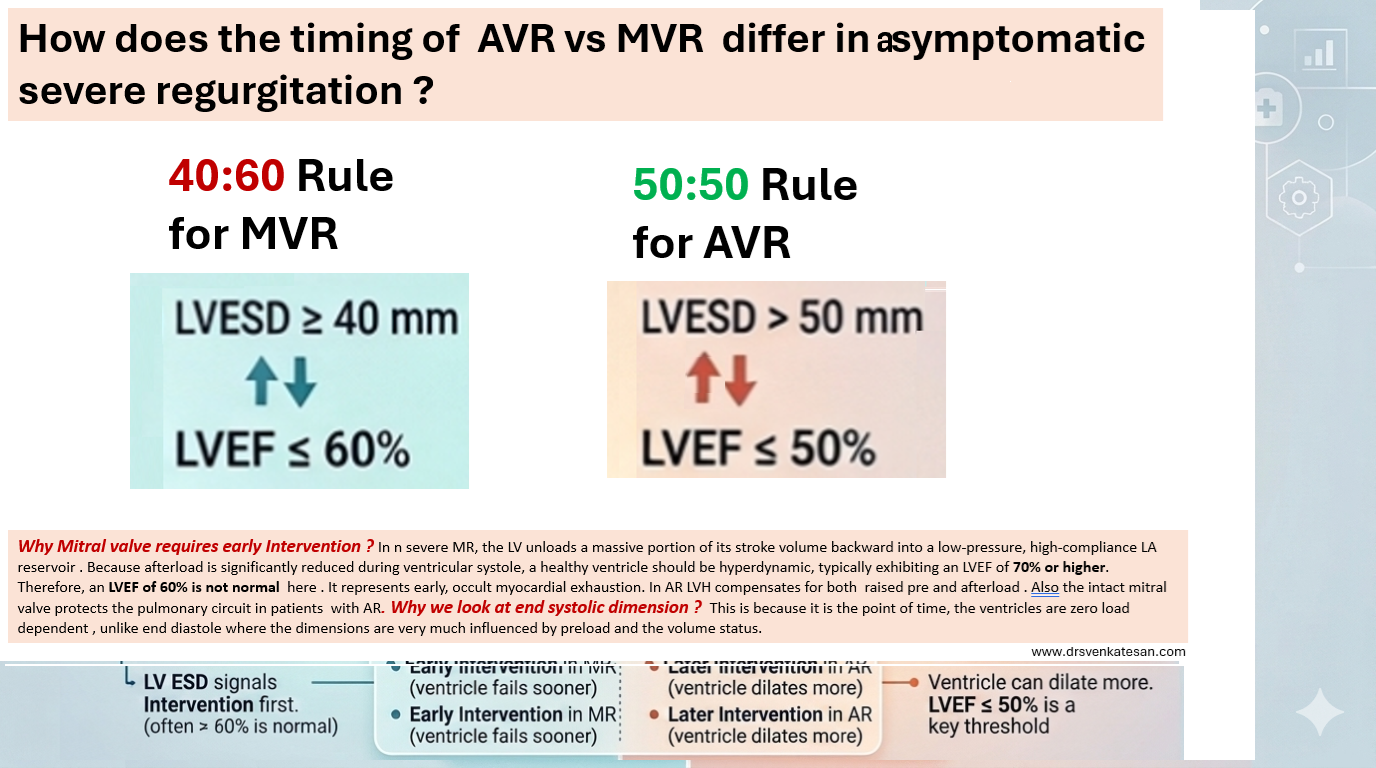
For mitral valve the threshold is lower. It needs early MVR for same degree of regurgitation as AR. It is proposed by the author for the benefit of fellows to easily remember the cut offs. It may be called as the 40:60 rule for MVR and 50:50 rule for AVR. The former denoting LVESD and later referring to LV EF %. The difference in echocardiographic thresholds arises because the two lesions cause fundamentally different hemodynamic loads on the left atrium and ventricle .(Read the legend in Image)

Final message

With mortality and morbidity for valve replacement steadily falling, it is likely more and more patients will be taken for surgery early. Of course, intervention guys are raring to go with percutaneous TAVR and TMVR, so the thresholds are expected to fall.

Read Full Post »

Journal Club Review : LACC for AF

What is the brief conclusion of this important study from EUROPACE ?

This meta-analysis aggregates data from 6 randomized controlled trials involving over 7000 patients to answer a burning question. Should we aggressively plug LAA in lieu of OAC or DOAC in AF ?

The short answer from this meta-analysis is a resounding “no” if our primary clinical goal is preventing ischemic stroke

Link to the article

Some observations and comments about this study

For all those cardiologists and physicians who have huge trust on LAA closure for AF , this study delivers a remarkable reality check:

  • The Stroke Incidence : The very purpose these devices are implanted is reduce it. But what happens ? Patients randomized to LAA closure suffered a 41% higher relative risk of ischemic stroke or systemic embolism compared to those on oral blood thinners.
  • The Harm : In a 3-4-year follow-up period, the Number Needed to Harm (NNH) may not look significant. For every 100 patients given a shiny new implant, one additional patient will experience a stroke that a simple daily pill could have prevented.
  • The Bleeding Trade-off: To its credit, LAAC successfully reduce the bleeding risk associated with OAC/DOAC

Will this study change the FDA approval and guidelines ? What will be the future of LAAC ?

No, Absolutely not. FDA is unlikely to react. It reviews devices based on their safety profiles for specific, indicated populations. Because these devices successfully reduce bleeding rates for patients with an absolute contraindication to blood thinners, their approval is legally and clinically sound.

The medical gadget eco-system works seamlessly . A device manufacturer proves that a LAA plug stops bleeding in a high-risk group. The device gets approved. Most of us fall for it and naturally shifts toward “liberal usage” as an easy, high-revenue alternative for patients who just don’t feel like swallowing a pill every day.

Final message : LACC is clearly a useless device , still you can use, less

With an estimated 400,000 implanted annually , it is too hard for us to say LAAC is useless. The future will inevitably bring next-generation devices with fancier anti-thrombotic coatings or redesigned plugs. Meanwhile, thousands of low-risk patients will continue to undergo unnecessary procedures , forcibly accepting the risk of increased stroke for the sake of convenience & glamor.

Read Full Post »

Which one of the following is least important to become great doctor ?

A. Compassion & Caring

B. Sincerity & Hard work

C. Knowledge & Skill

D.Honesty  & Integrity

Trying to answer

Yes I agree, It is brutal to frame such a question. If healing, and taking care of the suffering  mankind, is the primary purpose of the noble profession , after 4 decades in to the medical profession, realised a possible truth . “Knowledge and skills are vital, but sadly lags behind the other three by considerable distance” 

Read Full Post »

CAD : It is been called IHD, CCD, CAHD, CCS, etc. Now there is a new proposal from European Heart journal (with an Impact factor of 48 ) to change the nomenclature of chronic coronary syndrome to NAMIS : Non acute myocardial Ischemic syndrome (Ref 1)

What is the purpose & need to change the current terminology ?

The proposed AMIS and NAMIS classification shifts the clinical paradigm from coronary anatomy to myocardial function. For decades, terms like “coronary artery disease” overemphasized epicardial stenosis, neglecting the reality that ischemia frequently occurs without structural blockages. Data shows that up to 40% of symptomatic patients lack macroscopic obstructions, suffering instead from microvascular dysfunction, vasospasms, or myocardial bridges. This new term NAMIS can bring in the phenotypes like INOCA and MINOCA into the CCS without any conflict. Further, replacing vague adjectives like “stable” or “chronic” with “non-acute” reflects the high long-term residual risk of cardiac events. I also think, this new framework is meant to unify the European and American guidelines, where they use differing terminologies.

Counter Point : Do we really need this ?

Purpose of any new classification is , it should have a clear impact on patient management. But, we often find, scientific committees periodically take pride in changing the nomenclature akin to make a academic fashion statement .Within a few short years, we migrated from stable CAD to SIHD, switched to CCS , CCD etc . The fact is , for the patients it is the same angina for which they seek relief. They simply don’t bother whether doctor label their condition a syndrome or disease .

Reference

1.Boden WE, De Caterina R, Kaski JC, Bairey Merz N, Berry C, Marzilli M, Pepine CJ, Barbato E, Stefanini G, Prescott E, Steg PG, Bhatt DL, Hill JA, Crea F. Myocardial ischaemic syndromes: a new nomenclature to harmonize evolving international clinical practice guidelines. Eur Heart J. 2024 Sep 29;45(36):3701-3706. doi: 10.1093/eurheartj/ehae278. PMID: 39211956.

Read Full Post »