Localising the infarct-related artery (IRA) In NSTEMI , when there is multi-vessel coronary artery disease (CAD) is a classic clinical dilemma. Because NSTEMI lacks the clear, localising markers as in STEMI, identifying the “culprit” among multiple chronic blockages requires a more careful approach. It requires combined analysis of electrocardiographic clues , Echocardiographic WMA, angiographic analysis, and finally as a last resort intracoronary Imaging. Note : In UA,without NSTEMI it can be called as ARA-Angina related artery.
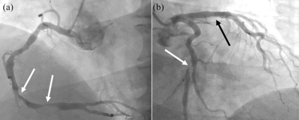
Image source : Baumann et al. Ther Adv Chronic Dis. 2020 Jul 1;11:2040622320938527
A meticulous review of the invasive coronary angiogram (ICA) will identify the culprit lesion in the majority of cases. Look for these specific features
- Plaque Morphology: Eccentric , hazy lesions, irregular borders, or filling defects indicative of an intraluminal thrombus
- Lesion Ulceration: Look for ulcerated edges, aneurysmal segments, or evidence of a dissection flap. Any vessel showing TIMI-1 or TIMI-2 flow, or prolonged contrast stasis during washout suggest microvascular plugging downstream from a fresh rupture.
ECG -ECHO correlation of WMA is the key
- Wall motion defect correlation: Matching the distribution territory of the suspected culprit vessel LADcs LCX or RCA can be most useful . Still extensive collaterals can prevents us, to make a rule of thumb
- Typical patterns like Wellens can help localise the IRA. Transient ST-depressions or deep T-wave inversions
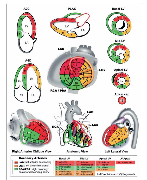
The most important illustration in coronary artery disease
Note* WMA can be deceptive as well, due to old MI, remote ischemia , or either due to collateral support or lack of it
Intra coronary Imaging
When angiography shows multiple severe lesions with no obvious acute features, coronary imaging like OCT/IVUS can be helpful. It visualises plaque rupture, thin-capped fibroatheromas (TCFA), red/white thrombi, and superficial plaque erosions. IVUS can help identify ruptured fibrous caps, and intramural haematomas.However, the temptation to do imaging should be resisited as performing this in ACS adds its own risk.
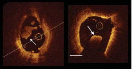
Thromotic lesion in ACS revealed by OCT . Image source Francesco Prati et al European Heart Journal 31(4):401-415
What about FFR & iFR ?
Physiological tools like FFR / iFR / iFR are less useful to identify culprit lesion rather it tell us about the flow across the lesion.
Final messsage
Identifying IRA in NSTEMI/UA is more challenging than in STEMI, relying on basic ECG and ECHO tests. It is good to realise, this effort may be redundant if we are considering only medical management.(Which is often the case in many low risk patients)
Reference




