Feeds:
Posts
Comments

Archive for September, 2008


                                                             Syncope by definition is a transient loss of consciousness due to cerebral hypo perfusion and loss of muscular tone, and the patient falls but  recovers fully and gets up either assisted or spontaneous.The cardiac and vascular counter response to syncope is most often intact .This makes syncope characteristically transient . If a patient does not recover from syncope it could either be a prolonged loss of consciousness( Stroke etc)  or if he never gets up he will be called a victim of cardiac arrest or  a SCD ! (Sudden cardiac death ) . So technically by defintion ,  all  patients  will  have to  survive  the  syncopal episode.

But the following questions need to be answered   

  1. How prolonged  a syncope can be ?
  2. Can syncope lead onto  sudden cardiac death ?(SCD)  
  3. What are  life  threatening syncope and non life threatening syncope ?                           

What is the link between, syncope and SCD in patients with ventricular arrhythmia’s ?

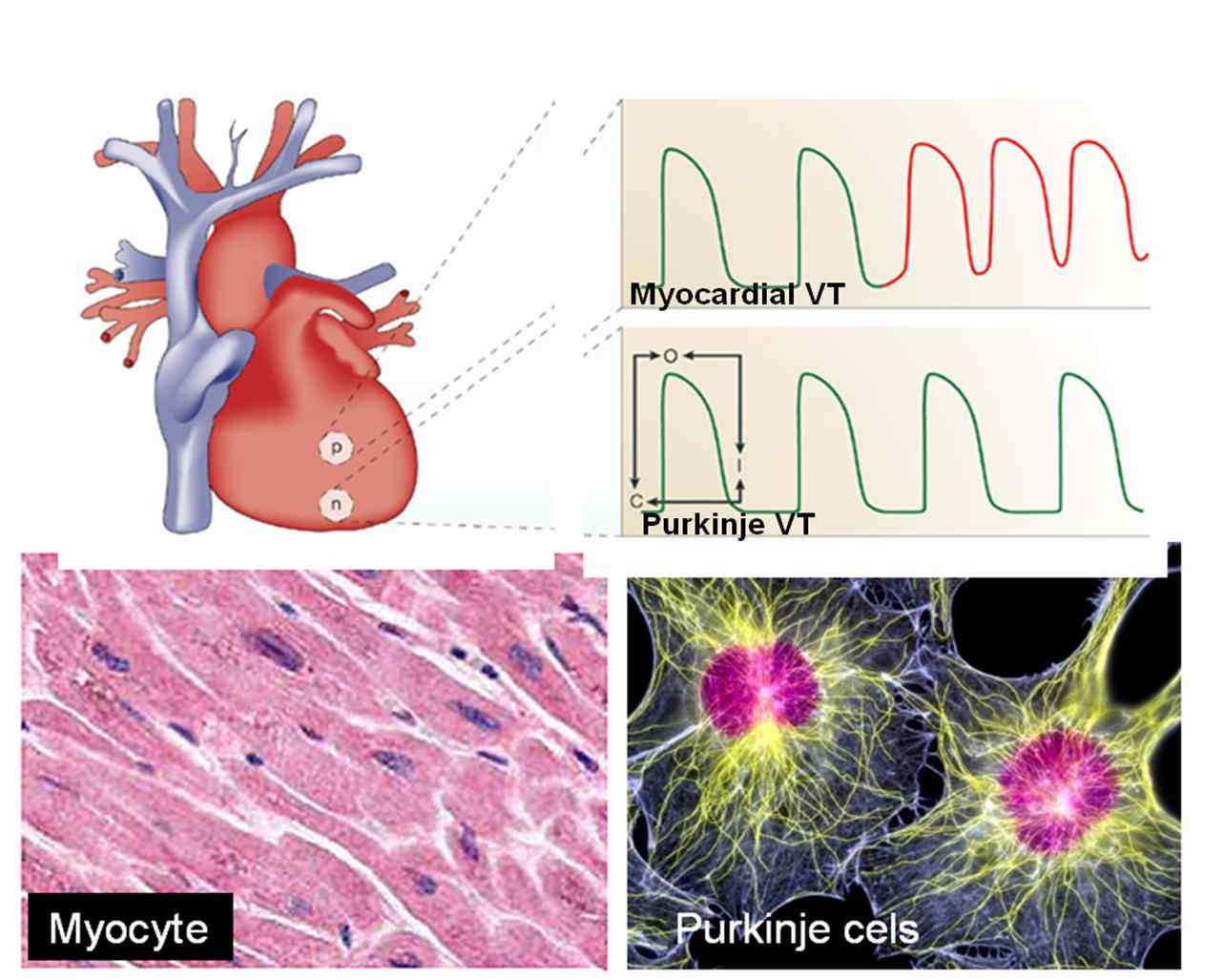
Some case of long QT syndromes could be life threatening especially in children as they inherit sudden death. A patient with a non sustained VT  may develop syncope  if  the  VT  becomes sustained especially  if there is underlying heart disease and LV dysfunction . Among this  few , may degenerate into ventricular fibrillation and patient may die.  

How common is syncope in acute myocardial infarction ? 
 Syncope is a very  rare presentation of acute myocardial infarction. 
 
Can syncope precipitate  or precede a  cerebro vascular accident  ?   


Prolonged syncope , TIA,  stroke in evolution and completed stroke   can be a continuous spectrum in patients with carotid and cerebrovascular  disese . But when a syncope evolves in to a stroke the   patient is not considered to be a victim of syncope but  they enter the stroke protocol.

There is a big list for the causes of syncope

But to put it simply

A.Cardiac

  •    Purely electrical ( Arrhythmic- Brady, Tachycardia)
  •    Mechanical( Valvular obstruction, and other structural heart disease etc)

B. Non cardiac

  • Vasovagal (Commonest 90% of all syncope)

C. Metabolic*

  •  Anemia
  • Hypoglycemia
  • Hypoxia
*Metabolic causes  coupled with simple  vaso vagal(Neuro cardiogenic)  constitute the bulk of causes of syncope .Siezure disorders are very  common and a close  mimicker of syncope and need to be ruled out.

How to work up  a patient with syncope ?

                   First ,  one need to confirm  it is indeed a syncope . If the initial examination is not clearcut   one  need to  go back to the  history and ask for  circumstances under which the syncope occured  and  details of prodromal symptoms  if any . Patient’s  family members who witnessed the event can give useful information . It  is the most  cost effective ( Comes free of cost infact !)  investigative tool available .Cardiac syncopes are usually sudden, vasovagal often have environmental or emotional factor. Apart from routine investigations , ECG, Echocardiography, holter are done generally, head up tilt test, Loop, event recorders may be reuired in few.

Final message

                                          Syncope is one of the common symptoms in cardiology and  general medical practice. Many times the diagnosis is easy . Common syncope is  never fatal but , ruling out dangerous  tachy and bradyarrhythmias is a key aim.  In a significant number (20-30%) identifying the cause could be really  difficult and  may never be made in spite  of the modern diagnostic tools. These syncope of unknown origin is grouped along with the neurocardiogenic category.

The one,  positive thing about syncope is (unlike chest pain) , it is rarely fatal in it’s first episode ,  gives the physicians to  investigate and correct the underlying problem.

Read Full Post »

   Dr. Venkatesan   Sangareddi  

AK 53/1, # 9

Narmada enclave

 7th main road

Anna nagar

Chennai -600 040

Tel:044 26209009


Age &  Date of birth

42 ,               25-5 -1964

Experience

1998–2007                  Madras  medical college                      Madras 

Asst. Professor  Of Cardiology

Work involves  intensive coronary care, invasive and non invasive cardiology.  Has special interest in  clinical research in Acute Coronary Syndromes

Has    publications in  various  Journals.


1997-1998                   Madras  medical college                       Madras           

Asst. Professor  Of Medicine

Worked  in intensive care medicine and  in

 Medical oncology for 6 months


1994–1996                    Madras   medical college                        Madras 

Resident   Cardiologist

Selected to resident programme   toping the state   in the super speciality  exam

Presented  papers in national conferences

Experience gained in invasive and non invasive cardiology


1991–1994                      Govt.  Health  Centre           Karur.       Tamil nadu

Medical Officer   

Worked in internal medicine  department

Family medicine and community health care.

And   socio economic aspects of health care.

Education

1994–1997              Madras  medical college                               Madras           

Doctorate  in cardiology (DM) 


1988-1991              Coimbatore medical college                          Coimbatore


Doctor  of Medicine (MD) Dr.M.G.R  Medical university  , Madras


Secured  three  gold medals for excellence in cardiology.


1987-1988            Coimbatore medical college          Coimbatore  India


Junior resident in  Medicine


1987                     Coimbatore medical college          Coimbatore


House  officer


1982-1986            Coimbatore medical college        Coimbatore


M B., B S.    Bachelor of Medicine and Bachelor of Surgery


                             Madras    University                           Madras     


Interests

Electro physiology, expert systems in cardiology., clinical research  in acute coronary  syndromes, preventive cardiology,  bio ethics, outcome analysis ,  logistics in cardiology and publishing  online journals. 


List Of  publications

Enclosed

Reference

Prof.V.Jaganathan. MD.,DM

Professor  &  Head of  Department

Institute of Cardiology, Madras Medical College  Chennai


Address  for   communication


Spouse


 Dr.Latha  Venkatesan  MD . Gynecologist,

 Sundaram  Medical  Foundation, Chennai  India.

AK 53/1, Flat no A- 9

TAS Narmada enclave

 7th main road

Anna nagar

Chennai -600 040

Tel:        044 6209009

Mobile : 9840059947

E.mail : drvenkatesans@yahoo.co.in>


List of publications by  S.Venkatesan


1.QTc  Interval  in atrial fibrillation.  The Tamilnadu Dr. M.G.R Medical university doctorate thesis 1991


2 .Thrombolysis in hyperacute MI.

 Indian Heart Jr  1999:51: 321


3. Circadian  Response To  Thrombolysis  In Acute Myocardial Infarction Indian Heart Jr  1999:51:686


4. Left Ventricular Mass in Pregnancy Induced Hypertension.

  Indian Heart Jr.  1999:51


5. Dissection of  interventricular septum by unruptured  right  sinus of valsalva    aneurysm  resulting in complete heart block.

Indian Heart Jr  1995 Nov-Dec: 605


6.Angiosarcoma  of leftventricle presenting as hemopericardium and  cardiac tamponade. Indian Heart Jr  1995 Nov-Dec:636


7.Asymtomatic multivessel disease  following  myocardial infarction

 Indian Heart Jr  1999:51: 686


8. Transmitral pulse doppler echo correlates of mitral regurgitation severity Indian Heart Jr  1999:51:636


9. Safety and efficacy of intravenous nicorandil  in unstable angina. Indian Heart  Jr  1999:51:704


10. Efficacy of nicorandil as monotherapy in ischemic heart disease Indian Heart Jr  1999:51:728


11. Left ventricuar function by angiogram in significant LAD disease. Indian Heart Jr  1999:51:687


12. Aortic root dimension in isolated rheumatic mitral stenosis

Journal of association of physicians of India abst: 1998


13. Serum phosphate in acute myocardial infarction

Indian J Physiol  Pharmacol 2000  44(2):225-8


14.Differential  Response  to  right  and  left  coronary  artery  thrombolysis   Indian Heart Jr  2000:52:715


15. Therapeutic  issues  in  Stable Ventricular  tachycardia: A coronary  care  unit  perspective Indian Heart Jr  2000: 52: 808.


16.Current   cardiology  practice: evidence  or  experience  based ?    An  analysis of  ACC/AHA  guidelines. World congress of cardiology   2002 sydney  Oral  presentation.(Published in Journal of American college of cardiology)JACC :2001.39:9 Sup.B 462B


17.Isolated  Diastolic Hypertension .World congress of cardiology  2002 sydney  poster  presentation..

( Published in Journal of American college of cardiology) JACC :2001.39:9 Sup.B 175B


18.Rescue thrombolysis in  acute myocardial infarction

Journal of association of physicians of India abst: 2002


19.Canadian   cardiovascular  society  classification of  angina:

 An  angiographic  correlation. Indian Heart Jr Abstract issue 2001


20.Non invasive management  of high risk unstable angina

Accepted for oral presentation in cardiological society of India annual scientific session Kolkata  Dec2003


21.Non dilated cardiomyopathy

Accepted for oral presentation in cardiological society of India annual scientific session  Kolkata  Dec 2003


22.Safety and efficacy of angiotensin-converting enzyme inhibitors in symptomatic severe aortic stenosis: Symptomatic Cardiac Obstruction-Pilot Study of Enalapril in Aortic Stenosis (SCOPE-AS).
Am Heart J. 2004 Apr;147(4):E19


23.Rheumatic heart disease occurrence, patterns and clinical correlates in children aged less than five years.J Heart Valve Dis. 2004 Jan;13(1):11-4.


24. Estimation of subjective stress in acute myocardial infarction.
J Postgrad Med. 2003 Jul-Sep;49(3):207-10.


25. Serum phosphate in acute myocardial infarction.
Indian J Physiol Pharmacol. 2000 Apr;44(2):225-8.


26. Canadian Cardiovascular Society classification of effort angina: An angiographic correlation.
Coron Artery Dis. 2004 Mar;15(2):111-4.


Coming soon :


List of  top ten  leading famous  cardiologist  in india

Read Full Post »

                                 The pleura and pericardium are very close anatomical companions within the thorax. Both contain minimal levels of physiological fluid.  It is quiet common to find combined pleural and pericardial  effusion. While the commonest explanation for combination of pleural and pericardial effusion is inflammation of both  in systemic disorders like polyserositis or malignancy . In cardiac failure also both effusions can  occur explained by raised venous pressure.

But there has always been a curious relationship between these two spaces.

                         Is there a antomical or physiological link between these two spacs ?  In fact a large pleural effusion some times result in sympathetic pericardial  effusion.  Tapping of pleural effusion may regress this pericardial fluid as well.

                         This is purely a clinical observation and needs an explanation .It is  believed , there is  some  non functional lymphatic channels shared between pericardial  and pleural spaces.This may get opened up in pathology of either of them.


Read Full Post »

Heart is externally covered by two layers of pericardium .  Pericardial space is formed between parietal and visceral layers of pericardium . It is a narrow space which is normally lubricated with pericardial fluid up to 25ml. When these two tissue surfaces  come into contact ,  pathological  rub takes place.It is heard  whenever the pericardium is inflammed . Pericardial rub is a distinctive but uncommon  clinical sign .

Common clinical conditions

  • Acute pericarditis
  • Uremic pericarditis.
  • Rheumatic pericarditis
  • Post myocardial infarction

Pericardium has two layers .

There are four  possibilities for pericardial rub to take place.

The rub can occur

1.Between the two layers of pericardium

2.Between the visceral pericardium and the epicardial layer of  heart*.

3.Between parietal pericardium and the  chest wall

4.Pericardium can rub with the adjacent pleura( Pleuro pericardial rub )

The second and third mechanisms are very rare.

An update

We have realized one more possibility . Diaphragm forms the floor of the heart on which the hanging heart  rests . Rubbing of pericardium over diaphragmatic surface is a beat to beat affair that lasts the entire life !. In inflammatory states of  diaphragm especially  the contagious  ones from abdomen  , can result in pericardio- diaphragmatic rubs .These rubs are almost impossible to hear clinically.

pericardial effusion rub plural pleuro pericadial

*The anatomic mystery : Is epicardium same as visceral layer of pericardium ?

Some anatomist feel that both are same entities. If that is the case myocardium can never split its relationship with visceral pericardium.But it is also a anatomical fact visceral pericardium engulfs the coronary artery and  are located sub epicardially.

How many components of pericardial rub are clincally heard ?

Pericardial rub  classically has three components. Systolic, mid diastolic, and pressytolic atrial components. Pericardial rubs are typically described as to and fro rub. Systolic component is most consistent. In atrial fibrillation mono component pericardial rub is heard.

Quality

Superficial , scratchy, high pitched ( Can also be low pitched)

Location

Left sternal border , left 2nd or 3rd space  .Best heard in  sitting , leaning forward in inspiration. Many times the rubs are transient and evanescent . Since it has multiple components it may be mistaken for added heart sound like S 3 or S 4.

What is the mechanism of pericardial rub in the immediate post MI phase ?

Presence of pericardial rub post MI indicate a transmural involvement or atleast significant epicardial involvement . Recognition of this is important as presence of pericardial rub increases the risk of rupture  and hemorrhagic effusion if anticoagulants are used.

What is the  relationship between  pericardial effusion and  pericardial rub ?

Generally it is said with the onset of effusion pericardial rub disappear.But this is not necessarily true.

Rubs after contusion chest and fracture ribs can be with the chest wall and may have  no relationship with effusion.

Is pericardial rub a painful condition ?

Pericardial  rub associated with acute inflammatory pathology is severely painful (like a pleuritis).But pericarditis associated with chronic inflammatory conditions are less often generate pain.The exact reason is not known.

What is pleuro pericardial rub ?

This  clinical entity is poorly defined , often taught by veteran professors  in clinical auscultation classes.It can be heard in the mid segment  or diaphragmatic pleuritis with or without pericardial effusion in patients with  atypical pneumonias.

Read Full Post »

No reflow is the terminology used primarily in cath labs where, even  after a successful opening and stenting  of a coronary artery the coronary blood flow is not  restored to myocardium . The point to be emphazised here is blood do cross  successfully the site of  the obstruction but fails to enter the muscle segment  to which the coronary artery is supplying. So the paradoxical situation of artery  being open but the  myocardium is closed to receive  blood flow  happens . This is termed as no -reflow.  Actually it is a  misnomer , and  ideally it should be called “no flow” because  normal distal flow  does not  occur (After PCI)  in the first instance  to get interrupted  later on  and be labeled as  no re-flow.  .The only positive effect of PCI in these situation is blood flow would have improved by few centimeters ie till it reaches  but falls short of myocardium . In fact no reflow , can be termed as  glorified and concealed  terminology  for  PCI failure . It needs urgent action . No reflow is also called as myocardial epicardial dissociation.

Mechanism of no reflow.

Curious case of open coronary artery and closed myocardium !

Coronary  microvascular plugging  is mainly  due to thrombus and atheromatous debri , myocardial  edema , microvascular spasm may also contribute.

Where can it occur ?

  • First described in cath lab, especially following primary angioplasty.
  • It can very  well happen following thrombolysis in STEMI.
  • Can occur in venous grafts.

How do you recognise no reflow?

In cath lab it will be self evident from the check angiogram. Some times it is less obvious and may  require, myocardial  blush score, TIMI frame  count, contrast echocardiography, PET scan etc. In post MI a very simple method to recognise this entity could be the observation of persistent ST elevation in ECG .

Treatment.

Extremely difficult. Almost every coronary vasodilator has been tried.(Nitrates, nicorandil, calcium blockers, etc).Success is less than 30%.  High pressure flushing with saline inside the coronary artery is advocated by some.Others believe it’s dangerous to do it. So prevention is the key. Avoid doing PCI in complex, thrombotic lesions. Use thrombus suction device like export catheter(Medtronic). Distal protective devices are double edged devices , useful only in experienced hands.

Unanswered question

What is the size of the particle (thrombotic and atheromatous  debri)  the   coronary microcirculation safely handle and push it into the coronary venous circulation and the coronary sinus for disposal ?

If we can lyse the thrombus into micro particles by some mechanism and make it traverse the coronary circulation this complication of microvascular  plugging can be treated and prevented .

What is the final message ?

  • No reflow is relatively common condition during emergency PCI done for ACS patients
  • More common in complex thrombotic lesions.
  • Can also  occur in STEMI
  • Treatment is often vexing . In fact the treatment of this condition is so difficult , it can be termed  almost synonymously with “Failed PCI” if flow is not restored.
  • Successful treatment of no- reflow  means not momentry restoration of  myocardial flow  by mechanical and pharmacological modalities ,but to maintain sustained myocardial   perfusion. This we realise, as patients who have had a no reflow during  a PCI, do not perform as well in the follow up  .
  • So prevention is the key.

Read Full Post »

Differential response of thrombolysis between left and right coronary system

  • Thrombolysis is the specific treatment for acute myocardial infarction. ( Privileged few , get primary PCI))
  • Failed thrombolysis occurs in significant number of patients ( 30-40%).
  • Persistent ST elevation  120 minutes after thrombolysis is best indicator of failed thrombolysis.
  • It has been a consistent observation  failed  thromolysis  is more frequent in anterior   or LAD myocardial infarction.

In a simple study we have documented  patients  with inferior MI  rarely had persistent ST elevation and thrombolysis  was   successful in vast majority  of  patients  ( Except in few patients associated lateral MI)


The mechanism of better thrombolysis in right coronary artery  is simple.The success of thrombolysis , apart from early time window , is directly correlated with pressure head  and the duration of contact between the thrombolytic agent and the thrombus. In right coronary circulation the  blood flow is continuous ,  occurs  both in systole and diastole that facilitates the maximum delivery of the thrombolytic agent . Further there is a favorable  pressure gradient  across RV myocardium  as the transmural occluding pressure across RV is considerably less then LV myocardium.

This paper was presented in the  “Annual cardiological society of India scientific sessions”

at Chennai, Tamil Nadu.India December 2000

Click to down load PPT full presentation

Read Full Post »

The cell of origin of ventricular tachycardia is rarely discussed at bedside. It is still in research labs !

                                    Ventricles are not made up off entirely myocytes. Apart from myocytes it contains specialised  purkinje cells , fibrocytes, interstitial cells and  some times primitive mesenchymal cells. Ventricular tachycardia can arise either in purkinje cells, the myocytes  or even the fibrocytes. The myocyte  VT  classically occur during ACS or post infarct VTs.They are  more often hemodynamically unstable and quickly degenerate into ventricular fibrillation. Myocardial VT is likely to be pulseless and require DC cardiversion frequently. Purkinje VTs are relatively less unstable. If VT arise proximally in the septum near the distal his, or in bundle branches (BBR) the VT is more stable.They  are likely to respond to be medical management.

What is the therapeutic implication of knowing  myocardial VT ?

                               In fact  ,simply knowing the cell of origin of VT is not suffice .The ionic currents inside the cell that trigger and sustain the VT is more important. There are few ionic circuits responsible for VT. Sodium , Intra cellular calcium, potassium , beta receptor mediated calcium current.If we know the individual ionic culpirit we can block that specifically  . Now we have multi purpose ion blockers  like amiodarone which acts like a broad spectrum antibiotic and terminates a VT.

                              So as of now there is no real purpose of breaking our head  in locating the cell  of origin  and the ions responsible for VT  at  the bed side ,( Researchers will do that for us !).  We have only few  antiarrhythmic drugs available in our crash cart  .Our job is to choose the optimal  drug  which will fit in for our patient. In electro physiology labs, radio frequency ablation is done .This is  nothing but shooting down the abnormal electrical  focus (Cluster of cells or a samll segment of myocardium).  In future,  a single abnormal  cell could be selectively neutralised with cell based therapy assisted by  nanopore robots !

Read Full Post »

                              Cardioversion with DC shock  offers immediate cure in many of the dangerous ventricular and atrial tachycardias.  It is often  taught ,  any hemodynamically unstable tachycardia  refractory to  medical therapy respond to electrical cardioversion.  One should also  remember electricity is in fact be called  as a drug !  and it should be delivered in proper form and dose. Here it is the paddle size, paddle position and the axis of current flow all are important. Now we have bi phasic currents for better efficacy.

                             While it is true, most of cardiac arrhythmias respond to shock,  there are few which do not respond or respond very transiently.There are few arrhythmias  in which ,DC shock is not only ineffective but may precipitate a ventricular  fibrillation.

                            Generally arrhythmias of reentrant etiology respond well to DC shock were interuption of  electrical circuit by external current is easily possible. In arrhythmia’s of enhanced automaticity ,  and ectopic tachycardia  it is difficult  to extinguish  the tachycardia focus with DC shock .

Arrhythmias where DC shock is not going to work are

A. Mutifocal atrial tachycardia(MAT)

B. Digoxin induced arrhythmias.Patients who are on digoxin,  has  enhanced ventricular  automaticity.These patients if they  get a DC shock will unmask the  ectopic foci.

C. In elderly with atrial fibrillation and sinus node dysfunction it may be dangerous to shock them with out temporary pacing support as sinus node goes for prolonged sleep mode.

D.In electrical storm with VT ,  if more than three shocks are required within a minute,  the VT will most often going to be permanent and the  electrical therapy can be termed as a failure. These patients will require intensive pharmacological management( Including magnesium, bretyllium etc)

E. And finally , sinus tachycardia (whatever the rate)  is an absolute contraindication for DC shock.

 Verapmil is often effective in MAT  but correction of hypoxia and acidosis may be critical.For digoxin induced arrhythmias phenytoin may be tried.

What to do when the DC shock fails?

  • It will be a  tricky situation and one wonder what to do next when the so called  universal antidote for cardiac arrhythmia fails !
  • Cellular internal millieu  is altered  by hypoxia and acidosis .It may prevent the  effectiveness of cardioversion.So try to correct them .
  • Over dirve atrial  pacing  is one option for automatic tachycardia.
  • And now ablation of arrhythmic focus is possible with radio frequency waves  in some of these patients.( Diffiuclt as an emergency procedure)

Read Full Post »

                                  Indication for thrombolysis in ST elevation MI  is mainly determined by clinical and ECG features. ST elevation of more than 1mm in two consecutive leads with a clinical suspicion of acute coronary event demands immediate thrombolysis.

                                 Early repolarisation syndrome(ERS) is a  is typical mimicker of STEMI . In ERS , ST segment elevation occurs in many leads especially precardial .This entity is estimated to occur in nearly 3-5% of population where a genetic variation in the potassium channel activation is reported.

                              If they  land in ER with some sort of chest pain , chances are high for labelling  them as ACS . It is not uncommon for  CCU physicians  to  witness  an  ERS being lysed . Even in many of the land mark trials (ISIS ) there has been many inappropriate thrombolysis , recognised later on.

What can really happen if you thromolyse them inadvertently ?

Generally nothing happens . But they are exposed to the risk of thromolysis. The ECG changes persist. And troponin will be negative and  echocardiogram will not reveal any wall motion defect.

Are we legally liable if a patient  with ERS was thrombolysed and he ends up with a bleeding complication like stroke ?

                        While the physician may feel guilty , there is no reasons for him to feel so.The guidelines are kept little lineant  for  the indication for thromolysis. When we are promoting  a strategy of early  thrombolyis  on a population based approach  in STEMI ,  there is bound to have a overlap with normality .The benefits out of early thrombolysis for eligible  patients for outweigh the few inappropriate thromolysis.

When you want to catch  a   real criminal  it is unavoidable,  one gets hold of all suspected criminals before letting them free . Unfortunately  in this exercise , some of the innocent  might experience   intimidation or even a injury  at the hands of law enforcers.

                               Similarly if a patient with ERS develop a severe esophageal spasm and typical  angina like chest pain he is absolutely certain to receive thrombolysis. (Troponin, CPK come later , and the results never veto the clinical and ECG criteria ,except probably in LBBB) .Many times critical  time dependent decisions are prone for errors in CCU.   So it may be  unscientific to ask why an ERS was  thrombolysed !

 How can one prevent inadvertent thrombolysis in ERS ?

                            Always ask for the previously recorded ECGs .If it is available and  look exactly similar to the current ECG  chances are unlikely  for ACS. In ERS ST segment is generally concavity upwards . ACC/AHA  guideline for STEMI  ,is  aware of this fact , but still  advices thrombolysis for all ST elevation irrespective of the morphology of ST segment elevation. This is propably intentional,   not  to incorporate morphology cirteria of ST elevation  for thromolysis .It would potentially  make many true STEMIs  diagnosed falsely  as ERS and deny thrombolysis.


What is the latest news about ERS ?

                       Now data are coming up, ERS is not entirely benign condition.Some of them ( Even a fraction of ERS population could be a significant number) can have a overlap between Brugada syndrome and they  could be prone for dangerous ventricular arrhythmia when challanged with ischemic or other stress.

Read Full Post »

The pressure tracing between two chambers of the heart are distinctly different .

 Apart from the magnitude of the  pressure ,(LV at systemic pressure ) The morphology also changes.

  •  RV pressure curve is triangular in shape,
  •  Upstroke is not rapid , (Low dp/dt)
  •  There is no sustained peak ,
  •  There is an early fall and
  •  The pressure falls to zero which  never happens in LV.

Contary to this LV pressure curve is bullet shaped,  with a rapid upstroke, sustained peak, fall later, and does not touch zero.

RV/LV pressure curves in normal persons .Adapted from , Curtiss 1975 Circulation

Note : The shapes of RV curve will change in pathological states.Example in TOF, large VSD there will be left ventricularisation of RV pressure wave forms. Also  in pulmonary hypertension RV pressure may mimic a LV curve.

Read Full Post »

Older Posts »