Feeds:
Posts
Comments

Posts Tagged ‘zipes’

Wide qrs tachycardia has a unique place in clinical electrocardiography .It is  a much fancied and glamorous entity for the simple reason , it continues to be the  cardiologist ever solved puzzle .For over three decades of research, clinical debates , symposiums , seminars have effectively failed to take away the uncertainties in decoding the wide  QRS  tachycardia . (Specifically ,  VT vs SVT with aberrancy)

Some wondered , should we really waste our efforts in differentiating the two . In emergencies it never matters , in fact one need  not attempt to do this often futile exercise !

Few dedicated criterias like Brugada etc have helped us .

While the difficulties in differentiating between VT and SVT with aberrancy remain over the decades .A less reported  , but more common issue is  confronting  us .

It is  the big question of  differentiating a  wide  QRS tachycardia from a narrow QRS  tachycardia

wide qrs tachycardia vt svt aberrancy

This  occurs  more often than we realise  ,because we define wide  QRS  tachycardia in a vague manner

  • Normal qrs width between Up to 80 / up to 100 ms acceptable  ?*
  • Narrow qrs tachycardia 80 ms?
  • Wide qrs tachycardia i> 120ms  ?
  • Definitely wide qrs >140msec

* The confusion is mainly because 20ms difference between limb leads and chest leads .

In reality one may not be able to all  tachycardia into narrow or wide .

There is big  overlap zone that need to be labeled a intermediate qrs tachycardia

If we can  triage the tachycardias into three instead of two it may help us arrive  fast  ,  to the  correct diagnosis

Narrow QRS tachycardia ( qrs 80ms)

  • Sinus
  • All svtS (avnrt etc)

Intermediate QRS tachycardia 90-120

  • Most of the SVT with  aberrancy  ( Except antidromic SVTs which are really to wide !)
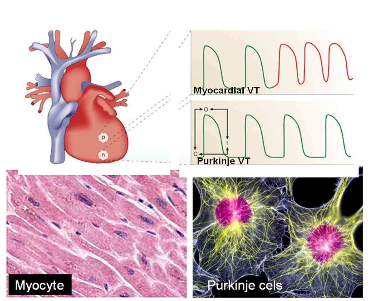
  • Septal VTs*
  • Fascicular VTs*
  • VT in PPM and ICD /CRT patients **

*  Any VT that arise near the major conducting system of ventricle conduct  fast and hence qrs are relatively narrow.

**These are rare entities where  base line wide QRS getting narrower with the onset of VT . (Ref : http://europace.oxfordjournals.org/cgi/content/full/eun254v1)

Wide qrs tachycardia >120ms

  • Most of the genuine VT (Ischemic , myocardial origin)
  • Post MI VTs
  • SVT aberrancy especially AVRT
  • Any SVT with preexisting BBB
  • Marked electrolytic disorders

Unresolved questions

  • Which lead we should look for measuring the width of qrs ?
  • Should we take the narrowest qrs or widest qrs or should we take the average ?
  • Should we calculate how much the tachycardia has widened the qrs from the baseline  width of a given patient ?  Is it not possible , what is wide for some may be normal for another !
  • If  there is no isoelectric line  and ST segment  blends with qrs complex  how to mark end of qrs ?
  • If  limb leads show a narrow qrs and chest leads shows  wide qrs what is the significance  ?
  • In precardial leads  if one lead alone shows a narrow qrs , what is the significance ?
  • Can a narrow qrs VT conduct  with aberrancy and making it  really  wide ?

Final message

When we are  able to solve   complex electrophysiological  problems  , we must also realise  even   simple  tasks can be demanding in medicne ! It is proposed to create a  new  group “Intermediate QRS tachycardia “that can help solve the issue where we have difficulty in labeling these  tachycardias which fall  in the  greyzone .We can try &  apply the modern EP based VT criterias  to this group and find out the hidden truths !

Read Full Post »

                                         Ventricular  tachycardia is considered as a dangerous electrical rhythm abnormality .It can immediately degenrate into ventricular fibrillation and result in SCD in many.Ironically, it is also a fact , a patient with VT can  present silently  without any symptom  .Some VTs are slow and recurrent without much affecting The hemodyanmics.


In chronic recurrent, beningn VT (Some may consider it , ” height of  absurdity ” to call a VT beningn ! but it  is a reality , the term beningn denotes –  very remote chance of converting into VF) ” Is there any other therapeutic option other than convertng into sinus rhythm. “(  Read related topics)


The following paper was presented in the Annual scientific sessions of  Cardiological society of India,  Kochi , seven years ago in  2002


VENTRICULAR RATE CONTROL  IN  VENTRICULAR TACHYCARDIA 

S.Venkatesan,,. Madras Medical College. Chennai


                           Mangement of  hemodynamically  stable  recurrent   ventricular tachycardia  remains a  delicate clinical problem. Reverting to  sinus rhythm  is  considered as  the only aim  of  treating  VT.While rate control is accepted as a therapeutic  option  in atrial fibrillation,  it is not  so,  for  ventricular tachycardia.In this  context  we attempted to analyse  the effect of  Amiodarone on   ventricular  rate  in stable ventricular tachycardia  which fail to convert  to sinus rhythm.


                            The  study cohort consisted of 49 patients with stable VT  who were admitted in the coronary care unit  of  Govt. General Hospital  between 1998 to 2002.The criteria for inclusion   were systolic BP>100mmHg and absence of  hypoperfusion of vital organs  The mean age was 52 years (range 26-68)  with a male female ratio  of 4:1.   Of the study group 36 patients  were either reverted with  IV lignocaine , Amiodarone ( 150-300mg   bolus )  or  DC  cardioversion . 13  patients  who did not respond to   either of these   were  followed up  with  Amiodaroneinfusion(1000mg)  for 24 hours.  The baseline  diagnosis were old MI (6)) DCM (3)  Arrhythmogenic RV displasia(2). Idiopathic VT was diagnosed in  2 patients.All these patients had  VT  during  most part of  the   24 hour  follow up.


                         The pre Amiodarone mean  ventricular rate was  152  (124 –196).  Post amiadaorne (at 24hrs) mean ventricular rate was 128(88-142). The time taken for   50% heart  rate reduction was  6.6h (4-24h).  The average  systolic blood pressure  improved from  100   to  112mmhg . These patients were  discharged  in stable clinical status with oral Amiodarone and  were  referred for  EP study.


                          It is concluded that Amiodarone, apart from it’s cardioverting ability , has a distinct ventricular  rate controlling  effect  which  can be of therapeutic value in  at least certain subset of chronic recurrent VT.

Final message


Some of  the patients  with VT carry a very low risk of VF  and SCD .In these  patients , the only  other major  aim is to prevent tachycardiac cardiomyopathy  that can be done with drugs which  controls  the ventricular rate whenever  VT occurs !

Corrrecting the primary cause like cardiac failire , revascularisation ,detailed EP study  ,tachycardia mapping , followed by RF ablation and ICD implantation is  the state of the art approch in the management of VTs.But this small clinical observation was made to  impress rate control could also be an option  in patients  in whom these procedures are  contraindicated  or not  available . 


Read Full Post »

The cell of origin of ventricular tachycardia is rarely discussed at bedside. It is still in research labs !

                                    Ventricles are not made up off entirely myocytes. Apart from myocytes it contains specialised  purkinje cells , fibrocytes, interstitial cells and  some times primitive mesenchymal cells. Ventricular tachycardia can arise either in purkinje cells, the myocytes  or even the fibrocytes. The myocyte  VT  classically occur during ACS or post infarct VTs.They are  more often hemodynamically unstable and quickly degenerate into ventricular fibrillation. Myocardial VT is likely to be pulseless and require DC cardiversion frequently. Purkinje VTs are relatively less unstable. If VT arise proximally in the septum near the distal his, or in bundle branches (BBR) the VT is more stable.They  are likely to respond to be medical management.

What is the therapeutic implication of knowing  myocardial VT ?

                               In fact  ,simply knowing the cell of origin of VT is not suffice .The ionic currents inside the cell that trigger and sustain the VT is more important. There are few ionic circuits responsible for VT. Sodium , Intra cellular calcium, potassium , beta receptor mediated calcium current.If we know the individual ionic culpirit we can block that specifically  . Now we have multi purpose ion blockers  like amiodarone which acts like a broad spectrum antibiotic and terminates a VT.

                              So as of now there is no real purpose of breaking our head  in locating the cell  of origin  and the ions responsible for VT  at  the bed side ,( Researchers will do that for us !).  We have only few  antiarrhythmic drugs available in our crash cart  .Our job is to choose the optimal  drug  which will fit in for our patient. In electro physiology labs, radio frequency ablation is done .This is  nothing but shooting down the abnormal electrical  focus (Cluster of cells or a samll segment of myocardium).  In future,  a single abnormal  cell could be selectively neutralised with cell based therapy assisted by  nanopore robots !

Read Full Post »