Feeds:
Posts
Comments

Archive for December, 2015

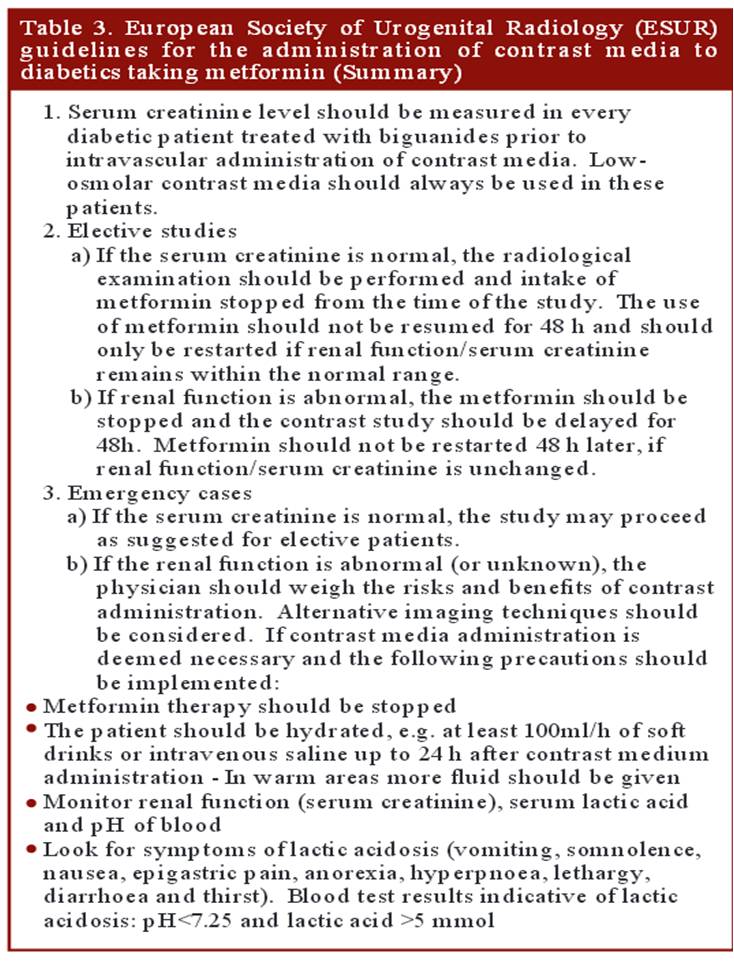
Metformin is one of most commonly used oral hypoglycemic drug listed by WHO as an essential anti diabetic drug. .It is a biguanide  which  blocks the hepatic gluoneogenesis . Since lactate is the major substrate for the process of gluco-neogenesis , excess of which  spills into blood .Lactate is swiftly cleared by the normally functioning  kidneys .Metformin is completely excreted by the kidney. Hence in patients  with compromised renal function (or when  contrast agents compete with Metformin in renal excretion ) high levels would not only cause lactic acidosis (> 5meq), it can also aggravate contrast induced renal injury resulting in a downward hepato -reno-metabolic spiral.

Though the incidence of Metformin induced Lactic acidoss  is low , the outcome can be bad , hence the concern. The European society of urogenital radiology  has provided clear cut guidelines regarding Metformin usage when contrast agents are being used.

metformin and contrast induced nephropathy guidelines cin european

What can be done in emergency situations 

  • Since the risk  of  lactic acidosis is very low , in case of emergency situations Metformin need not be discontinued prior to contrast usage.However it need to be stopped for 48 hours from the index procedure. (Fortunately Metformin is a dialysible drug that can be removed in case of toxic accumulation.)
  • Consider alternate mode of Imaging if renal function is really concerning


Reference

1.Pre admission metformin use and mortality among intensive care patients with diabetes: a cohort study.Christiansen C, Johansen M, Christensen SCrit Care. 2013 Sep 9;17(5):R192. doi: 10.1186/cc12886

2. http://ccforum.com/content/pdf/cc12886.pdf

contrast nephropathy

A good  article  from drug review Contrast induced nephropathy and metformin


Read Full Post »

Wall motion defect , in patients after CABG is fairly common.These  defects are difficult  to interpret  as the mechanisms can be multiple.Though the commonest wall motion defect appears to  involve the interventricular septum. it can occur anywhere in antero-lateral zone.

The mechanism attributed is  the effect of pericardiotomy , which surgeons as we understand leave it open after grafting  .This can cause lack of localised ventricular interdependence and results in a a brisk septal movement (bounce )It is an indirect effect .

post cabg wall motion defect

Note the, wall motion defects are confined to the exposed areas of the heart during cardiac surgery .In short axis echocardiography it correlates anywhere between 9 to 3 O clock position. Though interventricular septum is not covered by pericardium in the true sense , there is a indirect bounce effect over IVS due to interference with anterior ventricular interdependence .

More commonly a direct wall motion defect in the 12 to 3 O clock position in short axis is seen .This can closely mimic true wall motion defect as pericardial adhesions can tether these segments. Careful observation is warranted.Myocardial thickening is the key differentiating feature.

What is the physiological impact of these wall motion defects ?

It is generally considered benign (It is !) .Though in echo it looks awkward and suggest desynchrony. The real issue is , it can  mislead the echocardiographer to errors in calculation of that universally  sacred parameter called EF %

Importance of  knowing pre existing wall motion defect.

This has to be reviewed with old reports as it can wrongly create a new wall motion defect de-crediting the surgeons.

New pathological wall motion defect.

Of course it can happen due to peri-operative ischemic insult or infarct . However , It need to emphasised transient wall motion defects are common post CABG due to apparent hypoxia.This seems to be more pronounced with on pump surgeries than off pump .(Expected though) In my opinion, 2-4 weeks cooling off period is required before  a meaningful assessment of  wall motion post CABG.

Late pericardial reactions and localised constrictive features has been reported.

Disappearance of wall motion defect : How  common ?

Any disappearance of WMA is welcome . It happens rarely though . Some of the post ACS population (Both STEMI and UA/NSTEMI) can experience this ,  as they could harbor  zones of myocardial segments afflicted by  ischemic stunning rather than true  necrosis , that might  disappear.


Read Full Post »

Inferior STEMI is as  common as Anterior  STEMI .Unlike the anterior  STMI  which  auto localises  to LAD , inferior STEMI has to be fixed either RCA or LCX.

Following ECG features help localize Inferior STEMI  .

  • ST elevation in lead 3 > lead 2  suggest RCA (Not always true )
  • ST depression in lead V1,V2,V3 strongly suggest LCX. (More objectively the sum of  ST depression in V1, 2 , 3 divided by sum ST elevation in 2,3, AVF ,  if less than 1 indicate LCX.   Or simply ST depression  V3 > Lead 3 indicate LCX.)
  • ST depression in lead 1 indicate RCA
  • ST elevation in lead V6 strongly suggest LCX

Finally , and most importantly RV infarction as documented  by  ST elevation in V4R almost always localises the lesion in proximal RCA.

Role of Echo

If ECG  features  are not clear , a rapid bed side echo has a very good  localizing value. To fix RCA  look specifically for wall motion defect between “6 to  8”  O-clock position .It corresponds to  infero basal septum  that is invariably  supplied by RCA. For LCX involvement concentrate  on “3 to 6” o clock position.

stemi localisation by echo inferior rca lcx

Image source and courtesy http://www.aseuniversity.org

Which has better  outcome RCA or LCX STEMI ?

  • Though RV infarction  does not occur with  LCX , incidence  of MR is more with LCX and  can be truly troublesome. This probably negates the potential advantage of  “protected RV”  in  LCX  STEMI.
  • Since LV lateral free wall involvement  is extremely rare with RCA STEMI , it  has a lesser  impact on LV function while LCX STEMI can  give a double blow to LV   (MR and LV dysfunction)
  • On the down side ,coronary artery spasm and thrombus load are more with RCA .

Interventions in RCA is fairly straightforward ,while acute LCX PCI  has some  issues . Apart from technicalities of  intubating  the posteriorly  curving LCX ,realistically it involves fishing in troubled waters , as we need to cross the left main , likely physical contacts with LAD ostium , which is the sole supply chain for the injured and ischemic LV myocardium . Meanwhile ,  If RCA  is the culprit  , its a well cordoned crime scene where one can spend time liberally and fix the lesion.

Final message 

It is easier to localisethe culprit artery in inferior STEMI ,but its a tricky  to  predict outcome .Both can be troublesome .It depends on  dominance of the RCA/LCX ,proximal nature of lesion, the number and caliber of OMs, and PLVs and RV branch .However, it remains a fact  LCX STEMI has a  overall turbulent course.

Read Full Post »

Co-dominant coronary  circulation is defined as , when  posterior crux of the heart receives twigs from both right and left system making this water shed area with advantage of twin innervation.They essentially supply  inferior and posterior aspect of both left and right ventricle including the posterior aspect of interventricular septum.

Traditionally inferior and basal aspects of heart are perceived (wrong tough !) as less important  than anterior  surface of heart.Infero posterior MI can be extensive and cause significant LV dysfunction and poor outcome. Longitudinal function (AV grooval velocity) and Mitral valve function  is critically  dependent on  posterior circulation.

Is there an advantage for co-dominant circulation  with reference to ischemic mitral regurgitation ?

Obviously ,one would expect there is some advantage in co-dominant circulation when ACS occurs  either LCX or RCA.It could theoretically  protect against development of MR as posterior  papillary muscles could receive supportive twigs from its companion.

However , there is a caveat .The antero-lateral papillary muscle normally has twin blood  supply from LAD(Diagonal ) and LCX (OM) . But in co-dominant circulation this pap muscle is  at risk of becoming single blood supply as the dominant RCA has a trade off with OM with its large PLV branch. It is likely in   co-dominant circulations if LAD is the culprit outcomes are likely to be worse.

Final message

A rare  study involving  more than 200,000 patients which specifically addressed this issue  of dominance and outcome , threw some surprising  findings. In concluded  PCI outcomes  with left or co-dominance has a worse outcome than Right dominant system.

Reference

1.Left and Codominant Coronary Artery Circulations Are Associated With Higher In-Hospital Mortality Among Patients Undergoing Percutaneous Coronary Intervention for Acute Coronary Syndromes . Report From the National Cardiovascular Database Cath Percutaneous Coronary Intervention (CathPCI) Registry  Nisha I. ParikhEmily F. HoneycuttMatthew T. Roe, Circulation: Cardiovascular Quality and Outcomes. 2012; 5: 775-782  2012; 5: 775-782

2.Papillary Muscle Perfusion Pattern A Hypothesis for Ischemic Papillary Muscle DysfunctioPaolo Voci, Federico Bilotta, Quintilio Caretta,Circulation. 1995; 91: 1714-1718

Read Full Post »


Verdict ?

Only complicated or high risk  STEMI,  would require immediate anatomy based management. Please note, this population at worst is never beyond 20 % of all STEMI. Hence more than majority of  patients  can be managed effectively without CAG.

My reasoning tells me,though knowing the  coronary anatomy appear vital  , it is rather the physiological impact of those  anatomical lesions  that will determine the outcome. So,post STEMI, if at all , we need to investigate, it should be about the  adequacy of the over all blood supply to left ventricle.This is done by a pre or post discharge sub maximal stress /nuclear test .If it’s negative with a good exercise tolerance  CAG will never be required as any critical flow limiting lesion ( that would require intervention  )is excluded with near 100% surety.

Postamble :Try asking  any neurologist , how often they demand to know cerebral arterial  anatomy for managing stroke  ? You will get a real surprise answer !

Read Full Post »

Holter monitoring is the Initial test for all those with documented  syncope (or Pre syncope ) with suspected cardiac arrhythmia .It is a 24 hour ambulatory  ECG monitoring , expected to pick up any electrical abnormality and its correlation with the resultant symptom if any. Though the test looks  attractive , the diagnostic yield is far less. (About 10%) .The reason being the episodes can be rare  to be  missed by 24hr sample time. We have extended Holter (48hr) , Event monitors , Loop recorders and implantable devices that can record ECG for extended periods.(18 Months ,Reveal Plus Medtronic)  that improve the yield  up to 45%.

One common issue that often confuse us  while reporting  Holter is, the  pauses that occur during day / night .

What is the significance of these pauses * ?  Nocturnal vs Daytime

Pauses are obviously significant when the patient is awake . It is generally accepted pauses more than 3 seconds during day time  (ie Heart rate of < 20/mt ) is significant . This is logical , as pauses more than that,  is expected to cause syncope ( or atleast pre-syncope ).The problem comes when you document pauses more than 3 seconds without any symptoms . Then this  difficult  question comes up ,At what degree of pause syncope occurs ? How is that some persons mange  even prolonged pauses with just giddiness.(Good overall vascular integrity and tone ! )

We know such pauses are  especially  common during sleep. How does the brain react when pauses occur  during sleep ? as there is no question of fall as such and loss of muscle tone is non existing.

*Please note ,when we say pause we mean only Sinus pause , Pauses due to AV blocks are very significant

nocturnal pauses during sleep holter

Source : Brodsky M, Wu D, Denes P,et al.Am J Cardiol 1977; .

Dramatic  pauses during sleep do occur

There has been prolonged pauses reported  during sleep without fatality . A 35 second nocturnal pause resulting in seizures has been documented by implantable  recorders.(Mairesse 2003)

Causes for prolonged pauses

  • Sinus node dysfunction
  • Obstructive sleep apnea
  • High dose beta blockers therapy

Final message

 Most bradycardic episodes during sleep are benign.This is due to depressed autonomic control during sleep. Holter interpretation is primarily done with  awake rhythm data in most individuals .So, empirically shall we fix a  5 second pause as significant during sleep ? We don’t know.While this may seem applicable even with structural heart disease , one may be vigilant while interpreting the nocturnal pauses in this population .

Caution

** Please note,  all these rhythm monitoring extravaganza is meant for people  with equivocal symptoms .Patients  with well documented syncope with ECG features suggestive of  cardiac rhythm disorders would never require these tests and go for pacemaker straightaway.

Reference

4.Arrhythmias documented by 24 hour continuous electrocardiographic monitoring in 50 male medical students without apparent heart disease. Brodsky M, Wu D, Denes P,et al.Am J Cardiol 1977;39:390–5.
For Advanced readers
guidelines for syncope nocturnal pause holter

Read Full Post »