Feeds:
Posts
Comments

Archive for the ‘echocardiography’ Category


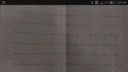
A 50-year-old man was referred for dizziness, bradycardia and dysphagia .He was very clear in describing his symptoms and  landed up in Gastro- enterology  OPD , from there was referred to my clinic for cardiac work up . His ECG showed a sinus bradycardia HR of 48 /mt.

Screenshot_2017-07-05-19-09-12

Echocardiogram revealed a structurally normal heart as we expected , but was surprised to spot suspicious shadow in para-sternal long axis view , beneath left atrium.

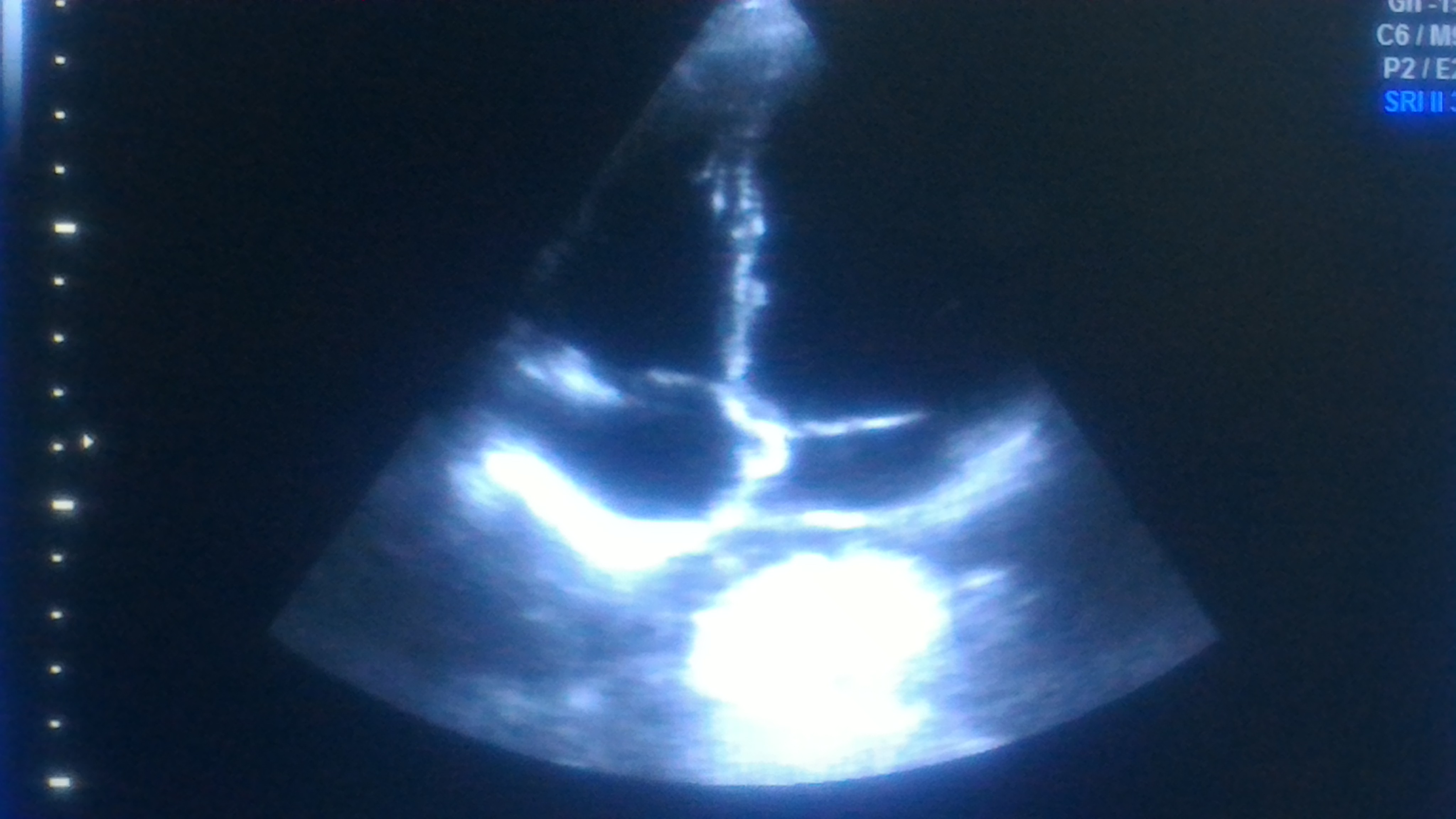
A well demarcated large mass compressing left atrium.  Trans Thoracic Echocardiography  may not be looking at the heart alone ,(Its technically Thoracic Ultrasound though we may refer it as Echocardiogram   )

  • Aortic aneurysm ?
  • Mediastinal teratoma?
  • Bronchial adenoma ?
  • Esophageal mass ?

The Answer is none of the above

As I was wondering what it was, the staff nurse in charge threw a heavy folder with well worked up gastro Investigations.

That moment , diagnosis became obvious , without a need for further scrutiny to my medical acumen.

Note: The barium swallow of the Esophagus reveals the Intimate relationship between the food tube and the heart as it descends vertically downwards posteriorly  . Realise , how the proximity of these two structures could  confuse a physician when symptoms spill over on either way. (I would have expected a lateral view to show the compressive effect of Esophagus on the left atrium the radiologists felt its not important !)

Yes , it is Achalasia of the cardia , dilating the lower end of esophagus with fluid /mass effect  , compressing the posterior surface of Left atrium.He underwent a myomectomy surgery.

Why bradycardia  ?

There is well described esophago-vagal reflex reproducible by stressful swallow or balloon inflation in the lower end of esophagus at D7 level.(Ki Hoon Kang,Korean J Intern Med. 2005 Mar; 20(1): 68–71.)

Achalasia cardia is known to be associated with symptomatic bradycardia, dizziness, and rarely swallow syncope,though this patient didn’t have a classical syncope.The bradycardia is probably due to high vagotonia, (Hugging effect on posterior surface of heart known for rich innervation of vagus.) . Complete reversal  of bradycardia after esophago -gastric surgery is expected.

Implication for cardiologists

There has been instances of patients with esophageal syncope and reflex bradycardia getting permanent pacemaker therapy. I think , clinical or sub clinical esophageal disorders should be included in the work bradycardia before labelling them as intrinsic sinus node dysfunction .(Ref 1,4)

Final message 

The field of Cardiology  is often referred to as a super specialty atleast in India . I disagree with it strongly. Cardiologists are neither super(eme) nor special .We need to be reminded  its afterall a sub-specialty of Internal medicine and each specialist should undergo retro-training in medicine periodically .This patient is a typical example of a gastric problem entering the domain of cardiac Imaging.Strong foundations in symptom analysis and some degree of medical  curiosity will enable an occasional cardiologist to make a correct diagnosis belonging to a remote foreign specialty.

Reference 

1. Palmer ED. The abnormal upper gastrointestinal vagovagal reflexes that affect the heart. Am J Gastroenterol. 1976;66:513–522. [PubMed]

2.Armstrong PW, McMillan DG, Simon JB. Swallow syncope. Can Med Assoc J. 1985;132:1281–1284. [PMC free article] [PubMed]

3.Turan I, Ersoz GBor S..Swallow-induced syncope in a patient with achalasia
4.Dysphagia. 2005 Summer;20(3):238-40  4.Basker MR, Cooper DK. Oesophageal syncope. Ann R Coll Surg Engl. 2000;82:249–253.

Read Full Post »

Hey , What’s that moving object over  AML ?  It looks odd,  it doesn’t look like a thrombus or a vegetation.

Yes, I agree , its moving  independently  but  I think , Its benign threads of fibrin attached to the valve .They are called as valvular strands.

Is it ?, I haven’t heard about it !  Can you please  tell me something about it.


Strands are  highly mobile, fine, filiform  threadlike excrescences that is seen arising from valvular structures. Synonym : Its same as Lambl’s excresceneces , the Czech physician who described  it over Aortic valve in 1860.

The following TEE clip shows strands attached to Aortic valve

Incidence

Reported Incidence of valvular strands  varies .Some reports suggested it may be up to 5-10 % .( SPARC study Mayo clinic 1999 its staggering 46 % !)The reason for  such high incidence  is,   many of us are still not clear what we refer to as strand.The imaging modality also has a say. With improving resolution of TTE and liberal TEE use more  strands are detected .A recent large study from Israel , suggest a good news , in large population based study (21,000) true strands are  observed in  just around  1 %.(Marina Leitman 2014 )

Is it Physiological or Pathological ?

The valve closure lines are physiologically stressed , some amount of denudation of endothelium is expected .This leads to a thrombus formation along with the exposed mucopolysacchride  layers of the valve form a filiform ,filamentous structure. .To call it physiological or pathological is left to our wisdom and  perception. The size however matters. It could be  the reason behind many  unexplained strokes.

What is the natural history of these strands ?

Its difficult to believe It may persist for lifetime.If its truely fibrinous strands it may have a life cycle and disappear.

Size

Should be less than 1 mm.

Length varies between  3 mm to 5 mm

Location 

Can be seen in any valve or even in aortic root.

Attachment : Atrial side of mitral valve and ventricular side of Aortic valve.

Strands over prosthetic valve is also reported.

Clinical significance 

It has three common issues.

One: Getting confused with other more pathological entities.

Two : Risk of stroke.

Three: Nidus for normal native valve endocarditis ?

Strands may closely mimic 

  • Vegetations
  • Bland thrombus
  • Redundant leaflet /Chrodae (Marfan and variants)
  • Disrupted chordae (After MVR)
  • Flail leaflet
  • Fibroelastomas

Risk of dislodgement and  stroke 

These strands are minute. It seems plausible dislodgement need not necessarily result in stroke or other organ ischemia.We don’t know whether it gets dissolved on transit.However the risk of stroke is increased in most reports except few studies(Roldan).

Management

First question to ask is , Should we inform our patients about  these ubiquitous accessory valve  tissues if detected incidentally ?

Largely benign and can be ignored in most.A follow up echo may be adviced once in a year or 2. (I have one anxious patient  after I reported such strands in Marfan syndrome )

In patients who has h/o stroke presence of these strands gains importance and  is an indication for anticoagulation.

Surgical excision of large strand is a dramatic option and is rarely performed.

Queries with no answers 

Is it accessory valvular (mesenchymal) tissue ?

Does Atheromatous plaque contribute to these strands in Aortic valve ?

Strands , if  disappears  by natural means , do they regrow from the same spot of raw surface  ?

Final message 

Fibrous strands detected  over the valves by routine echo are uncommon .However , It may give considerable  anxiety if  documented and reported to our patients and physicians .Though these have negligible clinical significance , the risk of stroke is increased in those with large strands.

Reference

Links

Read Full Post »

Inferior STEMI is as  common as Anterior  STEMI .Unlike the anterior  STMI  which  auto localises  to LAD , inferior STEMI has to be fixed either RCA or LCX.

Following ECG features help localize Inferior STEMI  .

  • ST elevation in lead 3 > lead 2  suggest RCA (Not always true )
  • ST depression in lead V1,V2,V3 strongly suggest LCX. (More objectively the sum of  ST depression in V1, 2 , 3 divided by sum ST elevation in 2,3, AVF ,  if less than 1 indicate LCX.   Or simply ST depression  V3 > Lead 3 indicate LCX.)
  • ST depression in lead 1 indicate RCA
  • ST elevation in lead V6 strongly suggest LCX

Finally , and most importantly RV infarction as documented  by  ST elevation in V4R almost always localises the lesion in proximal RCA.

Role of Echo

If ECG  features  are not clear , a rapid bed side echo has a very good  localizing value. To fix RCA  look specifically for wall motion defect between “6 to  8”  O-clock position .It corresponds to  infero basal septum  that is invariably  supplied by RCA. For LCX involvement concentrate  on “3 to 6” o clock position.

stemi localisation by echo inferior rca lcx

Image source and courtesy http://www.aseuniversity.org

Which has better  outcome RCA or LCX STEMI ?

  • Though RV infarction  does not occur with  LCX , incidence  of MR is more with LCX and  can be truly troublesome. This probably negates the potential advantage of  “protected RV”  in  LCX  STEMI.
  • Since LV lateral free wall involvement  is extremely rare with RCA STEMI , it  has a lesser  impact on LV function while LCX STEMI can  give a double blow to LV   (MR and LV dysfunction)
  • On the down side ,coronary artery spasm and thrombus load are more with RCA .

Interventions in RCA is fairly straightforward ,while acute LCX PCI  has some  issues . Apart from technicalities of  intubating  the posteriorly  curving LCX ,realistically it involves fishing in troubled waters , as we need to cross the left main , likely physical contacts with LAD ostium , which is the sole supply chain for the injured and ischemic LV myocardium . Meanwhile ,  If RCA  is the culprit  , its a well cordoned crime scene where one can spend time liberally and fix the lesion.

Final message 

It is easier to localisethe culprit artery in inferior STEMI ,but its a tricky  to  predict outcome .Both can be troublesome .It depends on  dominance of the RCA/LCX ,proximal nature of lesion, the number and caliber of OMs, and PLVs and RV branch .However, it remains a fact  LCX STEMI has a  overall turbulent course.

Read Full Post »

Aortic stenosis is diagnosed by 2D valve morphology, area ,and pressure gradient across the aortic valve.Though anatomical 2D images and indices are good enough to diagnose severe AS , we are obsessed *  with pressures  which are subjected moment to moment hemodynamic and contractile variables. To record a good gradient we need a normally contracting ventricle and good flow across the narrowed LVOT. If any one of the is critically compromised  gradients can’t be picked up by Doppler.(A new entity of AS was recently included , which fails to generate the gradient in spite of good LV function and the AS being significant.)

So ,whenever one records a “Low gradient AS” there are 4 distinct possibilities.

  1. Truly mild AS
  2. Technical inadequate Doppler alignment , with possible true moderate /severe anatomical  AS .
  3. Low gradient AS due to LV dysfunction, with true moderate /severe anatomical AS
  4. Low gradient AS with Low flow but normal LV function, with true anatomically moderate/severe AS

Echocardiographer should rule out 1 and 2 before going to the complex world of low gradient severe AS.In my personal opinion , the entity of Low flow , Low gradient with Normal LV function appear  redundant ( or is it beyond my understanding ) .One should look at the valve morphology and decide in such situations.

Then , one will shortly bump into this query  is it 2 or 4 ?

How to differentiate a  technically defective  recording  of low gradient AS  from  true low flow due to narrowed LVOT.(Low gradient for me , high gradient for my professor !)

Now, basic readers  may please leave ,

Few inquisitive may ask   ( naturally though)

Does the ” low flow -low gradient AS”  is an exclusive phenomenon  that can occur only with normal LV function  or can it  occur in  dysfunctional left ventricle as well ,  who also have small cavity size and narrow LVOT  ?  (Within the low gradient AS due to LV dysfunction subset ,  How much is attributable  due to anatomial low flow  and how much is related to depressed LV contractile force ?)

Another googly . . .

Why can’t  Doubutamine* stress test  routinely  undertaken in the subset of patients with  with subjects with Low gradient /normal LV function to augment the anatomical low flow and find whether it is true  low flow or not ? *This would mean , a most impractical situation wherein every patient  with even mild AS should need to undergo dobutamine testing to rule out significant AS.

Final message

As of now ,this new concept   “Low flow , Low gradient, with Normal LV function” appears an  intellectual excess with little impact on patient outcome.The proposed new entity ultimately increase the likelyhood of over diagnosing  severe AS.Iam still expecting  more clarity  on the issue. ( or else for the moment forget the pressures and  simply fall back on  a meticulous assessment of 2D valve morphology and take a call , you will be surprised how often we get into man made scientific traps. )

Reference

1.Low-flow, low-gradient aortic stenosis with normal and depressed left ventricular ejection fraction.Pibarot P1, Dumesnil JG.J Am Coll Cardiol. 2012 Nov 6;60(19):1845-53

Read Full Post »

Mconnell’s  sign is a distinct echocardiographic sign that occurs in Acute pulmonary embolism , where RA and RV dilates. RV shows a distinct regional wall motion abnormality in which RV free wall shows akinesia (or severe hypokinesia ) with well-preserved RV apical contraction.This is visible in apical 4 chamber view.

This sign is explained by  both anatomic  and hemo-dyanmic reasons.

  • RV when exposed to  sudden pressure overload  it not only dilates , it’s wall stress increases (Laplace law : Wall tension = P x Radius  )   and end up mechanically stunned . But , since the RV has a complex shape the distribution of this stress  is not uniform .As the RV assumes more spherical  shape the apical  part is not exposed to this stress as it tend to abut under LV.
  • RV apex is anatomically tethered with LV apex and share significant amount of circumferential fibres .In patients with acute pulmonary embolism ,  LV usually is hyperkinteic  due to tachycardia .This pulls the  RV apex  along with it for a proxy contraction .
  • Rarely , primary RV ischemia  due to RCA under perfusion* may be responsible for this unique  wall motion defect . Since RV apex  is mostly supplied by LAD it is free from ischemia . (*Acute elevation of RV intramural pressure due to PHT , compromising RCA perfusion pressure  )

Reference

1.McConnell MV, Solomon SD, Rayan ME, Come PC, Goldhaber SZ, Lee RT. Regional right ventricular dysfunction detected by echocardiography in acute pulmonary embolism. Am J Cardiol. 1996; 78: 469–473.

2. Rachel P. SoslandKamal Gupta,McConnell’s Sign circulation. 2008; 118: e517-e518

3. Link to the Echo clipping of McConnell sign in echocardiography


Read Full Post »

Meticulous Aortic imaging is vital for assessing  atherosclerotic plaques in stroke evaluation ,  aneurysms of aorta ( Both dissecting and non dissecting.) and during aortic surgeries. Peri  procedural  aortic imaging has become mandatory in many of the complex aortic endovascular repair as well .

TEE is an extremely useful investigation and has revolutiolised our appraoch to aortic disorders .

However , we have an issue .

blind spot for tee in aortic imaging distal ascending aorta and proximal arch bracho cephalic trunk

How to overcome it ?

During peri-operative TEE a simple but innovative idea is to displace  the tracheal air with saline filled balloon and capture the aortic arch with ultra sound . What a way to un-blind our vision deep inside the thorax  !

A specific catheter is available for this purpose .

a view endo tracheal balloon catheter how to overcome the aortic blind spot in tee

Product catalog

Read Full Post »

TVI  (or VTI)  is a hemo-dynamic  echo parameter measured from Doppler spectrum  across the valves ,usually in the outflow.This parameter is used to calculate cardiac output .VTI times the cross sectional area gives the stroke volume.


time velocity Integral  TVI

A correction: TVI is multiplication of velocity into time not division as suggested in the cartoon   

Time velocity Integral

What is time velocity integral  TVI echocardiography

Summary

*VTI is a Doppler parameter described in cm , it can be referred to as Stroke distance.This stroke distance when multiplied by  valve area gives the stroke volume from LV/RV  (or flow volume across AV valves or through any conduit)

Read Full Post »

The right ventricle  is considered as a docile cardiac chamber with passive filling and  emptying  properties .

This belief  was reinforced when Fontan  in early 1970s suggested a principle in the management of  cyanotic heart disease  when  the right side of the heart is underdeveloped. He  proved  RV can be by-passed safely , with  great veins  (IVC/SVC)  by  themselves  take care of filling the pulmonary circulation  without the need of RV pumping function.

While it is true for few complex cyanotic heart disease, largely this a misleading  concept. In clinical cardiology practice  ,sudden or non sudden  RV deaths happen every day in the form of . . .

  • RV Infarction
  • Acute RV dysfunction in massive pulmonary embolism
  • COPD with RV dysfunction
  • Most cases dilated cardiomypathy  the terminal event is due to RV  failure.

So , RV function can never be dispensable in day to day cardiac hemodynamics.

RV has some unique properties in terms of shape , size and  hemodynamics . We are getting more insights from  modern blood pool imaging by MRI , about  how the RV handles the blood volume .

We know RV has a unique shape  triangular ( partially  pyramidal ) . It can be inferred the RV cavity is formed by fusion of  many  eccentric spacial planes. We have always believed  RV handles the blood it receives from right atrium in a unique way .Now we are beginning to understand it .It is now documented the RV segregates the blood it receives into 4 components.


right ventricle physiology anatomy hemodynamics

It is curious  to know  RV inflow is connected to the outflow by an invisible   physiologic Bridge . About 44% of  blood traverse the RV in this fashion.


RVOT blood flow right ventricle

Note : RV blood flow preferentially enters the RVOT with out transiting RV body and apex.Image courtesy http://ajpheart.physiology.org/


Which is the most important part in RV ? (Among Inflow, Body, Apex, Out flow)

After reading this article it seems to me , the mechanical  function of RVOT could be most  vital. If it fails to handle the first increment  which  comes directly from  RV inflow, stasis  is likely in RV body and apex , elevating RVEDP and later promoting stasis leading to clinical events.

Clinical implication of this study

  • Differential dilatation RV chambers to pressure or volume  overload is observed .
  • We need to analyse why RV dilates in some   but   goes for hypertrophy in others when confronted with pressure overload (VPS vs PAH)
  • RV apical clot in restrictive cardiomyopathy  is a direct consequence of stasis  of blood  in RV apical zone .
  • RVOT pacing  may have a hemodynamic advantage  over RV apical pacing  . However , for anatomical reasons RV apical pacing  is  far safer than RVOT pacing where the lead  is subjected to constant life long strain due to this busy RV inflow to outflow express  high way !

Final message

Traditionally we have labeled  RV  as a  passive venous chamber .It is clearly a misnomer.It  has to handle both the venous and pumping function beat to beat with precision  without  back log .Obviously ,  RV has to think and work  more than it’s  big brother !

Reference

I wonder , if  there is  any other site other than APS . . . to  find crucial  answers in cardiac physiology  !


Right ventricle physiology blood flow  3d 4d analysisAfter thought

  • There is huge gap between physiologists  who work in research labs and the physicians at bed side .
  • I appeal all young cardiologists  to visit  APS  once in a while ,between your busy cath lab schedule and help narrow this gap.
  • Without understanding the physiology properly how are we going to intervene the pathology ?


Read Full Post »

Many modern  day cardiologists  consider  doing echo ,  a mean job and leave it to  technicians and fellows . Final report  often ends up with a cursory glance. The culture of reporting an  important aspect of LV  function is reaching a new low. It is common  to find the following terminologies  in  the  echo reports in many  parts of the country*     (Guess it is not used elsewhere ! )

  • LV function good
  • LV function adequate
  • Good bi-ventricular  function
  • LV function fair

what is adequate LV function

Among these , the term adequate LV function has caught  the  imagination  of young cardiologists ! Especially , this description  often appear in pre- operative  screening echo for non cardiac surgeries .

Recently ,one of my patient asked me what do we mean by adequate  LV function . I told  him it means nothing . . . it’s all  fancy words  !  but , generally  it is used to imply  normal LV function . . . I  clarified .

Think over for few minutes   . . . what do we  want to convey  by calling LV function  as adequate ?

Does it mean normal ?  or  Just less than normal  ?

If adequate LV function  is accepted ,  what is inadequate ?

Adequate for whom ?  For the patient ?  or  for the physician ?

Adequate  for daily activity  ?  or  Adequate to  with-stand  the proposed  surgery ?

Final message

Even  learned cardiologists indulge in this  term  frequently . This is  rather a fancy and unprofessional  way reporting LV function . They pass this  style to their residents  as well para medics .Adjectives  in medical science are not banished . . . but should be   judiciously used . In my opinion  the term adequate LV function should be removed from all echo labs .   Youngsters please  watch out.

Related links in this site .

LV ejection fraction fallacies

What is LV dysfunction ?

Read Full Post »

Anterior mitral leaflet  has a classical M  shaped motion. Infrequently , M mode echo will record a triphasic pattern .

Triphasic AML motion

The exact  answer is not known . I guess it is a normal variant.

Often  it is recorded  when there is a long  and redundant AML , especially if the M-mode cut is too close to  the tips.

Though it is not common , I have seen in few the triphasic gets converted into classical M shaped pattern if the cursor is moved slightly away from the tip of AML.

Relationship to Heart rate

Some times it appears in slow heart rate and tends to disappear with tachycardia .

Triphasic Doppler filling vs Triphasic M-Mode

We do not know yet ,  how  the  triphasic AML motion  correlate  with triphasic Doppler filling pattern which  is considered a fairly good evidence for  LV dysfunction.

Read Full Post »

Older Posts »