Feeds:
Posts
Comments

Archive for the ‘Cardiology -Hemodynamics’ Category

(This post is about some basics in echocardiography meant for fellows, and echocardiographers. Others can skip please ) 

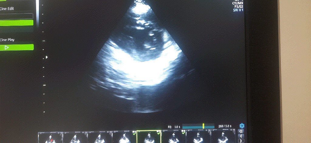
This is a 27-year-old woman who was referred for routine* cardiac evaluation. What do you see?

What is the diagnosis?

This echo clip is from a woman who is 8 months pregnant. What you are seeing is perfectly physiologically and normal. On lying down there is a mechanical push of the diaphragm altering the LV shape and contraction. In the short axis, the left ventricle is contracting well, but the shape is not spherical in systole implying some desynchrony. Further, the  IVS arena is contracting vigorously, which makes, the other segments appear to be poorly contracting. (Someone could report it as a wall motion defect in antero- lateral segments inviting temporary panic)

It is worthwhile to go through this list of non-ischemic WMA and find the pregnancy at the bottom of the list.

Few more conditions, that can be added to this list

  • Though LBBB is the classical cause for WMA, we have seen even LAFB showing the bumpy motion of IVS and the anterior wall.
  • Some patients with ERS and some patients with Brugada show wall motion defects due to repolarisation heterogeneity. 
  • Regioanl pericarditis
  • Intracardiac scars. Localized fibrosis.
  • Extracardiac tumors 

iFAQ on this topic 

Is this wall motion defect in pregnancy, really an artifact or real? 

They are true artifacts in the sense, the heart is an innocent bystander in this pulsating fight between intra-thoracic vs intrabdominal pressures. A similar situation happens in ascites. 

Any other mechanism other than mechanical push?

WMA due to RV volume overload of pregnancy may also contribute. 

Does this WMA affect cardiac hemodynamics?

Logically it should, but it doesn’t. The normal heart has enormous resilience, it just ignores these subtle pushes from below and keeps working normally. Still, enormous distension of the abdomen especially in twin pregnancies, in small body habitus, can make some women breathless, or orthopenic. I am sure, one of the mechanisms could be this geo-mechanical encroachment.

Final message

Wall motion defects are not synonymous with CAD. There is an important list of non-ischemic conditions that can cause WMA. Cardiology fellows and echo technicians are encouraged to go through the above list one more time. While this knowledge can prevent false alarms, at the same time it is always wise to ask for the ECG before doing echocardiography, and not to miss the omnipotent CAD.

Postamble 

*DIscerned readers might wonder why a routine echo was done in a normal pregnancy. I am surprised to note there is an ongoing fad in this part of the world, to do echocardiographic screening on every pregnant mother to rule out cardiovascular disease. (A luxury even the world’s richest country can’t afford) I am told, this echo is meant to rule out peripartum cardiomyopathy for legal purposes. A spot echo at term can never be going to either predict as an event that is mainly going to happen postpartum. This newfound epidemic of anxiety among obstetricians is unwarranted. 

Reference  

A well-written focused review specifically on this topic 

Yavagal ST, Baliga VB. Non-Ischemic regional wall motion abnormality. J Indian Acad Echocardiogr Cardiovasc Imaging
2019;3:7-11.


Read Full Post »

Syncope is one of the common, yet difficult symptoms to evaluate especially in the elderly. Post-prandial syncope is one condition likely to be missed out.As the name suggests It has a distinct relationship with food intake. Mild fall in postprandial BP is an expected response but if it exceeds a  limit* syncope is triggered. (*Highly variable)

Hemodynamics of Postprandial state

  1. Normally splanchnic circulation demands up to a 25%  increase in blood volume after a moderately large meal. 
  2. When this happens there must be compensatory vasoconstriction elsewhere especially in muscles. Lack of this response results in inappropriate falls in SVR. (The second mechanism is more constant and can be disproportionate to fall of BP)
  3. The mediators for this are either neurogenic or hormonal or both.
  4. Gastrointestinal mediator (Vasoactive Intestinal polypeptide dysregulation) is thought to play a major role. 

From Jansen et al  Archives of Internal medicine 1995

When does it occur?

It can manifest as early as 15 minutes, up to 2 hrs. The fall in systolic  BP is around 20mmhg. More common with large, hot meals. The fact that it can occur up to 2 hrs post meals, there is a likelyhood we might overlook it in history.

Other differential diagnoses 

Management 

There is no specific therapy. Some of the following might be effective.

  • Caffeine,
  • Somatostatin,
  • Acarbose,( α-Glucosidase Inhibitor ) 
  • Avoiding acute high carbohydrate intake.
  • A psychogenic component can be noted in a few that is attenuated by cognitive-behavioral therapy.
  • Midoridine, an Alpha¹ receptor stimulant  can be surprisingly more effective in some who have overlap with orthostatic hypo  (Cleve Clin J Med. 2010 May; 77(5): 298–306.)

Final message

Postprandial hypotension/syncope is a less recognized entity. As always, history is the most important diagnostic tool in the evaluation of syncope, which comes free of cost as well. The diagnostic yield is much greater than sophisticated Holter and event monitors.

Please note, there is a much more prevalent, lesser version of this condition, ie postprandial dizziness or giddiness. However, as already stated there is a significant overlap between orthostatic hypotension and postprandial syncope. It’s worth ruling out diabetes and autonomic dysfunction, (even subclinical Parkinsons) in elders with such symptoms. 

Reference

Here is a  comprehensive and elegant study (I think, It is only one of that kind on this topic )

1.Jansen RWMM, Connelly CM, Kelley-Gagnon MM, Parker JA, Lipsitz LA. Postprandial Hypotension in Elderly Patients With Unexplained Syncope. Arch Intern Med. 1995;155(9):945–952.

Postprandial hypotension Jansen1995


Read Full Post »

This cartoon succinctly  depict all the options we have in our fight against end stage heart failure .We know , a failing heart is often compared to a sick , aged and tired horse.

cardiac failure cartoon tired horse whicpping lionel opie book

Image courtesy Heart Physiology: From Cell to Circulation :Lionel H. Opie Lippincott Williams & Wilkins, 2004


1.Don’t whip the horse (Except in emergency)

  • Avoid all Inotropics ( Doubutamine and Milrinone were shown to improve quality of life marginally but  with dramatic reduction in quantity of life ! However , the same thing does not apply for Digoxin as it is the  the only Inotropic with a soothing para-sympathetic comfort  !
  • Please be reminded, CRT wires could act as  “multiple whip equivalents” right inside the heart , especially in advanced class 3 or just recovered class 4 patients. Beware!

2.Unload the horse

Vasodilators

  • ACEI/ARBS

3.Slow the horse

  • Never exert too much (Not more than 70% of capacity)
  • Beta blockers
  • Ivabradine (Slow the sinus node and expect a reduction in MVO2 )

4.Change the horse

  • Heart transplant may be the best solution

5.Switch to an Artificial Horse(Tractor )

  • ie  LV assist device

6.Finally try to heal the horse (Still largely in research labs!)

  • Genetic engineering
  • Tissue repairing
  • Stem cells
  • Holistic and spiritual healing etc (Has really  worked in few )

Read Full Post »

The right ventricle  is considered as a docile cardiac chamber with passive filling and  emptying  properties .

This belief  was reinforced when Fontan  in early 1970s suggested a principle in the management of  cyanotic heart disease  when  the right side of the heart is underdeveloped. He  proved  RV can be by-passed safely , with  great veins  (IVC/SVC)  by  themselves  take care of filling the pulmonary circulation  without the need of RV pumping function.

While it is true for few complex cyanotic heart disease, largely this a misleading  concept. In clinical cardiology practice  ,sudden or non sudden  RV deaths happen every day in the form of . . .

  • RV Infarction
  • Acute RV dysfunction in massive pulmonary embolism
  • COPD with RV dysfunction
  • Most cases dilated cardiomypathy  the terminal event is due to RV  failure.

So , RV function can never be dispensable in day to day cardiac hemodynamics.

RV has some unique properties in terms of shape , size and  hemodynamics . We are getting more insights from  modern blood pool imaging by MRI , about  how the RV handles the blood volume .

We know RV has a unique shape  triangular ( partially  pyramidal ) . It can be inferred the RV cavity is formed by fusion of  many  eccentric spacial planes. We have always believed  RV handles the blood it receives from right atrium in a unique way .Now we are beginning to understand it .It is now documented the RV segregates the blood it receives into 4 components.


right ventricle physiology anatomy hemodynamics

It is curious  to know  RV inflow is connected to the outflow by an invisible   physiologic Bridge . About 44% of  blood traverse the RV in this fashion.


RVOT blood flow right ventricle

Note : RV blood flow preferentially enters the RVOT with out transiting RV body and apex.Image courtesy http://ajpheart.physiology.org/


Which is the most important part in RV ? (Among Inflow, Body, Apex, Out flow)

After reading this article it seems to me , the mechanical  function of RVOT could be most  vital. If it fails to handle the first increment  which  comes directly from  RV inflow, stasis  is likely in RV body and apex , elevating RVEDP and later promoting stasis leading to clinical events.

Clinical implication of this study

  • Differential dilatation RV chambers to pressure or volume  overload is observed .
  • We need to analyse why RV dilates in some   but   goes for hypertrophy in others when confronted with pressure overload (VPS vs PAH)
  • RV apical clot in restrictive cardiomyopathy  is a direct consequence of stasis  of blood  in RV apical zone .
  • RVOT pacing  may have a hemodynamic advantage  over RV apical pacing  . However , for anatomical reasons RV apical pacing  is  far safer than RVOT pacing where the lead  is subjected to constant life long strain due to this busy RV inflow to outflow express  high way !

Final message

Traditionally we have labeled  RV  as a  passive venous chamber .It is clearly a misnomer.It  has to handle both the venous and pumping function beat to beat with precision  without  back log .Obviously ,  RV has to think and work  more than it’s  big brother !

Reference

I wonder , if  there is  any other site other than APS . . . to  find crucial  answers in cardiac physiology  !


Right ventricle physiology blood flow  3d 4d analysisAfter thought

  • There is huge gap between physiologists  who work in research labs and the physicians at bed side .
  • I appeal all young cardiologists  to visit  APS  once in a while ,between your busy cath lab schedule and help narrow this gap.
  • Without understanding the physiology properly how are we going to intervene the pathology ?


Read Full Post »

IVC filter usage has increased many fold in recent years.Please note , it is not indicated in every case of recurrent DVT/or PE. There are specific indications.

Permanent IVC filters

  1. Patients at risk for DVT /PE  with  absolute contraindication to anti-coagulants.
  2. Recurrent DVT/PE in spite of adequate  anti-coagulation

Temporary /Retrievable filters*

  1. It is used during high risk periods  for DVT following major trauma or Bariatric/Spinal /Neuro surgery (PREPIC 2 study ) .*Some of the retrievable filters can be kept for months ,years or even permanently. (If the risk period extends or it has trapped a huge clot.)


indication for IV filter prepic study

Outcome  of IVC filter (PREPIC  -8  year follow up study )

  • Reduces risk of PE
  • Increases risk of DVT
  • No impact on long term  survival
  • Clogging of IVC remain  an important Issue

Reference

Read Full Post »


A 40 year old  women came  with acute dyspnea who had a prosthetic mitral valve implanted 2 years ago for RHD  .

An emergency  echo showed  a peak gradient of 35mmhg across the valve .She was on warfarin regularly and her last INR was 2.2.Heart rate  was 138/minute, lungs showed congestion .LA,LV were dilated. LV function appeared mildly compromised  but could not be accurately quantified as the  patient  was in distress.


The fellow on duty had no hesitation  in diagnosing prosthetic valve thrombus .He Initiated Inj streptokinse bolus followed by infusion  along with diuretics . After few hours the gradient regressed .Patient felt better .Every one was happy . The consultant congratulated the fellow for  the good job done .To recognize prosthetic valve obstruction early and  successfully lysing it too !  The fellow  felt gratified .

prosthetic valve obstruction thrombus 002

Since I  was hanging around the CCU , watching the proceedings , I Initiated  a debate which was  curious to the team that handled the  patient !

Was it really thrombotic obstruction that caused his symptoms ?

  • No one has visualized the thrombus
  • TEE was not done
  • Fluroscopic evaluation of disk motion was not performed
  • There was no documentation of raising FDP that would Indicate clot lysis.

All  we have  is an unexplained  tachycardia with raised  trans prosthetic gradient . . .

Why we are tuned to think  raised gradients  to be  synonymous with thrombus ?

There has  been lot of assumption here . Subsequent analysis of history and  clinical presentation revealed the patient had a febrile illness which triggered an  atrial tachycardia  that possibly  resulted in transient  LV dilatation and dysfunction.

Once the failure is controlled the gradient has come down , I argued !

Of course, this again could be  a guess work , How can you  still rule out a thrombus ? They wondered !

I told them ,  question here is not ruling in or ruling out prosthetic valve thrombus.

It is an important  lesson to learn , raised  prosthetic gradient is not equal to thrombus  in many  acute hemodynamic situations* .

Many factors other than prosthetic valve obstruction  can elevate the gradient.

After all ,  prosthetic valve orifice is inherently stenosed  .(MVO is  never >2.5sqcm in any prosthetic mitral valve) . So at times of tachycardia the gradient is bound to be elevated .

Other factors that can elevate trans – mitral gradient includes

  • LV dysfunction
  • Acute diastolic dysfunction
  • Acute peri-valvular MR
  • Loss of LA compliance

 *One of the  commonest (yet not recognised) cause for elevation of trans mitral prosthetic gradient is acute left ventricular failure due to any cause.It  can be either acute diastolic dysfunction or a sudden raise in  blood pressure that result in  after load mismatch.

Final message

Please remember flow across prosthetic valve is governed by  delicate  local hemodynamic rules .The gradient  is  critically dependent on heart rate, LA  size and compliance , LVEDP and after-load mismatch if any !

Transient raise can occur at times of tachycardia and falling LV function (Mitral valve has to push hard, in the process elevating the gradient)

Simple raise in trans-valvular gradient should be carefully interpreted. Since visualising thrombus in routine TTE is  difficult  in an acutely  dyspnic  patient  many of us have taken this  granted !

Nothing wrong in playing  guess games in medicine . . . but we  need to acknowledge it!

*Note:Other causes for chronically elevated gradients like pannus formation, mechanical defects of valve, degenerated prosthesis  should be addressed separately.


Read Full Post »

Can we close an  ASD in a  25 year old women  severe  pulmonary hypertension ?
Volumes of literature has been written  on the subject.Dedicated cath studies  have been done with multiple parameters .
Still , there is a lingering doubt !
Here is  a  3 minute  practical solution  based on 5 easily available parameters. (* Also referred to as  unscientific  in medical parlance !)
1.  O2 saturation
2. Pulmonary artery diastolic and pulse pressure
3. RV function,
4 .Systemic pressure
5. Functional class
  • If O2 saturation is > 90 % consistently  there is likely to be significant  left to right shunt  .Closure is to strongly considered
  • If 02  saturation is near 95 % there is absolutely no contraindication at any level of PVR.
  • Systolic pulmonary artery pressure derived by TR jet is least useful index.Pulmonary artery diastolic pressure reflects true vascular  reactivity of the pulmonary  circulation.A wide swinging pulmonary arterial pulse indicates dynamism in circulation and hence operablity.
  • If pulmonary artery  pulse pressure is  wide (>50)  , or PA diastolic BP is < 30 one can safely presume irreversible damage to pulmonary vasculature has not occurred and these patients would  benefit  from surgical closure .
  • RV  function should be assessed carefully in every patient.This is as important as PVR .Significant RV dysfunction is an absolute contraindication.
  • Never close the shunt in patients who is in class 4  symptoms.
  • Never close a shunt if the systemic blood pressure is low( 90mmhg)
  • Some believe  PDA may be closed at any given PVR , while  worst outcomes occur with ASD as supra-systemic pulmonary pressure is possible.
Always monitor these patients meticulously especially  in the initial days following surgery  for deterioration .Most patients will do well if they cross the first 30 days. The RV  learns to adopts with  new  pulmonary hemodynamics !


Read Full Post »

Normal  left atrial  (LA) volume is about  22ml/sq.meter body surfacearea  at all ages.Maximum LA volume in physiology is  about 46ml in  females and 56 ml  in males( Average 35 ml)

LV stroke  volume  for each beat is  about  70  ml . . . so where does  the remaining 35ml come from ?

Answer .

  • Pulmonary veins ?
  • Residual LV end systolic volume ?
  • Mix of the two ?

It is logical to assume about 35 ml of fresh  blood  from 4 pulmonary vein*  rushes into LV with every diastolic cycle  .It  never stays in LA  .It just uses LA as a transit  route ,

*In diastole the four PVs,LA  and LV all act like one single chamber .


Is this reasoning correct ?.

If we believe the continuity equation this explanation is correct . However still what  we need to know the fate of residual  LV volume (End systolic LV  volume which is also  about 35 ml that would be  in queue for ejection into Aorta  for the next beat !)

Further , we know the LV end systolic volume is not constant .During exertion it  can reach  negligible levels (<10 ml) .At times of vigorous contractions  it can touch near zero as well . Then , It become vital for the  pulmonary venous reservoir  to be act as a  major donor for  LV blood volume for  every ensuing beat.

If the hemodynamics of pulmonary vein LA interface is tricky even in physiology ,  one can imagine the complexities  if the LV diastolic function and left atrial compliance  is affected

Debit and credits of  LV end -diastolic volume .

Let us assume LVEDV is about  1o5  ml .LA blood volume is  roughly one third of LV volume .For every beat equal amounts of fresh blood  from pulmonary vein . These two (LA+PV)  adds to the  residual  blood in LV  to make LVEDV 105   ml . From this 70 ml is ejected as stroke volume leaving behind 35 ml.


lvedv lvesv stroke volume wiggers cycle left atrial volume pressure volume loop residual diastasis

Image template from http://www.cvphysiology.com

 Further questions

LA Chamber volume and blood volume need not be same .What  I struggle to understand is , total anatomical  LA volume  measures  35ml , while the amount of blood it is supposed to hold is also about the same .Does  it mean the LA is completely filled with blood . . . air tight !

Will the LA compliance make it accommodate twice or thrice the blood volume during exercise ?

What is quantum of residual end diastolic  LA volume ?


Reference.(Normal LV and LA volumes )

echopedia


Read Full Post »