Feeds:
Posts
Comments

Archive for the ‘Syncope’ Category

Syncope is one of the common, yet difficult symptoms to evaluate especially in the elderly. Post-prandial syncope is one condition likely to be missed out.As the name suggests It has a distinct relationship with food intake. Mild fall in postprandial BP is an expected response but if it exceeds a  limit* syncope is triggered. (*Highly variable)

Hemodynamics of Postprandial state

  1. Normally splanchnic circulation demands up to a 25%  increase in blood volume after a moderately large meal. 
  2. When this happens there must be compensatory vasoconstriction elsewhere especially in muscles. Lack of this response results in inappropriate falls in SVR. (The second mechanism is more constant and can be disproportionate to fall of BP)
  3. The mediators for this are either neurogenic or hormonal or both.
  4. Gastrointestinal mediator (Vasoactive Intestinal polypeptide dysregulation) is thought to play a major role. 

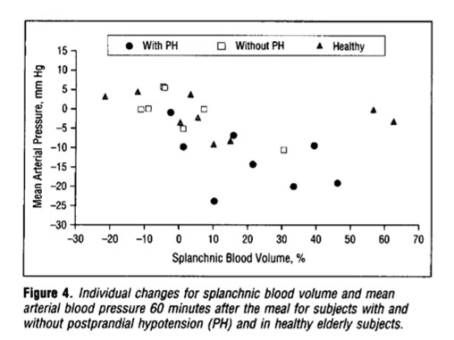
From Jansen et al  Archives of Internal medicine 1995

When does it occur?

It can manifest as early as 15 minutes, up to 2 hrs. The fall in systolic  BP is around 20mmhg. More common with large, hot meals. The fact that it can occur up to 2 hrs post meals, there is a likelyhood we might overlook it in history.

Other differential diagnoses 

Management 

There is no specific therapy. Some of the following might be effective.

  • Caffeine,
  • Somatostatin,
  • Acarbose,( α-Glucosidase Inhibitor ) 
  • Avoiding acute high carbohydrate intake.
  • A psychogenic component can be noted in a few that is attenuated by cognitive-behavioral therapy.
  • Midoridine, an Alpha¹ receptor stimulant  can be surprisingly more effective in some who have overlap with orthostatic hypo  (Cleve Clin J Med. 2010 May; 77(5): 298–306.)

Final message

Postprandial hypotension/syncope is a less recognized entity. As always, history is the most important diagnostic tool in the evaluation of syncope, which comes free of cost as well. The diagnostic yield is much greater than sophisticated Holter and event monitors.

Please note, there is a much more prevalent, lesser version of this condition, ie postprandial dizziness or giddiness. However, as already stated there is a significant overlap between orthostatic hypotension and postprandial syncope. It’s worth ruling out diabetes and autonomic dysfunction, (even subclinical Parkinsons) in elders with such symptoms. 

Reference

Here is a  comprehensive and elegant study (I think, It is only one of that kind on this topic )

1.Jansen RWMM, Connelly CM, Kelley-Gagnon MM, Parker JA, Lipsitz LA. Postprandial Hypotension in Elderly Patients With Unexplained Syncope. Arch Intern Med. 1995;155(9):945–952.

Postprandial hypotension Jansen1995


Read Full Post »

Syncope and seizure are most dramatic symptoms that rarely fails to call the attention of the patient and family.Syncope is primarily evaluated at medical or cardiac units. However ,when syncope presents as convulsions (often It is ! ) the patient lands up in a Neuro unit as a case of epilepsy.Some how, many of them are prescribed anti convulsants without being evaluated for what triggered the seizure.

heart_and_mind

Cardiac seizure and Neural syncope : Require a balanced approach ! (Image courtesy http://3.bp.blogspot.com)

Real life experience now suggest, a bothering  number of patients in epilepsy clinic might harbor a primary cardiac disorder in the form of either brady or tachycardia which is often inherited due to defect in ion channels of cardiac cell.

The issue is two fold. 

  • Cardiac patients mis-diagnosed as seizure
  • Primary seizure patients suffer a cardiac death (as seizure induced arrhymias or acute pulmonary edema )

Incidence of sudden cardiac death in patients with seizure disorder though rare is being increasingly recognised. Mechanical problems like valvular Aortic stenosis can also result in syncope followed by seizure.

Final message

Cardiologists do have a major role these situations.It may be wise to advice basic cardiac work up in  every seizure disorder.  As we are beginning to understand the neurogenic triggers in sudden cardiac deaths , the need for Neuro-Cardiac units is real.(Some of big university hospitals do have such departments)

Reference 

1.Zaidi A1, Clough P, Cooper P, Misdiagnosis of epilepsy: many seizure-like attacks have a cardiovascular cause. J Am Coll Cardiol. 2000 Jul;36(1):181-4.

2.Leestma JE, Annegers JF, Brodie MJ, Brown S, Schraeder P, Siscovick D, et al. Sudden unexplained death in epilepsy: observations from a large clinical development program. Epilepsia. 1997 Jan. 38(1):47-55.

3.Kloster R, Engelskjøn T. Sudden unexpected death in epilepsy (SUDEP): a clinical perspective and a search for risk factors. J Neurol Neurosurg Psychiatry. 1999 Oct. 67(4):439-44

4.Leestma JE1, Walczak T, Hughes JR, K A prospective study on sudden unexpected death in epilepsy.Ann Neurol. 1989 Aug;26(2):195-203.

Read Full Post »

Dizzines , giddiness , light headedness , fear of fall or true fall (Syncope) are the  most common symptoms beyond middle  age .They usually end up with consulting physicians , neurologists and cardiologists .Cervical disc is commonly blamed for this.While all these symptoms can be a manifestation of cervical disc , true syncope seems to be rare with cervical spinal disorder.

how common is suncope in cervical spondylosis Why true is  syncope rare with  cervical spinal narrowing ?

There is a fundamental ignorance here. We are not yet clear  whether giddiness /dizziness is neural or vascular .(Or combinations of both ) in cervical spinal disease.

Many of the patients experience momentary unsteadiness and feel like falling but very rarely end up in a fall or syncope.By definition syncope requires global transient hypoperfusion of brain to trigger a fall .

Cervical canal carries only the vertebral artery .Both internal carotids are no where near the spine. So, however severe the vascular compromise within the cervical canal , syncope is unlikely as anterior cerebral circulation compensates at the level of circle of Willis. Is that the correct way of reasoning ? Unilateral vertebral compromise rarely matters as other side takes care.It is very unlikely both vertebral artery get compromised by cervical spinal spurs simultaneously.

How many seconds of vascular compromise is  required to produce syncope ?

Brain seems to be a funny and sensitive organ .In erect posture it is very sensitive. It reacts  in a  fraction of time for any transient reduction of blood flow.The same cardiac events which can cause symptoms of impending syncope in erect posture is quiet comfortably tackled in recumbent posture .

In erect posture , blood pressure need to much higher to get it pumped above the heart against the gravity. Though , it seems absolute blood pressure and cerebral perfusion that matters, there is something more we are missing.  The brain-stem -vestibular system perceives differently when a recumbent person develop cerebral ischemia than when he is erect . The reflex circuit that activates on/off switch  for abrubt loss  generalised muscular tone is not yet been identified .

It is a biologically  heartening irony that syncope is a natural counter mechanism by which  the organism assumes instant recumbent position to maintain perfusion of brain .A fall promptly does this.(Ofcourse with a risk of Injury )

We think more than 3 second pause will initiate a  syncope circuit .But it is not a fixed number. Some can tolerate up to 5sec or even more especially in lying posture.

Final message

Syncope , as such in isolated cervical spondylosis is rare even though it can potentially interfere with  the vertebro basilar system .Why the vascular compromise almost always fall short of syncope and end up with just momentry unsteadiness is not clear.

Further queries to be addressed

Is dizziness, giddiness , presyncope  are vascular or neural  event?

Can vertigo occur with cervival spine disease ?

Read Full Post »

Syncope is a classical feature of LVOT obstruction especially with valvular aortic stenosis.The mechanism of exertional syncope in Aortic stenosis is traditionally attributable to the fixed obstruction .This fixed obstruction is not able to cope up with increased cardiac output as demanded by the exercising muscles . But exercise  induced reflex as well as local vaso-dilatation mechanism is intact . The consequence is predictable. A critical fall in SVR amidst a obstructed LVOT precipitating a syncope.

However , If the above mechanism is the sole reason for syncope in Aortic stenosis , we have a problem to explain why syncope is  rare even in critical mitral stenosis which is also fixed LV inflow obstruction ?

Is there some thing unique in LVOT obstruction that causes syncope ?

No, it is nothing to do with LVOT .To generate a true pathological syncope, reduction in cardiac output per-se may not be enough . It appears there should be an inappropriate systemic vasodilatation as well to precipitate a syncope.This can happen only if the parasympathetic system gets activated by some means . The trigger is located in the mechano- receptors of left ventricle . Hypertophied left ventricle with high Intra cavitory pressure (Often above 200mmhg) generated due to LVOT obstruction activates the syncope circuit.The same rule may apply for RVOT as well .One could get syncope with critical valvular PS or severe pulmonary hypertension when RV mechanical receptors get a triggered.

What happens in mitral stenosis ?

In mitral stenosis , LV is under- filled ,  wall thickness is normal .There is little likely-hood of LV mechno-receptors to get stimulated as the LV wall stress is normal. This is the reason syncope is less common in mitral stenosis. However , this is not  absolute rule , syncope can still occur in severely narrowed orifice of mitral valve due to low flow state alone or a ball valve thrombus and paroxysms of arrhythmia .

Read Full Post »

Recently , I came across a   young women  who underwent the following three tests for one episode of syncope after witnessing her pet dog bleeding with  an Injury !

  1. Carotid doppler
  2. Holter monitoring and event monitors
  3. Brain MRI /MR angiogram

This was followed up  by Head up tilt(HUT)  in a premier hospital

After 1 week of investigation ,a diagnosis of  Neurocardiogenic syncope was made and she was reassured and no drugs were prescribed.

(The collective yield of the above three investigation in fixing  a specific diagnosis is  less than 10 % of all known causes of syncope )

Syncope  approach  evaluation

To diagnose  common syncope . . . we need common sense !

Syncope is a dramatic  symptom.It is one of the commonest symptom in ER as well . Life time incidence of syncope is at least one episode in 50% all human life ! The definition  of syncope until recently , was liberal .Any transient loss of consciousness with spontaneous recovery  was termed syncope.

This includes

  1. Hypoglycemia
  2. Anemia
  3. Siezure disorders
  4. Structural  neurogenic (Including ,  brain tumors , Dural hematomas etc )
  5. Panic attacks (psychogenic)

Cardiologists wanted to fix syncope as an exclusive disorder of  circulatory insufficiency.By bringing in a modification in the definition  , ie  syncope is  now defined as a transient loss of consciousness due to   reduction in cerebral perfusion  .

This definition helped cardiologists  to exclude the above entities . Still many would include all in single basket as patient should be seen as a whole and we can’t expect them to  land according to our convenience and classification.

Here is an incomplete* list about causes of  syncope (* 99% complete ?)

Vascular

  • Vaso- vagal syncope in young ( Neuro-cardiogenic , Common , Benign)
  • Autonomic dysfunction of elderly ( Including postural hypotension )

Cardiac

Arrhythmic ( Sinus node dysfunction /CHB/Idiopathic VT/Long QT syndromes)

Structural heart disease

  • Valvular  heart disease  (LVOT/RVOT obstructions)
  • Myocardial disease
  • Rarely ischemic heart disease

Miscellaneous

  • Severe pulmonary hypertension (Including PPH ,  pulmonary Embolism )
  • Paradoxical embolism.
  • Aortic arch disease -Takayasu related arteritis .

Investigation

We have a sophisticated array  of investigation for syncope .It can be a never ending exercise , ranging from  spinal cord evoked potentials to diagnose Shy-drager syndrome ,   . . .  to implanting long-term loop recorders to decode  heart beat behavior.

However , evaluation of syncope is the ultimate wake-up call  to all current generation cardiologists  . . . Why clinical cardiology  should  never  be allowed to die (and  it  will not ! )

Common sense begins with answering  few simple questions . Is it really syncope ?

If  you ask this question three times and with  specific leads to the patient  and the witness ,  truth will come out  . 90% of times it may not be syncope at all (Near syncope, accidental  fall, dizziness ,extreme blurred vision, drowsiness  etc)

If it is syncope , Is there a non cardiac cause ?

It may related to the Hypoglycemia / Anemia /Panic attacks.Get a neurologist opinion , it would be terrible mistake if you miss a space occupying lesion  within the brain. (Missing chronic silent sub dural hematomas is  frequent   in the evaluation of syncope of elderly !)

Ruling out  cardiac syncope is relatively easy

In the remaining  patients  basic investigation like routine blood tests,ECG, ECHO   will help us  rule out most serious cardiac disorders.Similarly  bulk of the electrical cardiac syncope can be diagnosed.(Holter , carotid study in selected few )

Need for neurologist -cardiologist interaction.

Syncope due to VBI,  transient Ischemia attack , Senile vascular dementia  is a grey zone . Many have complex neuronal -vascular mechanisms . What is Consciousness ?  and  What is LOC ?  :Is it the lack of blood or severely depressed nerve signal in the reticular activating system? Lots of interaction between cardiologist and neurologist is required to clear our ignorance.(I  have one such  elderly patient who is intermittently awake ! I call this chronic syncope !)  .

Undiagnosed syncope is not  a crime

Realise the most important lesson in Medicine . If you  have ruled out all serious  causes of syncope you should have the courage to be satisfied with that !

Scientific pursuits has a limit. Searching for the mechanism of a psychogenic  fainting attacks with intra cerebral electrodes is a clear case of  physician acquiring a psychotic  behavior !

Final message

Syncope is not only a dramatic symptom for the patient , it also unfolds a drama of costly  investigations  . .  . many  with  dubious value.

Talk to the patient personally for  10 minutes in a quiet room, try to apply that elusive  clinical sense  . . .   it would rarely let you down !

After thought

What is the true clinical value of * Head up tilt Test (HUT)?

Will be posted soon

Read Full Post »

Syncope in CHB is due to unsafe escape rhythm, changing focus of VPDs,  extreme bradycardia, (<20 /minute),  pause induced VT, (Usually polymorphic and torsades is quite common .)  ultimately may end with convulsions,  ventricular fibrillation, and death.

Syncope in SND is due to extreme slowing of SA node . Sinus pauses or even arrest can happen resulting in ventricular standstill. Fortunately, a stable escape rhythm ensues more often than in CHB. (It may just be around 20 or 30/mt. still, ventricular arrhythmias are uncommon. ) This implies an important fact that stability is more important than slowness.Fatality is rare in SND.However, the mechanism of syncope in  SND is influenced by the integrity of AV conduction also. If it is severely impaired it can trigger ventricular arrhythmias as well as the escape focus becomes unstable infra hisian location.

Paradoxically, in patients with SND, an episode of palpitation due to AF  or sinus tachycardia precedes the episode of syncope. An intelligent patient may recognize this as a warning and can take lying posture after runs of palpitation.This is because of tachycardia-induced suppression of  SA node prolong the sinus node recovery time still further.

How to differentiate cardiac syncope from simple vasovagal syncope?

Cardiac syncope  is differentiated by common vaso-vagal syncope (VVS) as the latter occurs during erect posture . It may be entirely due to vascular component and hence it may simply represent hypotension without a true cardiac limb .(Vasodepressor syncope)

Hence the pulse rate and volume may take some time to recover in VVS, while Stokes  Admas of CHB  usually have a well-formed bounding pulse in the recovery phase, as the rate is low and systemic hypoxia is a consistent feature.

How is the respiration during Stokes – Adams syndrome ?

Intact. Oxygenation in the lungs goes on for time being. The pooled pulmonary blood gushes after the termination of syncope and causes  the classical flushing. Since the hypoxia causes systemic vasodilatation the flushing is more obvious.(Unlike vasovagal syncope where they are often pale)

History of stokes Adam’s syndrome Morgagni is the  one who gave credit to their  discovery

Though Morgagni first described the clinical picture of this syndrome in 1761,  It was published much later by Two Irish Physicians  Stokes, Adams. Wish this entity is referred to as Morgagni-Stokes-Adam’s syndrome

Reference

1.R. Adams. Cases of Diseases of the Heart, Accompanied with Pathological Observations. Dublin Hospital Reports, 1827, 4: 353–453.

2.W. Stokes. Observations on some cases of  permanently slow pulse. Dublin Quarterly Journal of Medical Science, 1846, 2: 73–85

Read Full Post »