Feeds:
Posts
Comments

Archive for the ‘Clinical cardiology’ Category

William Heberden first introduced the term angina to the medical community in 1772. His descriptions became immortal. Still, no one would ever know what was the angina-related artery, Heberden was alluding to.

Now, some jobless cardiologist is asking this question after 200 years. How is angina from the LAD system differ from the RCA  system? or let me put it another way, How does angina of anterior circulation (LAD) differ from posterior circulation (RCA/LCX)? Though there is distinct hemodynamic profiling of RCAvs LAD ACS, surprisingly, cardiology literature does not answer the chest pain aspect of it. One rare study, done  4 decades ago throws some light

Here is a curious little study, with a simple & crisp conclusion.

chest pain and IRA localisation angina LAD angian RCA

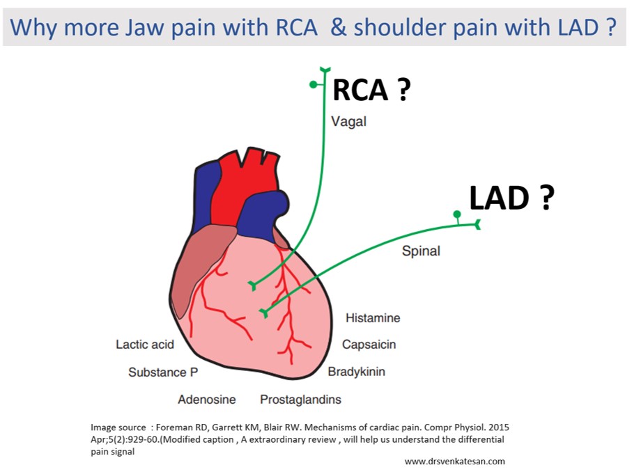
It concludes, that LAD angina rarely radiates to JAW or epigastrium. While RCA angina relay radiates to the left shoulder.

So, why does this happen?

What I could guess is the ubiquitous vagal fibers that travel in the posterior aspect of the heart, and carries pain signal directly up to the jaw whenever these areas become ischemia. LAD is less likely to irritate the vagus. Of course, there can be a definite overlap.

OMG, give me some time to keep in touch with  basic science 

Now, fellows of cardiology, please take a  pause from your regular aggressive cardiac cath lab workouts and get a break at least once in a while. How does the ischemia of myocardial tissue generate pain? Why it is severe in some, trivial in others, and even dead silent in some, 

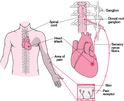
The chest pain genesis is initiated by sensory electrical neural action potential, that captures the epicardial neural plexus first, switching over from somatic to the visceral pathway and trespassing the para ganglionic plexus and traveling further to the spinal cord. Where it may collide with other incoming sensory signals ascends in specific myelinated and non-myelinated neural cables, reaching the brainstem, interacting with local nuclei, and finally reflecting on subcortical and cortical pain matching centers.  We haven’t yet located the exact center for anginal pain. (Perithalmic and amygdala could be closer to real centers) 

So, it is a really complex sensory world yet to be understood fully. Mind you, I haven’t touched upon the neurophysics of referred pain, linked or clandestine angina.

  • What is the effect of cardiac denervation, autonomic neuropathy, or on the perception of chest pain(Does a quadriplegic feel angina ? or post-transplant heart immune to angina ? (Gallego Page JC,Rev Esp Cardiol. 2001) 
  • Is it biochemical or neural, can substance P in blood cause pain hitting the amygdala? 
  • Will hypoglycemia and anemia cause angina due to lack of glucose and oxygen?
  • Finally, how is Infarct pain is different from ischemic pain (Ischemic)

Where do get the answer to these questions?

This paper from Dr. Robert Formean(Ref 2) university of Oklahoma is just the best source I think, to explore and understand the topic. (Reading time 60 minutes: Let me tell you, it is worth more than a time spent on an insignificant angioplasty of painless PDA lesions)

Final message 

So, what have you learned from this post? Does this question about angina matter at all? Surely not. in this space-age cardiac care where we are right inside the coronary even before we listen to the patient’s complaint properly. We are always at liberty to do what we want( or love) to do. But, the urge to understand the foundations of clinical science is the last remaining hope, that will keep the specialty of cardiology enchanting. 

Reference
 
 
A comprehensive reference for the genesis and signal processing  of chest pain 


Read Full Post »

Read Full Post »

GettyImages-865142952-5b5eef884cedfd0050112fa6

Charles river esplanade ,Boston* : A healthy middle-aged man who was jogging quietly, while his heart was under intense scrutiny by the bionic eyes of Apple i-watch’s smart patch electrode. Suddenly, it detected some bizarre ST segment fragmentation (Seems it can predict in advance , Ischemic signals 10 minutes prior to onset of ACS ) The built-in cosmos direct GPS instantly alerted & summoned a titanium powered Space X drone that pulled the patient from the riverside to the nearest human wellness port .

EHANG 184

It dropped him through a remotely accessed split glass roof right inside the hybrid heart lab, to find , men and women chatting with flattish Artificial intelligence panels who readily allowed the robotic arms to hug the patient which engaged the coronary artery pushing radiation free magnetic gas found nothing inside and what would become a perfectly normal human coronary artery .

An amused resident robot gently plucked the patient from the cath table with sheepish laughter and called for another drone to drop the patient exactly in the same place from where he was picked up.The healthy hearted patient thanked the doctors profusely and continued his routine evening jog across the Charles of course with a 16-minute delay!

Next day . . .

Event auditing firm medi-logic mind congratulated the entire cardiac team and its digital health hub for the quality of the network and completing this daring coronary rescue mission in 16 minutes. While the drone to hospital roof time was 3 minutes, the coronary artery visualisation time was perfect.The auditing team had a special mention about the astonishing capability of Apple time watch algorithm that made sure that the patient’s evening routine was unaffected in spite of this life-threatening non cardiac pseudo-emergency. The crowning glory was, the entire expenses amounting to 250000 dollors (after a special money back discount coupon for the first false alarm) were taken care by the patient’s virtual insurance blockchain payment gateway.

*You have just read the news that wasn’t – January 2030 AD

Now, back to reality,

Stumbled on this news clip from pages of Times of India, (20-6-2019) months after I wrote the above piece. I wondered the chase between fact and fiction is becoming  really a close race.

Read Full Post »

Evening cardiac clinic is generally a relaxing place until an occasional patient  with vague chest discomfort present with this sort of an ECG .

He was a 68 y old hypertensive and was on Tablet Amlodipine .After a cursory look at his ECG , eyes wandered to look for some specifics. Suddenly ,my  ECG reading skill was stretched . Is it really LVH ? or Ischemia ? I asked for any old ECG which he couldn’t provide. I had to blink  more than a moment , before asking him to describe his chest pain in detail one more time . I got almost convinced it was not ACS  since he was having only localized pain over left side of chest. Still , I didn’t have the courage to send him home. An Echo was done.

Curious to know what the Echo showed ?


Yes , you also probably guessed right . It is LVH and there is no wall motion defect either.

How to differentiate between Ischemia and LVH ?

This is a common question asked in the board exams.There are number of ECG clues to differentiate the two .Mostly it will help  fellows  to pass exams. Academics rarely comes to your rescue when patients land with  chest pain  especially at odd hours.

Find the answer in this link

https://drsvenkatesan.com/2009/12/12/how-to-differentiate-lv-strain-pattern-from-primary-lv-ischemia/


Now comes the real twister , Does presence of LVH  exclude Ischemia in any way ?

Why can’t be ACS in a patient with LVH ?

When I posed these questions , some one suggested global longitudinal strain  with speckle  tracking to rule out ischemic wall motion defect, my resident suggested  high sensitivity Troponin and Ischemia modified albumin.

Whatever is the technological assistance , one thing  is certain,  we need to finally fall back  on patient’s symptoms . Unstable angina is neither an ECG diagnosis nor biochemical  or Echo diagnosis . (Its all about patient description about his angina , that clinches the  diagnosis !)

It remains a fact normal spot Troponin can never rule out  ACS on time ,( Even patients with unstable angina who  harbor  tight  LAD lesion can be both ECG /Enzyme negative )

When we are not sure ,the traditional coronary care dictum  shall operate .It demands admit, observe, with serial ECGs and enzymes. This protocol cant’t be followed strictly for a variety of reasons . We may have to rely entirely on our clinical Intuition accrued over the years.

A modern-day cardiologist might have a different dictum .The simplest solution in such situations is a diagnostic radial snap shot coronary angiogram .It is an easy way out  . . . , and avoids the trouble of spending wasteful minutes of personal conversation with the patient .The ethics of outsourcing history taking , patient record review or even clinical examination has pushed the definition of professional competence in coronary care into murky grey zone.

One more reason we should hesitate to rush these patient to cath lab is the detection of  incidental insignificant CAD (Which will loom larger than life when they are wheeled in from  ER)  that will  lead on to  further inappropriate chain of events.

How relevant is clinical acumen in modern era ?

Clinical acumen in medicine can never be taught in class rooms or read in text books. It is the innate ability to  combine  knowledge, experience ,skill  and  lastly (and most importantly) courage to ignore conflicting and pervasive data from new generation Investigations. Instead of helping us ,they often directly affect our increasingly vulnerable native medical cognition.

Even if some one is blessed with a good clinical acumen it seems to have little value many times as power of Investigations and fear of missing a event will prevail over it ! I have been victim of this phenomenon many times and not able to follow what my mind preach me !

What happened to this patient ?

I had to admit him against my  wish (Of course I was safe!)  He too got admitted reluctantly and was observed till morning , spent Rs 5000 for pack of investigations and stay , was discharged without any issues with a diagnosis of simple Hypertension and LVH.

The non academic bug  didn’t stop there  . . . again  contrary to my conscience  I had to suggest  optional coronary angiogram to rule out true CAD as a precautionary statutory advisory !


Counterpoint 

How do you know this is really not CAD  ? I won’t believe unless and until I see the CAG and its normal.

Ok, Let me post his angiogram if he decides to  undergo it.

Read Full Post »

lv apex radiological clinical anatomicallv apex radiological clinical anatomical 002


Read a related article

https://drsvenkatesan.com/2008/07/02/how-do-you-define-apical-impulse/

https://drsvenkatesan.com/2008/07/02/how-do-you-define-apical-impulse/

Read Full Post »

Syncope is a classical feature of LVOT obstruction especially with valvular aortic stenosis.The mechanism of exertional syncope in Aortic stenosis is traditionally attributable to the fixed obstruction .This fixed obstruction is not able to cope up with increased cardiac output as demanded by the exercising muscles . But exercise  induced reflex as well as local vaso-dilatation mechanism is intact . The consequence is predictable. A critical fall in SVR amidst a obstructed LVOT precipitating a syncope.

However , If the above mechanism is the sole reason for syncope in Aortic stenosis , we have a problem to explain why syncope is  rare even in critical mitral stenosis which is also fixed LV inflow obstruction ?

Is there some thing unique in LVOT obstruction that causes syncope ?

No, it is nothing to do with LVOT .To generate a true pathological syncope, reduction in cardiac output per-se may not be enough . It appears there should be an inappropriate systemic vasodilatation as well to precipitate a syncope.This can happen only if the parasympathetic system gets activated by some means . The trigger is located in the mechano- receptors of left ventricle . Hypertophied left ventricle with high Intra cavitory pressure (Often above 200mmhg) generated due to LVOT obstruction activates the syncope circuit.The same rule may apply for RVOT as well .One could get syncope with critical valvular PS or severe pulmonary hypertension when RV mechanical receptors get a triggered.

What happens in mitral stenosis ?

In mitral stenosis , LV is under- filled ,  wall thickness is normal .There is little likely-hood of LV mechno-receptors to get stimulated as the LV wall stress is normal. This is the reason syncope is less common in mitral stenosis. However , this is not  absolute rule , syncope can still occur in severely narrowed orifice of mitral valve due to low flow state alone or a ball valve thrombus and paroxysms of arrhythmia .

Read Full Post »

We know cardiac pain is often  referred to Jaw and neck .

What prevents the neck pain of cervical spinal disease to be referred over the  heart ? Can pure spinal lesions mimic angina ?

The answer seems to be “Yes” . The neuronal  circuit is  there .Only , the traffic has to be reversed. Medical logic is always puzzling. There is indeed an entity called cervical angina.

The cardiac pain  can be  referred any where between  dermatomes  C3 to T 10 It is generally  believed cervical radicular pain  can go only one way . . . ie towards the nape of neck and  arms .Dermatomal overlap ,neural cross talks  thalamic inputs and cortical  reflection and perception always make the subject of referred pain too  complex.

Now,It seems possible ,the neck  pain can  spill over into the anterior chest wall ,mimicking  angina .Imagine the  confusion  if the patient  has both  cardiac and cervical entities ! Does the pain signals from the two sites  collide in the local spinal network ? Does one extinguish or amplify the other ?


refered pain

This article which was published  in the Spinal Cord .

cervical angina  reverse referral pain

Read also linked angina

http://www.nature.com/sc/journal/v44/n8/pdf/3101888a.pdf

 1.Guler Net al.Acute ECG changes and chest pain induced by neck motion in patients with cervical hernia: a case report. Angiology 2000; 51:861–865.
2.Wells P. Cervical angina.Am Fam Physician1997;55 2262–2264.
3.Jacobs B. Cervical angina. NY State J Med 1990;90: 8–11.
 4.Baba H et al. Late radiographic findings after anterior cervical fusion for spondylotic myeloradiculopathy. spine 1993;18: 2167–2173.

Read Full Post »