Feeds:
Posts
Comments

Archive for October, 2023

Cardiologists have always struggled to classify, assess, and grade one important valve disease, which is Mitral regurgitation. While valve replacement is the ultimate treatment, the timing of MVR is still a big debate. Apart from valve replacement, valve repair is a strong contender in selected patients. In recent times, cardiologists have made great strides to grab MR patients from cardaic surgeons. MitraClip, a percutaneous edge stitching , is possible with a varying degree of success.

Mitral valve edge-to-edge repair (MEER) is an interventional clone of Alfieri surgery that has shown conflicting results in the MITRA-FR and COAPT studies. The reason for this discrepancy in the MITRA-FR population is that they had larger-sized ventricles, which continued to pose challenges for the clip, which is focused only on the leaflets.

A new subdivision of secondary MR

Now, some of the cardiologists want to classify SMR/FMR into Proportionate vs Disproportionate MR. It may not be a great innovation, but it sub-divides secondary MR for optimal therapy. It simply says if LV dilation is significant, clipping the leaflet alone will not be sufficient; it would rather need an annular restriction either at the time of the index clip procedure or in due course. While Disproportionate MR implies, it is more of a leaflet coaptation defect, dominating over annular contribution.

Who proposed this ? What is the implication?

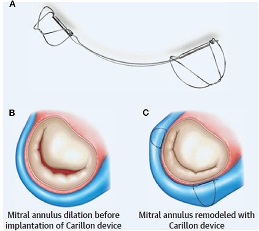
It is an afterthought, I think, from the makers of annular restriction device makers. MEER is found to be less effective in proportional MR.The Carillon device is a new arrival to tackle secondary MR .It is actually a wire that forcibly tightens the AV annulus inserted through coronary sinus . This modality takes advaantage of the aantomical proximity of coronary sinus to mitral annulus. Coronary sinus encircles and from a virtual wall along significant circumference of mitral annulus.

Unlike mitraclip, the Carillon device is claimed to tackle secondary MR irrespective of whether it is proportional or disproportional. It also has the potential to reduce LV dimensions in the long run. We have another device called IRIS-Millipede (to compete with Carillon).

Front. Cardiovasc. Med., 20 November 2020
Sec. Structural Interventional Cardiology
Volume 7 – 2020 | https://doi.org/10.3389/fcvm.2020.576058

Final message

We are free to have as many classifications in MR (Primary, Secondary, Functional, Atrial MR, & now Proportionate Disproportionate.) It is not the aim to bring up a rivalry between leaflet vs annular intervention. Ultimately, the most powerful component of the mitral valve apparatus, i.e., the LV muscle that matters.

I would request the esteemed researchers in MR ,not to keep EROs, regurgitation fractions, or chamber dimensions as primary markers of success of a device. Having strong clinical outcomes as the endpoint should be made mandatory, i.e., prolonging good quality of life and survival (But, the reality can bite hard. Someone told me, walking 20 meters extra in a 6-minute walk test is enough to get device approval from the authorizing entities.)

Final message

1.Grayburn PA, Sannino A, Packer M. Distinguishing Proportionate and Disproportionate Subtypes in Functional Mitral Regurgitation and Left Ventricular Systolic Dysfunction. JACC Cardiovasc Imaging. 2021 Apr;14(4):726-729. doi: 10.1016/j.jcmg.2020.05.043. Epub 2020 Aug 26. PMID: 32861653.

Postamble : A surgeons perspetive

While we debate about devices, the true benefits may lie elsewhere. A good MVR done by a mitral heart surgeon is the need of the hour. Says this paper from a top heart surgeon in India. Yadava OP. Disseminating valve repairs – a clarion call. Indian J Thorac Cardiovasc Surg. 2020 Jan;36

Read Full Post »

Read Full Post »

Recently, I visited a seven-star cardiology center. It had a fascinating lab with a near-360-degree roaming arm and floor (even the roof, I think!). The lab also had provision for optional thoracotomy support by surgeons and a CT scan. What’s more, I was told that an adorable robot will soon be added to the cath lab workforce to assist in delivering and maneuvering catheters. It’s truly amazing to see the advancements in science!

I asked the chief staff nurse how many procedures are being performed per day. She pulled out an Excel chart from a gesture-controlled touch panel. It was clocking about 20 to 25 cases per day. The lab performed the best of all procedures in our country. Great, well done, and congratulations, even as my lips uttered, my mind was thinking the opposite. Do we require such labs when the majority of the population struggle to get even basic medical care in emergencies?

Quantity vs quality of procedures 

The said lab went on to achieve 2000 cases recently. It was crowned with glory in a stunning anniversary bash. We all take pride in numerical accomplishments. Quantity is easily measured. It’s simple mathematics. Assessing quality is a tricky one. Even professionally well-accomplished interventions may be of low quality.

Low quality and low impact may not be synonymous; still, it conveys the same meaning if we look at it from the patient’s perspective. The quality of the procedure shall be assessed with reference to the cost involved, clinical outcome, procedural as well as a perpetual chain of risk, finally, and most importantly the test of time.

Overall, the quality check of a cath lab is not only for the machines, it is also about the operators  &  what they do. 

Postamble

Whover wants join this cath lab award session. Welcome .

Top 10 low-quality (Impact) cardiac Interventions” 

There were 10 nominees

  1. Distal bifurcation lesion
  2. LAA closure
  3. Delayed PCI in STEMI
  4. Valve in valve 
  5. VSR closure post-STEMI
  6. Septal ablation in HOCM
  7. His bundle pacings & CRTs 
  8. Poorly planned TAVRs
  9. Clumsy Paravalvular leak blockage
  10. Complex EVARS

 The winner was selected without any difficulty: No 3 : Its delayed PCI beyond 24 hours after STEMI (This accounted for 30 % of all procedures put together)

Meanwhile following competes for “high-quality” intervention 

  1. PTMC
  2. Left the main/ proximal LAD stenting in UA/NSTEACS
  3. Permanent pacemakers in CHB (Not all in SND)
  4. ICD implantation for the truly deserving
  5. RF ablation for AVNRT/ Some VT
  6. Selected cases of Primary PCI
  7. Some life-saving palliation in the newborn(Ductal stenting/IAS septostomy)
  8. Diagnostic coronary angiogram in a strongly suspected ACS.

Choosing the winner was really tough, just leave it to the readers.

Final message

It’s worthwhile to go for such an analysis periodically to improve procedural quality & Impact. It’s wiser to allow our brains to prevail over hands. Global health is all about technology optimization, outcome analysis, comparative efficacy. The future sounds exciting though, with deep machine learning, data mining, skill transfer. However, there seems to be a definite risk. In the name of artificial intelligence, we might contaminate machines as well with humanness.

 Let us restrict funding and stop Insurance coverage for low impact procedures. Instead, may use the resources for a truly life-sustaining & healing process.

Reference & Further reading

Posted in Uncategorized | Tagged comparative efficacy trialscost effectiveness in medicineethics in medicineEVO;LUTE PRO SAPIENS XThealth economicsHYBRID CATH LABTAVR TAVI VALVE IN VALVEtop 10 best and worst interventions in cath lab |

Read Full Post »

An unusal conversation with a flamboyant student fellow,


May I know , What is the normal Pericardial thickness, ?

Which pericardium you mean sir ? ,Parietal or visceral?

Would like to know both .

It is variable sheet of tissue .You want the thinnest part or thickest part ?

Don’t beat around the bush .Tell some answer.

You want, autopsy thickness, or real life thickness as surgeon sees it ?

I want it in practical Imaging modality

Do you want Echo, CT or MRI thickness ?

I should say , its too much for a first-year fellow in reverse questioning!

Ok relax sir, shall I tell the systolic or diastolic thickness?

So, you decided to spoil my day.

Ok, one final clarification sir,, you want thickenss over anterior RV aspect or over posterior atria ?

You are getting on my nerves. Thankyou, will you please leave the class .


Yes, pericardial anatomy ,can indeed make us crazy. Let us try to get some answer .

Whenever some one is asking about pericardial thickness, it generally mean the parietal,thicker fibrous outer layer. It’s about 1 to 4 mm*, never more than 3 -4 mm .As we can see, it is a highly variable sheet of folded tissue with different adherent surfaces. The visceral pericardium is very much less than 1 mm, in fact,it need to be measured in microns. It is called the epicardium and adheres seamlessly to the myocardium with intervening fat. (Sub epicardial fat)

Ref: Normal pericardium measures 0.4–1.0 mm thick in autopsy studies but normal pericardium measures 1.2 mm in diastole and 1.7 mm in systole on MRI studies [1].It is sort of surprising finding, to note pericardium also thickens in systole.

Mind you, echo over estimate the thickness, and its crudest form of measurement and should not be relied upon.Even CT over estimates.MRI is currently closer to true thickness.

In constrictive pericarditis, which pericardium compresses the heart?

In effusive constrictive pericarditis, organized fluid makes the two layers stick together and makes it thick, making it difficult to differentiate the two layers in any imaging modality. Hence, the answer is that both layers together compress the heart, but the fibrous layer is primarily responsible and exerts more pressure per inch.

Can epicardium per-se thicken with normal thickness of parietal pericardium?

We are not clear about it. The rarity of constriction in viral myo-pericardiits comparred to tuberculous suggest isolated epiicardial constriction is rare as epicardium is sngle sheet of cell .In wonder inflitrative pericarits might occur arsoing pure feom epeicardium following myo or epimyo caritis. . I dont know , may occur in some myopericardits.We need to ask Dr Renu virmani.

(*Answer to this question might alos explain why rheumtic pericardiits doesnt cause constriction as it doesnt involve fibrous pericardium )

Does sub-epicardial fat make epicardium appear thick ? How to differntiate it ?

This is a tough one to diffferentiate . There have been cases of constrictive physiology due to epicardial fat (Effusive-constrictive pericarditis with massive epicardial fat A Goto, M Lancet September 01, 2001)

One final question: Is it the thickness or the elastance /resistance of the pericardium that determines the likelihood of constriction?

Pericardial histology and elastance are more important. We know, that transient constriction with normal pericardial thickness is a very well recognized entity. (Haley JH,JACC 2004)

Final message

Never under-estimate the importance of pericardial anatomy. It can go as deep as its recesses and the tortuous sinuses. Try reading this wonderful review by Rodriguez.(Ref 1) I guess it might have taken tons of time to write such a great article. Don’t excuse yourself to waste 20 minutes in between your angioplasty times.

Reference

1.Bogaert J, Francone M. Cardiovascular magnetic resonance in pericardial diseases. J Cardiovasc Magn Reson. 2009 May 4;11(1):14. doi: 10.1186/1532-429X-11-14. PMID: 19413898; PMCID: PMC2685792.

2.Haley JH, transient constrictive pericarditis: causes and natural history. J Am Coll Cardiol. 2004 Jan 21;43(2):271-5. 147

Read Full Post »

AV valves exist to divert blood to the outflows when ventricles are contracting. If they leak significantly, obviously there are major consequences. What happens to the net cardiac output when these valves leak?

Clincally , we know ,fatigue is sign of reduced forward cardaic out put , that is more common in MR. But, edema , congestion, muscle fatigue is eqaully if not more common with TR. One importnat diference is MR jet is mainly a hemodynamic trouble in pulmonary circuit while TR jet , has one more component hits on liver function making it metabolic consequnce as well.

Can’t escape, tell us your answer for the question

*I am afraid, anyone has done a specific hemodynamic study on this. It should be clear for everyone, that RV stroke volume, is not only the preload of LV, and it is going to bcome LV stroke volume, a few seconds later. Hence TR equally reduce * (if not more ) the forward cardiac output as does MR. In fact, the compensatory mechanism of LV with its reserve work might maintain the forward output for a longer time. RV reserve functions are less.Fellows must analyze this. It is not a difficult one. Measure LV stroke volume by echo ( LVOT area X Aortic TVI) in patients with TR vs MR pre and post-correction.

Final message

RV & LV are like conjoined twins. One’s function can’t be decoupled from the other. Now we realize, TR plays a more central role in both symptom generation and the overall outcome in cardiac failure. Hence, aggressive interventions are being attempted to plug this leak. (We will come to know whether it is truly beneficial or not only later, as arresting TR puts more burden on RV, as TR has a pressure cooker release/relief effect on RV)

Reference

Unterhuber M, Kresoja KP, Besler C, Rommel KP, Orban M, von Roeder M, Braun D, Stolz L, Massberg S, Trebicka J, Zachäus M, Hausleiter J, Thiele H, Lurz P. Cardiac output states in patients with severe functional tricuspid regurgitation: impact on treatment success and prognosis. Eur J Heart Fail. 2021 Oct;23(10):1784-1794. doi: 10.1002/ejhf.2307. Epub 2021 Jul 28. PMID: 34272792.

Rebecca T Hahn, Luigi P Badano, Philipp E Bartko, Denisa Muraru, Francesco Maisano, Jose L Zamorano, Erwan Donal, Tricuspid regurgitation: recent advances in understanding pathophysiology, severity grading and outcome, European Heart Journal – Cardiovascular Imaging, Volume 23, Issue 7, July 2022, Pages 913–929https://doi.org/10.1093/ehjci/jeac009

Read Full Post »

STREAM trial (New Engl J Med 2013) was a sort of paradigm-creating study, that made Pharamco-Invasive approach – PIA an authentic via-media strategy, trying to accure benefits from both lysis and PCI in the management of STEMI.

However, one question remained. Does PIA work safely in the elderly, where bleeding risks are higher? Do they tolerate the double whammy of P and I ? Both procedures keep the blood clotting process tentative for a prolonged period of time.

Emprical usage of half dose lysis is not new, is existingn, for more than 4 decades, right from streptokinase days.

Now the multi-center STREAM-2 trial , released from Belgium , Published in the current issue of Circulation, asked this specific query on the efficacy of half dose Tenecteplace in PIA.

Both strategies had almost similar outcome with 87 % TIMI flow, implying elderly people , are reciveing twice the dose of lytics without any true benefits, but enhancing other risk.

Final message

The conclusions are not surprising. Our empirical thoughts have now, become acceptable sort of evidence. So , let us go ahead with this renewed PIA strategy and proceed with half dose of Tenecteplase ln elderly ( A liberal cut off of >60 years) without any guilt.

Meanwhile, the STREAM team has one more important job to do. So far, no one has the guts to test “P” vs “PIA” in the current era, in a large scale ( ie proceed with “I” if and only if “P“has failed. After all , if P is successful, that has resulted in good TIMI 3, and normal LV function there is no need for I )

Hope, somebody or institution get that courage and funds to design a STREAM- -3 trial comparing three distinct strategies, ie stand-alone TNK vs PIA, & pPCI and make it a reality. I am sure, it will bring surprise and very likely a pleasant one for all stake holders.

Read Full Post »

Are you still on warfarin? Come on, switch to DOAC./NOAC This seems to be a fashion statement among cardiologists and their patients with chronic AF. (By the way, NOAC has a new name, i.e., DOAC. NOAC stands for non-vitamin K anticoagulants. Now, N is replaced with D, direct-acting oral anticoagulants.)

NOAC does have some advantages. the major one is better patient compliance, and ease of administration as it doesn’t need routine monitoring. (One may wonder how this transforms into a real advantage? ) The drug acts in a dark invisible mode. Bleeding risks are either less, equivocal, or high. So we can’t conclude conclusively on true bleeding risk.

(We do have some cumbersome monitoring methods for NOACS)

(Rahmat NA Monitoring the Effects and Antidotes of the Non-vitamin K Oral Anticoagulants. Arrhythm Electrophysiol Rev. 2015 Aug;4(2):90-5. )

Switching games with DOAC

A detailed review of how and when to switch is in this article Adelakun BMJ open 2023

Switching to DOACs should be based on some solid scientific principles. (Of course, we have it !) But, getting rid of INR monitoring should not be the only reason. Still, the current thought process among us is to consider DOAC whenever possible or (if not make it possible somehow). In this context, this study from Circulation September 2023 (Ref 1)helps to choose which side of the OAC-DOAC debate one should fall, especially in the elderly.

Final message

Dad, are you still using that frail iPhone 11? I feel bad, will get you an iPhone 15 Pro. Its just out in Apple stores. You know, it has dynamic island technology. Wish to gift it to you this Diwali.

Oh, thanks, but I’m fine with the oldy. I can comfortably communicate with whoever I want to.Don’t enforce me, I will stick on with warfarin, my dear son.

Reference

1.Safety of Switching From a Vitamin K Antagonist to a Non-Vitamin K Antagonist Oral Anticoagulant in Frail Older Patients With Atrial Fibrillation: Results of the FRAIL-AF Randomized Controlled Trial. Circulation 2023;Aug 27:[Epub ahead of print].

2.Adelakun AR, Turgeon RD, De Vera MA, McGrail K, Loewen PS. Oral anticoagulant switching in patients with atrial fibrillation: a scoping review. BMJ Open. 2023 Apr 25;13(4):e071907. doi: 10.1136/bmjopen-2023-071907. PMID: 37185198; PMCID: PMC10151984.

Read Full Post »

When we realise, even class 1-A indication often blinks at the bed side,while class III -C appear to pour more sense, practicing cardiology becomes "Tough and exhilarating” .

Read Full Post »

(Why should the number 65 bother us in TCFA-detected by OCT? Does this number really deserve that respect? Trying to find some truths from 8 questions with & without evidence.)

1. Does TCFA really make a plaque vulnerable?

A.Yes

B. No

C. Maybe

Answer: Yes & Maybe. But there seem to be more important factors other than TCFA for a plaque to become vulnerable making TCFA not really a big deal.

2. TCFP is more common in which lesions?

A.Flow limiting lesion

B.Nonflow limiting lesion

C.No relationship between TCFA and flow

Answer: No relation, rather a random relationship.

3. What is the natural history of TCFA?

A.Get ruptured

B.Static

C.Get thickened

D.Further thinning

Answer: If someone can answer this query accurately, he can be rewarded Nobel Prize, in Lipidology

There are some studies available though.

Ref : This 2023 paper adds some anxious content to those who ignore TCFA. Long-term outcomes of patients with normal fractional flow reserve and thin-cap fibroatheroma EuroIntervention 2023;18:e1099-e1107. DOI: 10.4244/EIJ-D-22-00306

 4. What to do with FFR-negative(FFR>.9), TCFA-positive lesions (tcfa<50mic)? Does stenting TCFA make it less vulnerable?

No. Stents and scaffolds were never invented to make a plaque less vulnerable.

5. How is TCFA different in stable CAD from ACS situation?

They are very different. In the ACS situation, TCFA (Whether the cap is intact or not ) needs immediate attention. Does it mean immediate stenting? No, it is not. (But unfortunately, it is what happens in the reality.) Thrombus and its lytic products add a new dimension of risk for recurrent ACS. High-dose statins are critical here. Stenting is so tempting, and logic and science are in direct conflict here.

6. Do statins increase the thickness of TCFA?

A.No

B.Yes

C.Possibly yes.

Answer: Statin does have some action on the cap. there is no such thing as if the cap increase from 65 to 100 micron the plaque is safe. More than thickness, it stabilizes and hardens the contents that are being capped. European Heart Journal – Cardiovascular Imaging (2017) 0, 1–8 doi:10.1093/ehjci/jex102

7. Which is possibly, the thickest cap over an atheroma?

Intimal calcification on the luminal side is the thickest cap (like a helmeted plaque) that protects from any traumatic injury from blood components. (Sub Intimal calcific nodules may behave differently though)

8. If calcium is the safest cap in chronic CAD,, why do cardiologists, attack it with all sorts of weapons?

This is where “excessive knowledge along with academic ego‘ in incognito mode plays spoilsport with our hidden wisdom.

Calcium is an uninvited guest that directly challenges our might and talent, and interferes with placing a stent. No surprise, we take up this fight personally, and destroy, shock, drill, or displace it at any cost. Of course, tackling calcium* is required in very selected patients; it should never be done just because it interferes with the deployment of a stent. (We need to periodically remind ourselves, that PCI is not synonymous with stent implantation; stentless PCI is also possible.)

*Calcium and coronary artery is not a simple topic to understand. How can one agree with the above content, when there are innumerable studies that say a high CT calcium score is directly related to plaque burden. Mind you CT calcium score has little correlation with intimal plaque rupture.

TCFA: Should we worry?

TCFA is a concept born out of cutting-edge coronary Imaging technology OCT. After a decade of experience, we realize we can’t hunt & count these caps among the vast plaque burden. The 65-micron cap is just an anxiety-provoking number. Forget the cap. They are often Innocent.

Looking beyond TCA

What is beneath the cap and the biomechanical forces in the pool of tissue underneath, ultimately matter. The fluidity of the necrotic core and the ferocity of the angry macrophages, MMPs, as well as the triggers from within the lumen like the shear stress of blood plays a major role. We also know that diabetic glycation injury widens inter endothelial gap. We have sufficient reasons to assume, that the sharp edges of LDL molecules, hit the endothelium at a high mean BP along with neural triggers from the peri adventitial sympathetic network that injects the final catecholamine blow to the endothelium.

Final message

No technology can answer the above queries or predict the events. So be at peace with all basic precautions and risk factor regulation.

Reference

1.EuroIntervention 2023;18:e1099-e1107. DOI: 10.4244/EIJ-D-22-00306

2.European Heart Journal – Cardiovascular Imaging (2017) 0, 1–8 doi:10.1093/ehjci/jex102

Read Full Post »

Image courtesey : Sekhar, S. et al Canadian Journal of Cardiology, 2015 (Ref 2)

Coronary stent infection (CSI) is no longer a rare and fancy diagnosis. It is increasingly recognized and is equivalent to infective endocarditis. Though CSI appear simple & practical terminology, Infective endo-coronary arteritis may be the ideal term for this device-related infection.

Any prolonged fever following PCI must be investigated with this condition in mind. Unlike other forms of infective endocarditis, vegetations are rare, or do not occur . Instead, the infection erodes endothelium, often leading to the development of an infective aneurysm. Staphylococcus infections seems to more common. Though host immunity is a factor, it is possible inadequate cathlab sterility plays a major role.

Diagnosis

Conventional IE criteria may not be fulfilled. PET scanning helps us to detect & map the active inflammation in the peri-stent area.(See the Image) The PET criteria to define significant inflammation are not yet standardized. One possible differential diagnosis to CSI is, acute or subacute allergic stent rejection.(Not Kuonis syndrome)

Treatment

Is complex, and carries a high mortality rate. It is best to manage it without any aggressive intervention. Physical removal of the infectious focus, along with the stent, may seem like an ideal option.

However, real-world scenarios often preclude this option as exceptional surgical expertise and team work is needed as in this report (Ref 3)

Reference

There is an excellent meta-analysis on this rare entity by Nagendra Boopathy and others from my place Chennai (Ref 1)

1.Ramakumar V, Thakur A, Abdulkader RS, Claessen B, Anandaram A, Palraj R, Aravamudan VM, Thoddi Ramamurthy M, Dangas G, Senguttuvan NB. Coronary Stent Infections – A Systematic Review and Meta-Analysis. Cardiovasc Revasc Med. 2023 Sep;54:16-24. doi: 10.1016/j.carrev.2023.02.021. Epub 2023 Mar 8. PMID: 36906449.

2.Sekhar, S., Vupputuri, A., Nair, R. C., Palaniswamy, S. S., & Natarajan, K. U. (2016). Coronary Stent Infection Successfully Diagnosed Using 18F-Flurodeoxyglucose Positron Emission Tomography Computed Tomography. Canadian Journal of Cardiology, 32(12), 1575.e1–1575.e3. doi:10.1016/j.cjca.2015.10.022

3.B.G.K. Sudhakar,Pseudomonas aeruginosa septicemia resulting in coronary stent infection and coronary artery aneurysm and acute infective endocarditis of mitral valve causing severe mitral regurgitation- A case report, IHJ Cardiovascular Case Reports (CVCR),Volume 2, Issue 3,2018,Pages 191-195,

Read Full Post »

Older Posts »