Can you guess how many drugs a busy physician on an average writes in their prescription in his office ?
Three ? , Five , Six , . . . . Nine,? There is no specific study available for this non-academic query. I have got stunned to see a maximum of 18 drugs in one prescription. So, it should be anything between 1 to 18. May be a mean around 6 or so (Make your own guesstimate)
There is strong evidence to suggest writing a drug prescription has become a (un)conditioned habit-forming act. My professor* used to say generally 2 to 3 drugs are sufficient for most of the common illnesses we encounter (Only in extraordinary situation one may need to go beyond this )
One evidence less estimate though a random observation among the physicians suggested the bottom half of any long list of drug prescription is redundant and it doesn’t really address the specific problem the patient is suffering. Meanwhile ,the concept of poly-pill is making drug compliance easier in many cardiovascular and diabetic diseases.
*William Osler
Final message
Number of drugs human body can handle simultaneously without any harm is often an ignored chapter in the Principles of clinical pharmacology and therapeutics.
Let us mind the length of our prescriptions and ensure less harm to our beloved patients.
Related material
This was my old presentation made about polypharmacy in CHF :Perils and pearls
Psychological factors both depression and anxiety do confer a significant risk for CAD. However, a distinction must be made between risk factors and triggers. It is highly likely, depression has more consistent correlation with chronic CAD than anxiety.
Primary anxiety per-se is of less of a risk factor for chronic CAD, while it can still be a trigger for cardiac events. (when it occurs over heightened baseline risk) Primary depression increases the CAD risk many fold by slowing the system making it sedentary and promote endothelial dysfunction, which is the key promotor of atherogenic CAD.
It is also noted, anxiety is less associated with obesity (when compared to depression). Further,catecholamine fluctuations that are so common in anxiety states may act like an exercise equivalent ( It’s my quirky hypothesis to be tested by future generations)
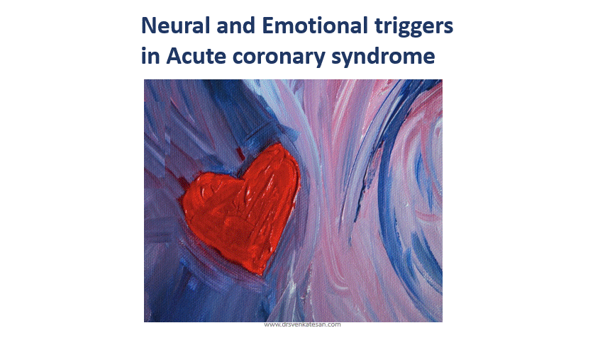
Emotions have a complex equation with neuro cardiac axis .Sudden emotional deaths due to possible arrhythmias or stress cardio-mypathies are important areas for research.
Sharing my presentation in one of the Annual physiologists conference held at Chennai in 2016.
No one would have Imagined a generally Innocuous entity called Diabetes will emerge into a “single disease sub-speciality” in medicine”. Thanks to the global authorities & pharma Industry for making this speciality a formidable one. The link between diabetes and cardiology is so strong, now with pharmacological strategies looking for overlapping Indications.
Let me share a presentation in one of the cardiology meet in 2017 at Thiruvananthpuram.TAN CSI meet , India.
The days are gone when anti-diabetic drugs were alleged to increase CVD mortality. New generation anti-diabetic drugs (SGLT-2 Inhibitors) are coming up that actively dictate and demand us to use it for reducing CVD risk.
(Am I crazy, to look ahead for stand-alone Indication for SGLT Inhibitors for cardiac failure in non-diabetic as well, as a powerful osmotic diuretic !)
Its almost like playing a billiards game in absolute blindness.
It is not an unusual scenerio, to see the balloon catheter delicately bending at IAS puncture site , dodging and deflecting with random jerks as it tries to steal a entry in a few diastolic milliseconds time window when the fish mouthed mitral valve opens in sub square cm areas of MVO trembling in fast atrial fibrillation.
Agree ?
Gathered some tips to cross a difficult mitral valve during PTMC.
This is a PPT presentation taken from archive (Made in 2012)
Often times Its noted we tend to struggle more at the mitral valve crossing than at IAS puncture during PTMC. Experience prevails over Image assistance. Assessment of LA size , IAS plane , and sub valvular disease seem to be critical. Probably the secret of success which I found out was , smart guys never hesitate to repeat IAS puncture site for optimal trajectory .Over the wire technique is not forbidden.
Unfortunately, TTE guidance is of little use to cross the mitral valve. Co-registration of fluro/3D TEE is promising , but most cardiologist continue to rely on their experience.
I don’t know whether you have seen this before. Surely , I haven’t seen a presentation such as this one.
Place: Annual scientific meet ASE 2013. Minneapolis
Presentor:Dr.Partho Sengupta, Mount Sinai hospital, New york.
Its a 3D presentation in “space” without a screen by Holography.
The stunning 15 minutes lecture take us into the myocardial architecture, with speckles , flow vortex echocardiography and fluid kinetic energy mapping.
Don’t miss, a dramatic live teleporting of ASE president on to the stage.
Can you Imagine , where does this technology take us to the future ?
Patients may reach doctor’s offices by holographic teleport for a medical examination or vice versa. Yes, it’s all going to happen someday.
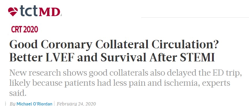
Coronary collateral circulation is one of the major determinants of symptoms and outcomes in chronic CAD. But, we generally shrug off the value of coronary collateral circulation in acute coronary syndrome. The fact is, it has a myocardial mitigating effect following sudden total occlusion.
When does it appear? We did a small analysis (PDF version)
We found it is noted in 25% of patients. With reference time of appearance, 6% had it within 12hrs and in few, it was noted as early as 6 hrs. One caveat is, we may not know whether its preexisting collateral due to chronic multivessel CAD. I am sorry to note this study did not address the outcome analysis. We however documented patients with good collaterals had negligible wall motion defect and near-normal function post PCI. Some of you can pursue research in this area.
Potential role of collaterals in ACS
It limits the infarct size
Keep the myocardium alive and give us time to intervene
Can converts a potential Q-MI to non-Q MI
Possibly prevent primary VT/VF and hence dreaded sudden death in early STEMI
Prevent early adverse remodeling of the left ventricle.
When these points appeared just my assumptions, Dr. Ali Aldujeli, (Lithuanian University of Health Sciences, Kaunas) in his presentation, at TCT 2020 confirms many of them are Indeed true
Final message
I agree, in the era of instant gratification with primary PCI, relying on coronary collaterals may appear a lesser professional virtue. Still, we may need to respect nature. Many times it bails us out.
Superficially, tissue hypoxia might look similar to Ischemia but differs in one important aspect. Though the hypoxic myocardium is short of oxygen, the respiratory excreta from cells ie Co2, lactic acid, and free radicles are promptly cleared and flushed as blood flow is normal. Hence, generally acute Ischemia of tissues is more cell threatening than regional hypoxia at any organ level.
How do you classify hypoxia? we need to go to physiology classes again.
The question we want to address here is the effect of systemic hypoxia on LV function.
We encounter this in different settings.
Chronic hypoxia :In COPD, there can be slow adaptation, still there will be some definite impairment of myocardial function.(Which may not be important in normal times but will tell at times of other stress ) Many studies have documented COPD to compromise LV function. In fact, DCM has a link with some severe forms of COPD (Personal observation, will try to get the evidence )
In acute hypoxia(Non-Ischemic) it causes organ dysfunction.(Acute pulmonary embolism, and sepsis.) We see this often in IMCU with ventilated patients with multisytem defects a poorly contractile ventricles. Some of us used to report this entity with a empirical term global hypokinesia due to hypoxia. The effect of systemic hypoxia on cardiac metabolism is complex as the heart has a unique ability to survive with ketones in anaerobic metabolism at least for a few hours to days.
It is truly fascinating to note, how the human donor heart makes a stunning statement (to thecoronary blood flow obsessed cardiologists) when it is shipped in ice bags with exclusive support of metabolic juices without a single drop of blood and come alive in the recipients without any damage. Is it not a wholesome proof? , for temporary survival, the heart just require ATPs and high energy bonds, not blood in the real sense.
How to study the effect of chronic hypoxia on LV function?
We may lack animal models but Ironically we have perfect human hearts to study the effect of hypoxia on the myocardium. They are cyanotic congenital heart disease. Here is one of the rare reports based on only two patients that address this issue. The authors (Neeraj Awasthy et al) to be appreciated for this pertinent observation.I think we need to look further. Even in TGA, the urgency to perform an arterial switch within a month or so is not only hemodynamic regression in LV mass facing pulmonary circuit but also early hypoxic injury to LV myocardium.
One more area of research
The corona pandemic gives us an opportunity to study the behavior of heart with extreme hypoxia all around.The presumed 5 % Incidence of global LV dysfunction with COVID pneumonia is hypoxia-related or viral myocarditis we are not clear yet.
Caution: Please don’t expect much scientific content in this post. I Hope, you can spare a minute to answer this hypothetical question.
What will be the shape of the curve, If you plot BMI in the X-axis and LDL/ total cholesterol in Y-axis from a thousand normal adult populations?
It will be linear for sure.
Maybe a little curvilinear.
Its likely J shaped with age
I think it is U shaped
No, it is Inverted U
Sorry, I don’t know.
No one knows.
Why BMI refuse to go along linearly with LDL (Cholesterol)?
Only an ultra-fraction of total body fat is represented as cholesterol within the vascular system. It’s worthwhile to note, total body fat store is about 8-10 kg, the amount of cholesterol in circulating blood is hardly 10 grams. It is presumed another 30 grams is present in cells. Body synthesise 1 gram /day.I don’t know whether I can make this statement. It may appear total body fat and cholesterol are almost unrelated things in spite of the dynamism of Intermediary metabolism exogenous and endogenous cholesterol levels are modulated to keep the intravascular cholesterol within an amazingly narrow range.
How justified are we to expect a good correlation between BMI and Cholesterol?
Apart from the presumed logic, there are other dynamic factors that dictate how any person deals with excess lipids/cholesterol.
Genetics profile(Hereditary dyslipidemia)
Dietary habits
Various hormone sensitivity to cells.
Physical activity
Age /Gender
Ethnicity
Most excess fat gets deposited within adipocytes. So it is scientifically impossible to guess the serum cholesterol level by just looking at overweight people.
Final message
Even after 50 years of vigorous research, we are clearly ignorant about this fundamental question in lipidology and clinical cardiology. This is directly reflected in myriad dietary guidelines that flooding in the academic and public domain. Let us be transparent to our patients about our knowledge or lack of it. At the minimum, we should stop confusing all those healthy, active people with borderline obesity.
Further, we need to come out of our villainous portrait of fat in general. Let us respect the fat as an essential building block of every cell and hence the whole body. We still need to pursue a long journey to identify & target only the high-risk population who have atherogenic dyslipidemia that impacts the cardiovascular outcome.
Counterpoint
There is indeed a correlation between body weight and serum lipid. Don’t be dogmatic with limited research and knowledge and confuse the academics. Go through the literature right from Framingham /MRFIT/Monica to Interheart study. There is scattered evidence to show BMI do have a reasonable correlation with blood lipids.
Yes, scattered is the right word, it can mean anything.
Reference
This well-conducted study suggests, within the normal BMI range there could be a correlation with LDL. I don’t know how useful is this data in clinical practice.
Coming next
There is one more question, which has not clearly answered. What is the relationship between seum cholesterol and Intra plaque cholesterol?
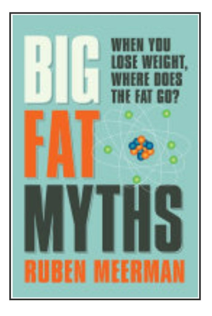
For lighter reading , Ruben Meerman is able to kindle the hidden science.
We can consider Jugular veins of the neck as a naturally present right heart catheter. It faithfully reflects the live pressure and waveform data from right atrium and ventricle .
Can JVP tell us anything about left heart pressures? Is there any relationship between JVP and PCWP or LVEDP ?
If you tell JVP reflects LV filling pressure in any graduate medical exams, you will be admonished. However in DM or post-doctoral exam, if you say there is no linkbetween the two, you are likely to be chided.(It is unfortunate the answers vary depending upon the level of training , which I feel is not academically correct )
Though the JVP-PCWP link, apparently appears Illogical, it does have a scientific basis. It is true, there is a huge (& multiple) anatomical barriers between the left heart and Jugular vein in the form of pulmonary arterial & venous circuits, the right ventricle and right atrium.Still ,the hemodynamic principles demand, whenever left heart filling pressure increases, the right heart pressure should increase correspondingly to drive the blood from RV across the pulmonary circuit.This raise should be in the mean pressure. (or diastolic pressure,) it’s rarely related to systolic pressures as RV systole normally generate more than twice or thrice the LVEDP.
This driving pressure across the lungs is called the transpulmonary gradient. (PA mean minus LA mean) The normal being < 7mmHg. So if there is a sudden increase in LV filling pressure to 20mmhg, there has to be elevated right heart pressures.(20 +7) This will be reflected in JVP as well. So patients with acute diastolic heart failure as in HFpEF must show elevated JVP. This can be documented elegantly In patients with positive responses during diastolic stress testing. (JACC: Cardiovascular Imaging Volume 13, Issue 1 Part 2, January 2020)
There are important caveats in JVP-PCWP link
If the PH is long-standing and precapillary (Reactive PAH) has set in the right heart pressure will no longer reflect the PCWP.
If there is any organic Tricuspid valve disease (Both TR/TS) JVP can reflect PCWP.
Finally, any cause of RV dysfunction will immediately elevate the JVP so biventricular dysfunction makes correlation of JVP with PCWP meaningless.(Acute pulmonary embolism, and RV infarction)
Further confounding can occur if we contemplate RV diastolic dysfunction as seperate entity. (At what level of RV systolic dysfunction, does the RVEDP begin to raise ? I think we don’t have an answer for this . Researchers please note.)
Some more mechanisms of elevated JVP with left heart disease
Bernheim’s effect and ventricular interdependence can make JVP elevated spuriously without elevating PCWP.
Acute mitral regurgitation left atrial V waves can “tide-back” all the way to PA and the right heart to elevate the JVP
In ASD and Lutembachers syndrome the RA pressure waveforms may reflect the LV filling pressure, though inconsistently.
Finally, and importantly in Fontan circuit JVP may exactly reflect the left heart pressure for the obvious reason, as SVC is connected to the pulmonary artery directly.
Final message
JVP will always tell what is happening to the right heart chambers only . It can, no way be taken as a direct marker of PCWP/LVEDP. However, there can be a correlation between JVP and PCWP/LVEDP in a certain subset of cardiac failure. (As in exclusive isolated left heart failure (typically HFpEF) the elevated JVP might just reflect the elevated LEDP provided there is normal RV function )
Reference
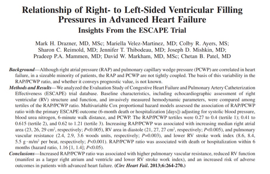
1.This study elegantly shows a correlation (or lack of it) in different subsets of heart failure. It tells us very clearly If JVP(RAP) is not correlating or disproportionate to PCWP, it implies RV dysfunction.
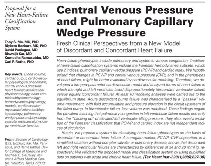
2. This paper suggests a really useful scheme to classify heart failure as concordant and discordant with reference to right and left heart.
It throws some interesting facts. I guess it will help us guide diuretic management and prognosticate chronic heart failure.
Covid has struck hard and this time it has consumed one of the Doyens of Neurology, from Coimbatore, India –Dr.M.B.Pranesh. Privileged to have him as my professor in Coimbatore medical college, my alma mater, watched him in close quarters during my undergraduate and MD days in the late 1980s.
Still recall, how he empathizes with the patient and their family in distress, practiced medicine in the best scientific manner at the same time with a humane and philosophical touch. I can’t forget, how the little genius standing beside the comatose patients In IMCU and tells so precisely the difference between metabolic vs structural coma without even asking for a CT or MRI scan.(We learned with awe, for the first time, how hyponatremia can cause havoc to the brain) I have seen him so tired in many days and sleeping in the ward chair for a few minutes and comes back fresh for the rounds. He used to say sleep is a luxury in our profession. What a statement to make for our generation next.
His favorite quotes are from William Osler and ask us to read the life history Harvey Cushings. He encouraged us, to learn the history of medicine. He was so emphatic to say “Unless we know how our past physicians toiled with their astuteness and hard work, we will not understand the value of clinical medicine”
One of the pure souls who showed us what is the true meaning of teaching, learning, and caring. Got this small clip, wherein he continues to wish us good.
The contents of the this blog is being published as Kindle E book , as per the request of many of the readers. Every article will continue to be open source in this site. Again I shall reiterate the book format is not aimed at any commercial intent. It is only to facilitate learning in a single book format Here is the link to book https://amzn.in/d/euhL5vu
Click below to see who is watching this website live !
This site will never aim for profit. Still ,this donation link is added at the request of few visitors who wanted to contribute and of-course that will help make it sustainable .
Please Note
The author acknowledges all the queries posted by the readers and wishes to answer them .Due to logistic reasons only few could be responded. Inconvenience caused is regretted.