Feeds:
Posts
Comments

Posts Tagged ‘acute coronary syndrome’

A brief conversation between an elite consultant and his fellow. (Caution: Grade 2 harsh language) 

Hey Doc, why is this guy’s name not found in today’s angiogram list? Any Insurance issues?

No sir, he has every requirement. Thrombolysis was very successful, ST has regressed well and it is nearly isoelectric and only T is inverted. His LV function is normal. In fact, I am not able to pick up any WMA.

Aren’t you aware, that being fine is never a contraindication for a PCI ? Which book teach you like that?

No sir, It’s already beyond 48 hrs sir. What is the purpose of knowing IRA status now? If it is open, well and good.If it is partially closed, again little to gain, right? 

Don’t expose your Ignorance. … haven’t you heard of the pharmaco-invasive strategy & open artery hypothesis. Always learn to respect science.

But sir, then why does late PCI of IRA in otherwise stable patients come under class 3 recommendation, if I understand the guidelines correctly, it is a contraindication, am I, right sir? But, this patient got stabilized by us still, why he is compelled to undergo another procedure exposing and adding further risk?  

That shows your immaturity. Doing an angiogram is never forbidden. It is the inappropriate late revascularisation of IRA that is the issue.

 Agreed sir, how confident are we, that we will stop just with an angiogram after visualizing a tempting lesion in either IRA or non-IRA? (My brief experience as a fellow doesn’t tell a fair story) 

 Now, you are trespassing into prohibited non-academic zones of cardiology practice. Instead, talk about FFR, OCT, multivessel angioplasty, and ( deferred or instant ) complete revascularization. Think like a true scientist don’t get spoiled at a young age in your career with all this ethical stuff. 

Final message 

Never allow an ACS to stabilize by medical management, if he is otherwise eligible and affordable for a procedure. You are not authorized to do that. 

Read Full Post »

When a culprit thrombus keep the  myocardium as hostage . . . don’t storm the coronary artery  indiscriminately   !

When a single gun men  keeps 100 innocent people as hostages , threatening their  lives, rescue mission should start .No can can afford to wait. But, without knowing  the  culprit’s true nature the process of rescue mission is always going to be tricky .There are so many instances Newton’s third law  was reversed , when reactions  evoke more chaos  than the index action.

In the recent world terrorist events ,  the  rescue missions  were so delicate and  it was very  unfortunate we  lost  many   innocent hostages !  The reasoning is ,there  is no way we can avoid these. I wonder is it really true ? !

rescue missionNot all culprit lesions  are true ones.They simply threaten  our myocardium with  thrombus and plaques  in various forms .Don’t show aggression to pseudo threats  you may  ultimately end up with more damage.(What I call as crazy culprits!)

(  Read here , why unstable angina even though thrombus is sitting right inside the coronary artery attempting to lyse it causes more  damage !)

After thought

Iam sure ,bulk of  the Interventionists wouldn’t agree with this thought . They would decry , watching a person  silently when the myocardium  is on  fire is a serious crime !

But . . . we  need to  remember the process of extinguishing  the fire  with some more fire arms is a delicate game played in undefined  philosophical turf.

The only way to introspect  such events in life is , to accept any eventuality    arising out of “not pursuing”  a  presumed rescue mission with vigor. No need to be guilty about that,after all , it can be a myth !

Modern human cognition , growing with a staple  scientific  feed  on a 24/7  basis  is  unlikely to realise , restraint can be an effective tool  even in critical moments !

Oh,is all that I have  scribbled so far  is just a repetition  of 1000 year concept of  “Primum non nocere”

Read Full Post »

Is “Non-flow limiting coronary  lesions  more prone for ACS ?

  • If  your  answer  is “No”, you can skip this article.
  • If your  answer is “Yes” , you need to read this article.

ACS is the commonest cardiac emergency .Thousands of patients are treated every day.Millions of dollars are spent.Bulk of the cardiologist’s life revolves around this entity.

Scattered atherosclerotic plaques in coronary artery lead to ACS either in a random fashion or in a predictable manner .

Still, we are  highly  uncertain about  which lesions are likely to result in ACS ! Some time in the beginning of  21st century, the main stream cardiology media were abuzz with the concept, that non obstructive , non-flow limiting lesions are more prone for ACS rather than more tight  stenosis.

atherosclerosis flow limiting lesion glagov plaque rupture vulnerable erosion fissure vs dissection

I fail to understand how a tight lesion is less  prone for ACS. Tighter lesions are  bigger and must be  prone for more complications . Image courtesy :http://upload.wikimedia.org/wikipedia/commons/9/9a/Endo_dysfunction_Athero.PNG

This reasoning was based on few studies, that lacked  solid scientific proof . In fact the initial  observation was  not made in living coronary arteries rather by autopsy observations .(Later live virtual histological studies came ,  but didn’t confirm this !)

Surprisingly the degree of  anatomical narrowing was conferred  vulnerability  , when we know plaque compositions , morphology and hemo-rheological  factors are many fold important in precipitating ACS . (Lipid content , fibrin cap  thickness, eccentricity , etc)

So where is the truth hidden?

Is it really possible, lesser the stenosis more  is the propensity for rupture ?

 We need to introspect .

“In all probability,  it is a meager statistical illusion”

For every tight lesion there are as many minor lesions scattered around in a given a coronary artery. These can progress into ACS  later.

It is basically wrong to assume non-flow limiting lesions are more prone for ACS than non-flow limiting lesions.To believe so , seriously underestimates  the  culpability of big lesions .It appears a coronary mockery to me  !

At best , we can conclude  non-flow limiting lesions  are not benign and can be an important source of ACS.

An unscientific chain reaction !

If we start believing non flow limiting (say  30%  stenosis ) is more prone for ACS , why we are not stenting all  those lesions ?

If the above concept  is  is applied in cath lab  routinely , the principle of  FFR   which relies solely on hemodynamic impact  will  crash into the dustbin !

Some  more truths

However , It is indeed true  when a plaque is hardened by severe sclerotic process or calcification it is less prone for  rupture and clinical ACS  but can be a source for stable angina.

Is it  justified to assume , larger the plaque the harder  would be it’s content  that  resists ACS ?

Meanwhile , we also know there need not be any lesion at all to cause an ACS.( In a young  smoker ,  100 % thrombotic STEMI  is possible  over an area of coronary erosion caused by endothelial dysfunction ! So , where do we go from here !)

Let us be clear

Are you confused more !   . . . after  reading this article, let us clear it by two-line summary !

As on 2014 ,

  • Symptomatic flow limiting lesion   are tackled by stents.
  • All non-flow limiting lesions  are treated by  high dose Statins  and vigorous medical management.

Final message

Contrary to popular  perception, tight lesions are  more complex, eccentric , soft and are at immediate risk of ACS.

Non flow limiting lesions remain static in most,  regress in many , still  carries  distinct  risk of progression into full blown ACS , at any time if conditions are favorable.

Fixed concepts and ideas in medical science do not help us  taking medicine forward. Especially so, when these are based on assumptions and approximations. If only we redo these studies with the currently available technology (FFR/OCT/NIR the conclusions would be dramatically different. !

Waiting for someone to nullify such false concepts in a more scientific way !

Reference

2.Glagov S, Weisenberg E, Zarins C, Stankunavicius R, Kolletis G. Compensatory enlargement of human atherosclerotic coronary arteries. N Engl J Med. 1987; 316: 371–375.

3.Fuster V, Lewis A. Conner Memorial Lecture. Mechanisms leading to myocardial infarction: insights from studies of vascular biology. Circulation 1994;90:2126-2146.

4.Ambrose JA, Weinrauch M. Thrombosis in ischemic heart disease. Arch Int Med 1996;156:1382-1394

Postample 
This post was written in 2014. Happy to find a scientific proof to this concept in 2018.
Source :  PROSPECT study

Retrospective angiographic studies and the prospective PROSPECT (Providing Regional Observations to Study  Predictors of Events in the Coronary Tree) study have shown that plaques with severe stenosis carry a higher per-plaque risk for producing clinical events than plaques that cause no or non severe stenosis.

However, such lesions are few, and overall, most ACS are precipitated by plaques  without significant stenosis on an antecedent angiography
weeks or months before. This epidemiology is consistent with the distribution of TCFAs, as shown by a combined angiography and optical coherence tomographic imaging study of nonculprit lesions.

Lesions that caused severe stenosis were twice as likely to be
TCFAs than lesions with only non severe stenosis, but the total number of TCFAs with nonsevere stenosis was three times higher than those with severe stenosis. The mild pre-existent stenosis of most TCFAs and ruptured plaques is explained by expansive remodeling, because such lesions are, on average, large.

The long-held notion that mild to moderate obstructive coronary lesions are responsible for the majority of MIs has been challenged by studies that described significant narrowing in the days preceding MI. However, significant narrowing shortly before MI may be a result of (rather than a precursor) for rupture.


Read Full Post »

A 60 year old man with chest discomfort and severe breathlessness  and  blood pressure of 160/110 was  wheeled into CCU. A diagnosis of  acute anterior  STEMI was made and he  was about to be  thrombolysed . Since  his blood pressure was high they were waiting for it to come down with IV  Nitrglycerin

I was called to see this  patient  .Here is his ECG .

Q-LVH INCOMPLET LBBB STEMI DIFFERENTIAL DIAGNOSIS 2

Though   ECG  suggested anterior  STEMI  , I  was  fairly  convinced  it  was  in fact  LVH and  incomplete  LBBB.

I confirmed with the  patient  about the onset of symptoms . It was primarily  breathlessness and only a  vague discomfort .Meanwhile , the troponin came as positive and CPK MB    was  normal. The combined troponin  positivity  and ST elevation  almost confirmed the STEMI ,  and  the  urgency for  thrombolysis was  intensified . One resident suggested  an  emergency PCI.

My self ,  in spite of  being a cardiologist was isolated among the physician team .  I  had to  urgently  prove to them it is indeed  not STEMI !  I did a bed side echo and showed  the  physician colleagues   a vigorously contracting  hypertrophied  left ventricle  with a EF of 68 % . There  was  negligible wall motion defect  . . .  if at all any !

They were still far from convinced ?  They  were  sort of  amused .There is   ST elevation ,  there is  troponin  positivity. . . what else you want  . . . they seemed to ask  ?

I asked them  . . . How can an  acute  extensive anterior   MI contract so well ,  without a trace of   wall motion defect ?

It took me considerable time and effort  to  convince them  that the whole thing was not a STEMI.  Finally they agreed .It was  a simple LVH with secondary ST elevation  due to incomplete  LBBB .  Troponin elevation  simply  represent minor myocardial  injury associated  with hypertensive  LVF . This  patient was discharged within 24 hours  in perfectly stable  condition . Since he had mild elevation of creatinine and was  sent for  nephrology  work up.

Final message

LVH with secondary  ST elevation in V1-V4 is a common situation that mimics  acute STEMI . Cardiac failure can result in non ischemic troponin  release .  Acute medicine is  an unique art . Some times it demands all your senses to be on alert mode . Realise ,  in the above case ,   in spite of the   the classical   triad of  chest pain ,    ST elevation , troponin positivity  it  almost led to a wrong diagnosis of  acute myocardial Infarction .

After thought : What  if they had thrombolysed this patient or taken for a PCI ?

When  the clinical suspicion is high and  circumstantial evidence  point to an ACS   ,   this error can be  justified . After all ,  5 % of   famous ISIS  study population were not suffering from STEMI  but got thrombolysis !

* One real possibility in this ECG is  old AWMI with re-infarction  or a dyskinetic septum lifting the ST segment .But both were excluded by the rapid bed side echo.


Read Full Post »

We know  acute coronary syndrome  presents* with  either  STEMI or NSTEMI. (*It actually doesn’t present , it is our understanding and interpretation ).  Bifurcating  ACS into two is more by convention and convenience .Does  the intra-coronary  plaque  dynamics  really  permit us to divide ACS in to two distinct ECG  entities ?

Are we oversimplifying it ?  Probably yes.

The following paper was presented in the cardiological society annual scientific session in New Delhi few years ago (2006)

It generated an intense debate  , finally the chair person  reluctantly concluded such events are  possible. . .

but need more proof   . What is your take on this issue ?

DUAL   ACUTE   CORONARY SYNDROME

S.Venkatesan ,G.Gnanavelu,V.Jaganathan,

Department of cardiology . Madras Medical College. Chennai

Acute coronary syndrome (ACS)  is  classified into  STEMI  and NSTEMI and has gained universal acceptance. The classification was done by   clinical & electro physiological   findings    with   some   pathological basis. The   classification   came into vogue primarily to simplify the decision making process of thrombolysis. ( STEMI –Thrombolysis eligible .NSTEMI  Thrombolysis ineligible.) The limitation of this classification is well   exposed   as   we   now know,    STEMI can evolve into NSTEMI and NSTEMI can evolve into STEMI .   Identifying the culprit artery in ACS is   not straight forward especially in NSTEMI. Adding further complexity   is   the newer   observations that diffuse vessel inflammation,  and  multiple active plaques(MAP) are responsible for many of the episodes of  ACS.

In this scenario   there   could be two are more pathological processes   one   resulting   in  a total occlusion   and other sub total occlusion resulting in both patterns of ACS simultaneously .(STEMI & NSTEMI  Dual ACS)

We   describe two  patients  who had   presented to our CCU  . Both had STEMI one in  lateral  other in anterior wall . They   were thrombolysed   as per  criteria. Both patients had gross ST depression (>4mm)  elsewhere. In one patient it  corresponded  to the reciprocal  leads .The outcome of  thrombolysis  was turbulent .Both patients worsened and one developed  recurrent VT . Paradoxically the ST elevation   regressed   indicating a successful   thrombolysis  in the STEMI  territory  even as the ST depression  was worsening in the other leads. Angiogram   revealed   multivessel CAD with   recannalised  LAD  lesion with eccentric , thrombus containing  lesion in RCA/LCX. One patient expired and other was referred for revascularisation.

We   believe   both of our   patients  experienced  Dual ACS.

When to suspect dual ACS ?

Dual ACS is likely , when  STEMI is associated with ST depression  in at least 5mm in any two leads  or   when there is disproportionate  reciprocal ST depression ( > 2mm of primary). The reason for the poor outcome could be due to a therapeutic conflict between   STEMI & NSTEMI as the former  is  thrombolysis friendly while the later is not . Role of   thrombolysis in  such situations were ACS wanders between STEMI & NSTEMI is not defined. Another possibility is the concept of reciprocal ST elevation,   where in the index event  could be NSTEMI and STEMI is a secondary response  and  thrombolysis is apparently  contraindicated.

We conclude that in patients with ACS,   two or more   plaques can simultaneously get  activated  and  present  as a combination  of STEMI / NSTEMI   in the same  patient  in two different coronary arteries.(Dual ACS) .We suggest   that in every  patient who present with  STEMI  a possibility of   dual ACS  is to specifically considered,  as  thrombolysis could be disastrous  and  instead  they  should  reach   the  cath  lab directly.  .

Read Full Post »

Acute coronary syndrome  is primarily a disease of blood vessel , which perfuses  the heart.  It can even be a disorder of blood, often called vulnerable blood which predispose  for intra- coronary thrombus .

Mind you  , heart is an innocent bystander ! to the onslaught of  coronary atherosclerosis !

Hence , we  often use two terminologies .

CAD : Pure vascular (Coronary )  disease without  any structural and functional impairment of heart  ( No Angina, No myocardial damage ) Most of the asymptomatic plaques  , non flow limiting  lesions, incidentally detected by the modern coronary imaging gadgets  fall in this category.

When does  CAD becomes CAHD ?

CAHD : Coronary artery heart disease .Here not only the coronary artery is diseased , but it has it’s mission fulfilled   ie target organ either damaged structurally (STEMI, NSTEMI ) or functionally (EST positive , Chronic stable angina CSA )

Does the heart does any wrong to suffer from Acute coronary  syndrome  ?

No, it is simply not .The fault lies in one or more  of the following   .Generally at-least two these factors are enough to impede blood flow )  . They  combine to produce an ACS.

  • Blood defect
  • Vessel wall defect
  • Slowing of flow (Stasis)

This is called as Virchow’s triad   suggested over 100 years ago . Still valid in the era of per cutaneous  aortic valve implantation.

* The concept of de-linking  disorders of  coronary  vascular disease  from myocardial disease  is vital  in understanding the implications of current modalities of treatment. 

Even though we PCIs target the culprit ie blood vessel , it need to  realised , we  always fall short of real target . . .namely the heart . In coronary interventions  the catheters and wires roam around superficially over the heart  and they never even touch the heart .This is the reason PCIs are struggling to prove it’s  worthiness over medical therapy in many CAHD patients , which can reach deep  into the vessel, heart  and even every individual cells of heart.

Many (or . . . is it most ?)  Interventional  cardiologists have a bad  reputation for ” failing to look  look beyond the lesion” .  It is estimated  a vast  number  of cathlabs  and CABG theaters worldwide  are engaged in futile  attempt to restore coronary artery patency after a target organ damage is done .This is akin to building flyovers  to dead and closed highways .

Salvaging a coronary  artery and reliving a coronary obstruction is an entirely unrelated and futile  exercise to  a patient who has a problem  primarily in  musculature .

The much debated concept of  documenting  myocardial viability  , before revascularisation  died a premature death as the concept  by itself , was not viable commercially . (Viability studies   , tend to tie down the hands of device industry further , some  interventional   cardiologists began to see this concept  as an  interference to their freedom to adventure  )

Of-course , now  we have  other parameters  phenomenon  like  FFR estimation by Doppler , epicardial  -myocardial dissociation, slow  flow , no re-flow are  gaining importance.

Final message

ACS is primarily a disease of blood vessel but it’s impact is huge on heart. We need to look beyond the lesion .Restoring  a blood vessel  patency  to an ailing organ (Heart ) is not synonymous with total  cardiac intervention  and protection . There is lot more to cardiac physiology other than it’s blood flow. Heart muscle is a too complex organ to be controlled by few balloons and wires  which beat around the bush.

Read Full Post »

Human body is  now  approached by many of the physicians as  collection of  multiple  organs . This is  the price we pay for modernity in medical science. The era  of great physicians  in general medicine has gone . Now, a  super specialist  of one organ  is  rarely concerned about what is happening to the patient’s  other organ ,  it is  considered    foreign to him  ! While ,  this is the dominant thinking pattern of   modern-day specialist

Let us  travel intime  and  go to the year 1954 . . .

Three  physicians from Michigan ,USA  published  one of greatest observation in clinical sciences , namely the ECG changes in various forms of stroke .

Now , a shrewd physician  , will  suspect a subarachnoid hemorrhage (SAH) by looking at the ECG when the clinical situation demands . But , what we need is every one should develop that skill . We have seen errors happening  even in big institutions (or is it because it is big ?)  when  an elderly person comes with deep T  inversions with or without  altered sensorium being rushed into  CCUs  & cath labs instead of  neurology units.

We  need to teach  our junior  colleagues  . . .  That ,  ECGs of patients with  acute neurological syndromes  (ANS)  can mimic as acute coronary syndromes (ACS) ( especially in elderly ) .

The following ECG changes * are observed during stroke

  • Deep  T wave inversion –   Sub arachnoid hemorrhage
  • Cerebral thrombosis   –      Prolonged QT interval, U WAVES
  • Cerebral hemorrhage –      ST segment  shifts /T inversion


The ECG changes tend to occur very early after CNS injury.May last up to 1 week. They are not useful to identify the type of stroke. But , deep T wave inversions strongly suggest SAH rather than ICH or thrombotic stroke.

What is the mechanism of these ECG changes ? 

It is a clear proof that heart and brain are interconnected by neural network. All the noted changes occur during myocardial repolarisation . (ie ST segment )  The current thinking is  (Ofcourse , it is same as our thinking  in 1950s !)  it is mediated by adreneergic surge  initiated by CNS insult  transmitted to  myocardium by the sympathetic system.

Why should SAH produce more  ECG changes than others ?

It is possible the net adrenegic drive from the brainstem and spinal cord will be greater in SAH as it  spreads the entire CNS  through the cerbro spinal fluid. While localised ICH and infarct is  likely to generate less adrenergic impulse. 

Reference

Read the link to circulation 1964 .With courtesey to circualtionaha.com

http://circ.ahajournals.org/cgi/reprint/9/5/719.pdf

This came 50  years  ago , we still quote their work and no one has improved their work . 

Final message

If  only  we make the  clinical bed side teaching as a  regualr habit ,  we  do  justice to   our  great  physicians of the past ,   who enriched  our  life  with their  clinical  skills  and  passion for knowledge  sharing .

Read Full Post »

Modern era of cardiology aims to treat ACS  as and when it develops .That is , as soon as the vulnerable plaque ruptures or a thrombus  blocks the victim’s coronary artery.

But this can be achieved only if the patient reacts to this event.We know 20% of ACS can be totally silent. Some produce very vague symptoms especially in elderly and diabetics. ECG and enzyme changes may help us in patients who do not have clear symptoms.There are variety of markers available for STEMI & UNSTEMI.(CPK-MB, Troponin T , myoglobin etc) Now we are working at finding a marker for ischemia without necrosis. Ischemia modified albumin is one such molecule that is showing promise.

The ER department world over have vigorous screening protocols to diagnose ACS  for  the patients with chest pain. There are thousands of triaging protocol in the  emergency management of chest pain.In spite of  the highest awareness and availability of  scientific expertise , knowledge base the error rate of diagnosing  ACS  stands at an astonishing 58%.  This may seem odd , but this is what  this land mark article in NEJM tell us  (Data from Boston , Milwaukee etc).

Out of 10500 patients with suspected ACS. Only 17 % had real ACS.  55% were admitted initially as ACS  later turned out to be non cardiac .This may seem  acceptable for many  even if it is  an act of unnecessary admission and investigation. It gives us , a sense of satisfaction for not missing a diagnosis of ACS. But it has it’s own risk of complication arising out of unnecessary investigations.It is a chain reaction of  suspicion that  may end up in a coronary angiogram in many ! .It is also a well recognised fact these patients    spend  atleast an average of  2 days  to get rid of the ACS tag over their  necks .

Experience has taught us  simple presence of a human being as a patient within an  ICU ( however short the stay  may be ) can be a health hazard and risk .  This  55 % error ,  which does exactly  this to  our  patients with chest pain  who reach the ER  never bothers us  This is because  we feel credited both academically as well as financially .

In the same study 2.3 %  (About 25 patients) with true ACS  were sent home  after a missed  diagnosis . Paradoxically  this 2.3%  has worried the medical professionals too much . . . This happens  ,  even as we  do not have proper data on  how many of them had a real adverse event after a missed  ACS.

So the message here is even in best centres both missed and wrong diagnosis are  rampant. while wrong diagnosis (25 fold more here  )  is easily accepted by the medical community .We can justify  this as a screening camp for ACS  ,  akin to arresting  a group of suspected  criminals in a  preventive raid ,  later releasing for want of evidence.

In the morals of  criminal judiciary  , it is often said one can afford to  lose  a real offender from the clutches of law  , but a  innocent should  never be punished in any circumstance .

In medical parlance this  goes something like this  . . . Thousand patients shall die because of his or her illness but not even a single healthy person should die due to unnecessary treatment.

The above thoughts  were in response to  the excellent original article on missed diagnosis  of ACS from NEJM.  http://content.nejm.org/cgi/content/full/342/16/1163?ijkey=652d8337709a8bf84c813f4c9d685863ee053162

Final message : (Sorry for the  lengthy message !)

Can we afford to miss an  ACS in emergency room ?

“Definitely not” . . .but do we succeed in that ?  The answer is same “definitely not “

When we are able to accept with pride every time  we make  a  wrong diagnosis of  ACS  in perfectly normal people , It may to provocative to say  we can  also  afford  to do  the same  when we occasionally   miss a  diagnosis of ACS  as well .  Law of statistics dictates for every correct diagnosis made there is many fold number of wrong or missed diagnosis takes place. May be , reducing that is the only aim of medicine.

We need to realise  with even with a 55% of false positive initial  diagnosis  2%  real ACS  escape net !The only  fool proof method  for  not missing  ,  even a single case of ACS   is to label every patient with chest pain as ACS .

In this vexing  issue , we should realise  , in field of  medical decision  making ,  errors  due to acts of commission  ( Making an  inappropriate drug/procedure /surgery  is easily accepted by medical professionals as well as   the court of law !) . But acts of omission ,   like missing a diagnosis or failure to prescribe  a  drug or perform a procedure  is rarely accepted   and  is  considered   a great negligence and  bring intense guilty feeling among the physicians .

This  perception is definitely  not warranted in this  greatest profession  of glorious uncertainties ! Both acts of commission and omission  cause significant damage to  patients . In this modern era  ,  we have clear  statistics  that   reveal ,  acts of commission  leads far ahead over it’ s counterpart in injuring our people .

Hippocrates got it right over 2000 years  ago .  First let us do no harm  . . .

Read Full Post »

Myocardial infarction (STEMI)  occurs in two distinct arterial  territories .The anterior LAD circulation and postero- inferior RCA/LCX circulation.The incidence is equally shared.

There has been some  learned and unlearned perceptions about Inferior MI.

Inferior MI is less dangerous than anterior MI.  True or false ?

Answer: Essentially true in most situations.

Reasons.

Inferior wall of the heart (strictly speaking there is no walls for heart , only surfaces , which blends with adjacent areas)  inferior wall  is formed by diaphragmatic surface and posterior surface.Inferior MI can occur by either RCA or LCX obstruction.The outcome of inferior MI is determined by mainly by  the extent  of   LV myocardial   damage it inflicts.To  quantitate this  we need to know , how much of LV is supplied by RCA , or LCX or combination of both ? This depend on the coronary dominance .It is estimated , the bulk of the LV is supplied ( up to 75%  ) by LCA. This becomes further high in left dominant circulations . In fact , it is believed LV can never get involved in non dominant RCA occlusions. This has brought in a new terminology  called “Small inferior MI”.Inferior STEMI due to PDA  occlusion or in a co -dominant circulation is not yet studied

Apart from the above  anatomical considerations the following clinical observations  have  been made regarding inferior MI.

  • When thrombolysis was introduced , many studies  suggested the the ST elevation in inferior  leads toched the isolectric levels  in most situations even without thrombolysis.Technically, this implies spontaneous , successful thrombolysis are more common in RCA. Among the thrombolysed ,persistent ST elvation is a rare phenomenon.
  • The well known difference in the conduction defect between anterior and inferior MI  is an important contibutor for better outcome in the later.(AV blocks in inferior MI , are often transient, non progressive, supra hisian location rarely require permanent pacemakers)
  • During acute phase cardiogenic shock occurs in a minority (That too , only if RV shock is included )
  • Even in the follow up the ejection fraction in inferior MI is  almost always above  40%. In many EF is not affected at all.
  • Progressive adverse remodelling of LV is rare

When can Inferior MI be dangerous ?

Anatomical factors

Inspite of the  above  factors  inferior MI can not be taken lightly . Especially when it  extend into posterior, lateral , (Rarely anterior) segments.

While  posterior extension  is often  tolerated , lateral extension is very poorly tolerated .This is probably explained as  the extension involves the vital free wall of LV and the laplace forces could precipitate LVF. Free wall rupture is also common in this situation.

Posterior extension , predominantly involves the surface of RV which is less important hemodynamically. Of course incidence of MR  due to it’s effect on posterior mitral leaflet can be trouble some.

inferior MI ECG

High risk clinical catagories.

Out of hospital STEMI  are at  equal  risk irrespective of the territories involved  .This is because,  primary VF does not differentiate , whether  ischemia comes from RCA or LAD .

  1. In elderly , dibetics and co existing medical condtions  the the established  benign   character  of  inferior MI disappear, as  any  muscle loss  in LV has equally adverse outcome.
  2. Even though  inferior MIs are immune  to cardiogenic shock  , a equally worrisome  prolonged hypotension due to high vagal tone, bradycardia, plus or minus RVMI can create trouble. Fortunately , they respond better to  treatment. Except a few with extensive transmural RVMI outcome is good.
  3. Presence of  mechanical complications of  ventricular septal rupture , ischemic MR can bring  the mortality on par with large anterior MI.

How different is the clinical outcome of infero-posterior  MI with reference  to the  site of  coronary arterial  obstruction   ?

The sequence of  outcome  From  best to worse  : Non dominant RCA* → Dominant RCA but distal to RV branch → LCX dominant with large OMs

* It is believed   an  acute proximal  obstruction of a  non dominant RCA may not be mechanically significant, but can be electrically significant as it retains the risk of primary VF and SA nodal ischemia. The ECG changes  can be very minimal or  some times simple bradycardia is the only clue. One should be able to recognise this entity (Non dominant  RCA STEMI)  as the outcome is  excellent and these patients  would never require procedure like primary  PCI

** A inferior MI due to a dominant LCX and a large OMs have comparable outcome as that of extensive anterior MI. The ECG will reveal ST elevation in both inferior and lateral leads.

***In patients with prior CAD  and collateral dependent  multivessel disease  the  inferior anterior sub classification does not make much sense as  entire coronary circulation can be mutually interdependent.

Final message

Inferior STEMI  generally lacks the vigor  to cause extensive damage to myocardium in most situations .Further they respond better to treatment. Risk stratification of STEMI based on the location of MI has not been popular among mainstream cardiologists. This issue needs some introspection as  the costly and complex treatment modalities like primary PCI  is unwarranted in most of the low risk inferior MIs.

Related posts in my blog:

1.Why thrombolysis is more effective in RCA?

Read Full Post »

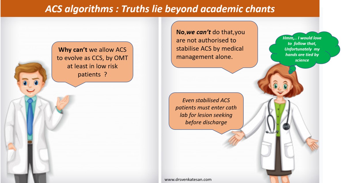
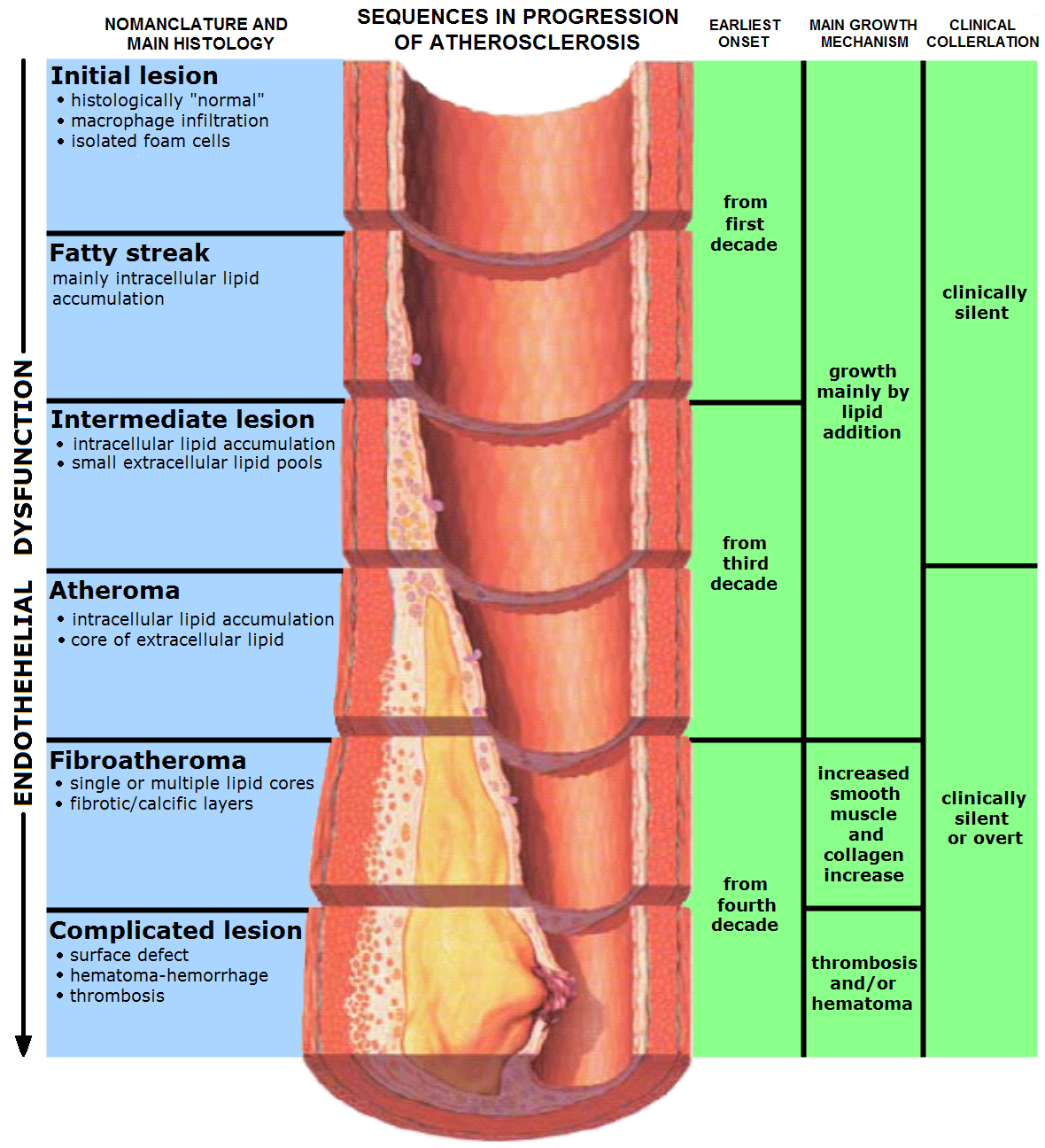
Coronary arterial spasm is a commonly discussed entity  in clinical cardiology. Originally described by prinzmetal decades ago .It was reported to occur only in coronary care units as variant angina .There is nothing called stable spasm every episode of spasm is considered as unstable angina.

How common is coronary artery spasm ?

This condition thought to be very  common during ACS , but  notoriously difficult to confirm.In fact many of episodes are clinically suspected and never confirmed. There has been lot of provocative tests for confirming coronary spasm like ergonavine etc.None of these tests were  neither useful nor practical.

What are the clinical situations where coronary spasm occur ?

Old concepts die hard.Most of us believe , the most common cause for coronary spasm is prinzmetal’s  angina ie , angina   in normal coronary arteries or  with minimal lesions .Clinical experience ,  has taught us coronary spasm can occur in all spectrum of acute coronary syndrome or even chronic coronary syndromes.

Some believe every episode of STEMI will have a component of coronary spasm.

In NSTEMI/Unstable angina spontaneous coronary spasm is an important pathogenetic mechanism

Now,  we witness transient coronary artery spasm in the cath lab due to catheter and other hard ware.The spasm  here  , is secondary to mechanical stress and this in no way related to the coronary spasm described by prinzmetal in the pre cath era.

What is the link between coronary spasm and electrocardiogram ?

The most common  fallacy among  coronary care physician is , if it is spasm  there must be ST elevation

The classical cardiology teaching all over the world , has been so powerful  this myth continues to prevail over . The fact  of the matter is ,  the  coronary arterial spasm  , can never have any direct impact on the ECG. (Unless , coronary smooth muscle generate ST currents !)  . So , whatever  be  the effect of spasm it  is through it’s effect on the myocardial  blood flow.

Hence ,  it is not the coronary artery spasm that will dictate  the movement of ST segment . It is the impact of myocardial blood flow  to the layers of the myocadium  namely , endocardium, epicardium  , epicardium (  or a combination of  the above )  that will dictate ST segment dynamics.

What are the  the ECG features of coronary artery spasm ?

It can be

  1. ST depression
  2. ST elevation
  3. Only with T wave changes (Tall or Deep T invrsions)
  4. Flattish ST segment  or  rarely , entirely normal ECG indicating balanced ST forces

Among this , the most common manifestation of coronary spasm is thought to be ,  subendocardial ischemia and resultant ST depression .This classically occur in many cases of NSTEMI/UA .

This makes  ST depression  the dominant manifestation of spasm  ,

How coronary spasm can elevate the ST segment ?

If it  can   induce a transmural ischemia  it  can elevate a ST segment . Does  all episodes coronary spasm result in transmural ischemia ? No,not at all .in fact it happens very rare  as we have already seen .

To result in transmural ischemia , the spasm should be total &  sustained ,   at least for few minutes   to   completely   occlude  the blood flow .

Milder forms of spasm can elicit only  a  sub endocardial ischemia  that  depress the ST segment.

*There is a rare variety of spasm where subepicardium is more affected than endocardium and hence ST elevation may occur.

What is the effect of Nitroglycerine on coronary spasm ?

This is a very powerful coronary spasmolytic agent.We can witness it’s action more vividly in cath lab .

It brings back the ST segment to neutral position , from either a elevated or depressed state  .Many believe , ( may it’s a fact ) much of the early  benefit of angina relief in UA/NSTEMI is  amelioration of coronary spasm

Is coronary spasm a neural event or a biochemical event ?

The exact  answer is not known .It should be chemicals as  neural  signals are also mediated by chemicals.

What are the biochemical mediators of coronary spasm ?

  • Smooth muscle calcium : Calcium flux is the immediate cause for spasm
  • Mediators .Neural :Catecholamine ,
  • Thrombus  secretes  Thromoxane A2  which is a powerful vasoconstrictor

Smooth muscle calcium

Is  coronary vasoconstriction  and  coronary spasm  are synonymous ?

Both terms are synonymous . Vasoconstriction is often used to refer  constriction of  microvasculature

while  spasm  often describes  the event in large  epicardial vessel. Some times the term coronary vasomotion is used to describe  fluctuating coronary arterial tone.

What are the determinants of coronary artery spasm ?

  • Location (RCA>LCA)
  • Lesion characteristics -Eccentric overhanging lesion can trigger a spasm by  the plaque ‘s weight.
  • Thrombus content
  • Acuteness of the event
  • Adrenergic tone and nerve activity
  • Concomitant betablockade  .  Theoretical risk ?

What are the types  of coronary spasm ?

  • Discrete ring or band like  spasm
  • Segmental
  • Diffuse long segment  spasm
  • Multilevel spasm in same coronary artery
  • Pan coronary spasm entire vessel (rare)
  • Remote spasm away from catheter tip

Unanswered questions in coronary spasm

Can a totally normal  coronary artery go for spasm all of a sudden ?

It is thought to be rare. Even in prinxmetal series some amount of atherosclerosis was documented in most patients.

What is the relationship between spasm and adjacent lesion ?

Spasm following PCI :Can spasm crush a stent ?

Can a  coronary collaterals go for spasm ?

Logically  Spasm  can  occur  coronary collaterals .But it is difficult to document .coronary collaterals eventhough  has three layes as that of artery (Intima, media, adventia) th medial smooth muscles are less sparse. Advential lack vasa nervorum and hence neural input is less.So spasm is a rare event in coronary collaterals

Read Full Post »

Older Posts »