Feeds:
Posts
Comments

Archive for the ‘Cardiac MRI’ Category

We learn from basic physiology  lessons that human body is made up of 60 % water. What about heart ? There is no reason for the heart should behave differently from rest of the body . If my  assumptions are correct when the normal heart weighs 300g  , 180g of which should be  be water. The same thing could be applicable for LV mass( * Reference requested)

Is there myocardial congestion in cardiac failure ?

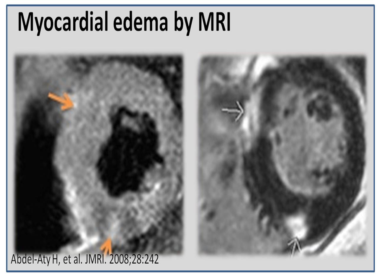
Genesis of edema in any tissue depends on local hydrostatic pressures, tissue resistive forces, osmotic balance, and cell membrane permeability. In the myocardium individual contribution of above factors are not known. Apart from total myocardial water content  , myocardial water logging depends upon the trans myocardial venous gradient and the coronary sinus exit pressure in right atrium.Technically ,any severe right heart failure should lead to myocardial congestion at least to some degree.Unlike the lungs , the myocardial edema fluid doesn’t produce crackle , (May cause S -3 gallop instead) . However ,we have modern technology to image water inside the myocardium. Yes, it is called proton / hydrogen imaging or simply called MRI .

This is especially evident In chronic kidney disease , where in the fractional water content within the myocardium is expected to increase further as the whole body is water logged.


myocardial edema by mri in ckd 2

We have seen time and again patients with CKD  improve in LV function immediately  after dialysis . It happens like a magic . The mechanism  is simple .The over-hydrated cardiac Interstitium  threatening to drown the myocytes  is promptly dehydrated by dialysis. This was my wild guess  until I came across this paper which proved the exact points.

myocardial edema in ckd chronic renal failure recovery of lv function after dialysis


Other situations  where myocardial edema may play a significant clinical Impact  (*Includes increased permeability of myocardial capillaries)

  1. Myocarditis
  2. Transplant rejection
  3. Stress cardiomyopathy (Takotsubo)
  4. Congestive heart failure
  5. Acute ischemic injury
  6. No Reflow situation after PCI

Final message

Myocardial interstitial edema in cardiac failure is a grossly under diagnosed  entity.  A water-logged myocardium is classical at least in CKD. We know it can severely compromise the LV function especially, the diastolic function that explains the all too common flash pulmonary edema in CKD.

The number of studies in this  topic (Myocardial Hydrology !)  is minuscule compared to other areas of research in cardiology literature.There is a need to involve both  Nephrologist and cardiologists to explore this curious concept of  dialysable  left ventricular mass in CKD/Cardio renal syndromes !

medical quotes new idea

One more area of research 

It is reasonable to believe,  cardiomegaly in cardiac failure  is primarily related to the increased end diastolic volumes .Still , we are not clear whether there is net increase in cardiac mass as the surface area of the heart increases with dilatation. (Even in DCM ? ) Whenever myocardial mass increases relative increase water is likely. Does the beneficial effect of diuretics in cardiac failure , and the restoration of  LV dimension is due to myocardial interstitial diuresis as well ?

Reference

myocardail edema water content congestion mri n myocardial diuresis interstitial

2.Andrés-Villarreal, M., Barba, I., Poncelas, M., Inserte, J., Rodriguez-Palomares, J., Pineda, V., & Garcia-Dorado, D. (2016). Measuring Water Distribution in the Heart: Preventing Edema Reduces Ischemia-Reperfusion Injury. Journal of the American Heart Association5(12), e003843. doi:10.1161/JAHA.116.003843

3.Cardiovascular magnetic resonance of myocardial edema using a short inversion time inversion recovery (STIR) black-blood technique: Diagnostic accuracy of visual and semi-quantitative assessment Darach O h-Ici, John P Ridgway, Titus Kuehne , Journal of Cardiovascular Magnetic Resonance 2012, 14:22  


Read Full Post »

The right ventricle  is considered as a docile cardiac chamber with passive filling and  emptying  properties .

This belief  was reinforced when Fontan  in early 1970s suggested a principle in the management of  cyanotic heart disease  when  the right side of the heart is underdeveloped. He  proved  RV can be by-passed safely , with  great veins  (IVC/SVC)  by  themselves  take care of filling the pulmonary circulation  without the need of RV pumping function.

While it is true for few complex cyanotic heart disease, largely this a misleading  concept. In clinical cardiology practice  ,sudden or non sudden  RV deaths happen every day in the form of . . .

  • RV Infarction
  • Acute RV dysfunction in massive pulmonary embolism
  • COPD with RV dysfunction
  • Most cases dilated cardiomypathy  the terminal event is due to RV  failure.

So , RV function can never be dispensable in day to day cardiac hemodynamics.

RV has some unique properties in terms of shape , size and  hemodynamics . We are getting more insights from  modern blood pool imaging by MRI , about  how the RV handles the blood volume .

We know RV has a unique shape  triangular ( partially  pyramidal ) . It can be inferred the RV cavity is formed by fusion of  many  eccentric spacial planes. We have always believed  RV handles the blood it receives from right atrium in a unique way .Now we are beginning to understand it .It is now documented the RV segregates the blood it receives into 4 components.


right ventricle physiology anatomy hemodynamics

It is curious  to know  RV inflow is connected to the outflow by an invisible   physiologic Bridge . About 44% of  blood traverse the RV in this fashion.


RVOT blood flow right ventricle

Note : RV blood flow preferentially enters the RVOT with out transiting RV body and apex.Image courtesy http://ajpheart.physiology.org/


Which is the most important part in RV ? (Among Inflow, Body, Apex, Out flow)

After reading this article it seems to me , the mechanical  function of RVOT could be most  vital. If it fails to handle the first increment  which  comes directly from  RV inflow, stasis  is likely in RV body and apex , elevating RVEDP and later promoting stasis leading to clinical events.

Clinical implication of this study

  • Differential dilatation RV chambers to pressure or volume  overload is observed .
  • We need to analyse why RV dilates in some   but   goes for hypertrophy in others when confronted with pressure overload (VPS vs PAH)
  • RV apical clot in restrictive cardiomyopathy  is a direct consequence of stasis  of blood  in RV apical zone .
  • RVOT pacing  may have a hemodynamic advantage  over RV apical pacing  . However , for anatomical reasons RV apical pacing  is  far safer than RVOT pacing where the lead  is subjected to constant life long strain due to this busy RV inflow to outflow express  high way !

Final message

Traditionally we have labeled  RV  as a  passive venous chamber .It is clearly a misnomer.It  has to handle both the venous and pumping function beat to beat with precision  without  back log .Obviously ,  RV has to think and work  more than it’s  big brother !

Reference

I wonder , if  there is  any other site other than APS . . . to  find crucial  answers in cardiac physiology  !


Right ventricle physiology blood flow  3d 4d analysisAfter thought

  • There is huge gap between physiologists  who work in research labs and the physicians at bed side .
  • I appeal all young cardiologists  to visit  APS  once in a while ,between your busy cath lab schedule and help narrow this gap.
  • Without understanding the physiology properly how are we going to intervene the pathology ?


Read Full Post »

Epicardial fat is increasingly recognised to be a coronary risk factor.
Methods to measure and quantify epicardial fat has not been standardised.
Nomenclature issues
There is some confusion in the literature regarding  the definition epicardial, pericardial , paracardiac fat etc.
Epicardial fat is the true visceral fat located in proximity with myocardium .Epicardial fat shares the same blood supply as adjacent myocardium and also show  para-crine functions.This is the risky fat that is metabolically active.
Paracardiac fat  is  the fat deposited outside the parietal  pericardium .It is also referred to as  thoracic fat .They carry less risk and behave more like somatic non visceral fat.
Pericardial fat is a loose terminology .Many  use epicardial and pericardial fat interchangibly .It is to be realised , fat either deposit inside the visceral pericardium or outside the parietal pericardium. It almost never deposits  within the pericardial space.

* Currently it is better to use  only two terms .Epicardial fat and  thoracic fat .Other terminology should be avoided.

How to measure and quantify epicardial fat ?
Distribution of fat over heart is not uniform .Though it appears a random distribution there app eras to be a pattern to it .It is largely loaded anterior surface of the heart, over the AV grooves, invariably hugging the coronary arteries and great vessel origins.Fat deposition posterior to heart occurs in gross obesity.
There is no accurate way to quantify it . A rough estimate can be arrived . The following investigations are useful.
  • Echo
  • Multi-slice CT  Scan
  • MRI
Epicardial fat pad thickness by M-Mode /2D  is simplest,  but considered crudest modality to quantify.  .Still , it is a useful screening test. In our experience , if there is distinct anterior fat pad > 5mm it is likely to be significant.
However, epicardial fat volume is ideal method to quantify it.(Read below)

Which part of cardiac cycle it is measured ?

Since epicardial  fat is a compressible structure , in diastole the myocardial mass encroaches the epicardium  any imaging modality the distinction between epicardial and para-cardiac fat  tends to attenuated.

Even MRI images obtained with  diastolic  gating ,  may  not be reliable .End systolic frames are now considered ideal.

(J Am Coll Cardiol. 2011 Oct 4;58(15):1640;Distinction of “fat around the heart”.Kaushik M, Reddy YM.)

What is the normal  epi-cardial  fat volume ?

There is no standard normal.In one large study from  Boston in people  with a BMI between 25-30 the  mean epi-cardial fat volume was 125 cubic cms

normal epicardial fat volume

(*Intrathoraic fat  same as para-cardiac fat located outside parietal pericardium )

How to differentiate fat from pericardial fluid ?

It can be a very difficult issue especially  when you expect both as in hypothyroidism .It needs proper  gain settings and interpretation.

Here is case report of how epicardial fat was mistken for PE even in TEE

Can we measure  the  net mass of  cardiac  fat , does it compress the heart ?

When we say average heart weighs 300 grams it includes fat embedded in it. I don’t think it is possible to measure the fat mass ,but clinically it may be important.Read this .Can-epicardial-fat-constrict-the-heart-an-authentic-yes-from-ulsan-korea ?

Final message

Estimation of epicardial fat volume is largely a research tool.In grossly obese patients it may  have some clinical utility.Increased anterior fat pad in routine 2D echo is an useful  screening test. However, it requires MRI/CT imaging for accurate quantification.

References

how to measure epicardial fat framingham study
2. Marwan J M, Achenbach .S  Cardiovasc Comput Tomogr. .Quantification of epicardial fat by computed tomography: why, when and how?2013 Jan-Feb;7(1):3-10
3. Sarin S, Wenger C, Marwaha A, Clinical significance of epicardial fat measured using cardiac multislice computed tomography. Am J Cardiol. 2008 Sep 15;102(6):767-71.

Read Full Post »

While many of us are preoccupied with wires and balloons ,( coronary  myopia ! )  , our radiology  colleagues are making rapid strides . Let us spend some  time  to understand  how  the myocardial segments  are inflicted the  final insult . We need to realize , there is a pattern  to  this myocardial  end game of scarring and fibrosis.

MRI is the  gold standard to assess the myocardial architecture . It has a role in both assessing the anatomy , function  , perfusion and viability .

how to differentiate ischemic dcm from idiopathic dcm myocardial scar epicardial transmural

  • LV function is assessed  by cine MRI
  • Viability  stud by  delayed enhancement MRI (DEMRI , also called as  LGE- Late Gadolinum enhancement  )
  • Myocardial scar best assessed by DEMRI*
* Why do you require DEMRI to identify scar ?
One can detect scars in plain MRI but contrasts make it better .Hence delayed enhancement in by DEMRI is used  to detect scars.
Is it ischemic  DCM or Non ischemic DCM ?  ( That is the question we commonly ask  
We rely too much on CAG anatomy for this. It can be misleading. Cine MRI with DEMRI  gives the answer straightway with high degree of accuracy  .  CAG is required in all  ,  but if it is normal , or  has insignificant lesions  , the dilemma  of ischemic DCM would continue !)
**Note ,there is one   simple algorithm proposed by the author   to  differentiate  Ischemic DCM from Idiopathic DCM  without MRI – Click here to  Link
Following  scar patterns in DEMRI help us to arrive a diagnosis.
Favors Non ischemic  DCM
  • Mid myocardial scar
  • Epicardial scars
  • Global sub-endocardial scars
  • No scar(Ironically if  no delayed  hyper-enhancement is noted it is likely to be non Ischemic DCM )
Favors ischemic DCM
  1. Regional transmural scars
  2. Localised sub-endocardial  scars
* Ischemic DCM will always involve subendocardium as ischemic wave front goes from sub-endo to epicardium.
examples for Non Ischemic DCM
  • Amyloidosis (Can be restrictive as well )
  • Chagas
  • Fabrys

Why is  scar localisation and Quantification important ?

Apart from differentiating various cardiomyopathies  it has  few clinical implication .

  • Since scar indicates irreversible damage , if extensive  it will  argue  against any re-vascularisation .
  • Scar location becomes vital if we plan CRT .It will be futile  to place a CRT lead over a scar.
  • Scars are often  form a macro re-entrant circuits for VT .Help us localize or zeroing in VT focus.
  • Scar quantification is helpful risk stratification of patients  with HOCM .and their family.
Final message
Myocardial scar location and quantification  is the new mantra in a  patient with dilated heart with cardiac failure.
It may be more important than even a coronary angiogram .MRI  will prevail over   any of the available echocardiogram modalities to assess the scar pattern.
Reference
myocardial scarring mri

Read Full Post »