Feeds:
Posts
Comments

Archive for the ‘myocardial disease’ Category

The right ventricle  is considered as a docile cardiac chamber with passive filling and  emptying  properties .

This belief  was reinforced when Fontan  in early 1970s suggested a principle in the management of  cyanotic heart disease  when  the right side of the heart is underdeveloped. He  proved  RV can be by-passed safely , with  great veins  (IVC/SVC)  by  themselves  take care of filling the pulmonary circulation  without the need of RV pumping function.

While it is true for few complex cyanotic heart disease, largely this a misleading  concept. In clinical cardiology practice  ,sudden or non sudden  RV deaths happen every day in the form of . . .

  • RV Infarction
  • Acute RV dysfunction in massive pulmonary embolism
  • COPD with RV dysfunction
  • Most cases dilated cardiomypathy  the terminal event is due to RV  failure.

So , RV function can never be dispensable in day to day cardiac hemodynamics.

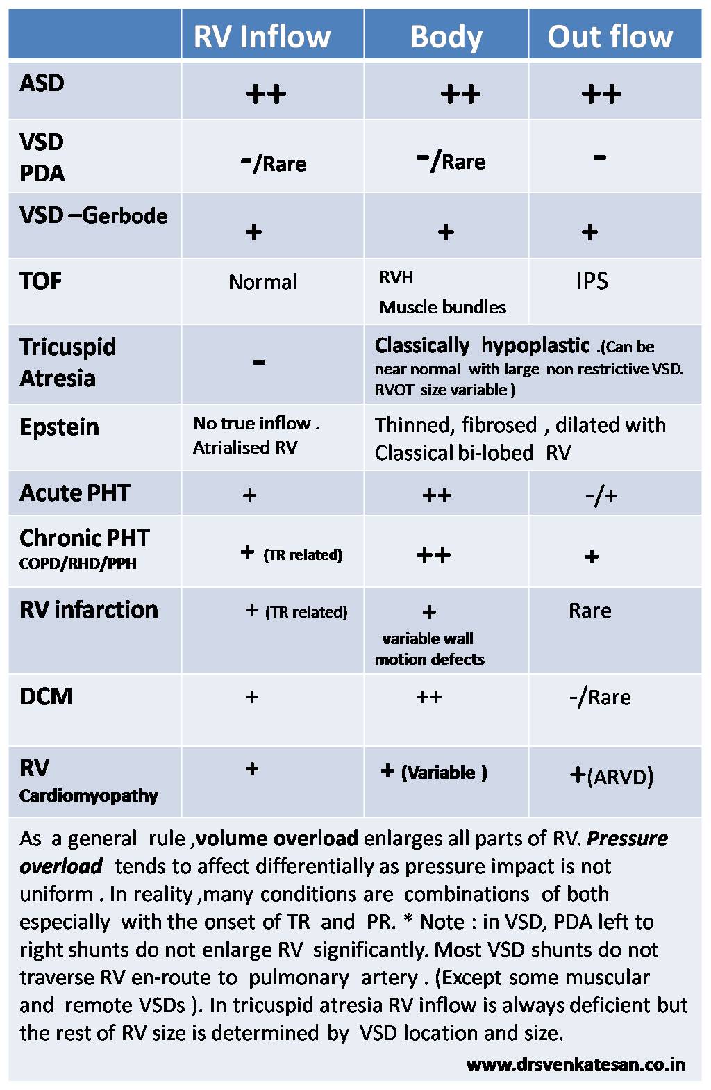
RV has some unique properties in terms of shape , size and  hemodynamics . We are getting more insights from  modern blood pool imaging by MRI , about  how the RV handles the blood volume .

We know RV has a unique shape  triangular ( partially  pyramidal ) . It can be inferred the RV cavity is formed by fusion of  many  eccentric spacial planes. We have always believed  RV handles the blood it receives from right atrium in a unique way .Now we are beginning to understand it .It is now documented the RV segregates the blood it receives into 4 components.


right ventricle physiology anatomy hemodynamics

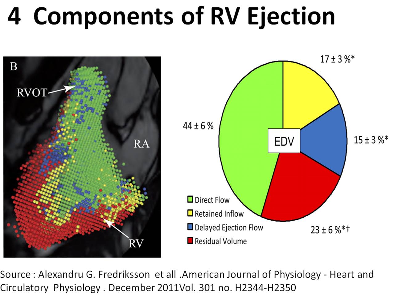
It is curious  to know  RV inflow is connected to the outflow by an invisible   physiologic Bridge . About 44% of  blood traverse the RV in this fashion.


RVOT blood flow right ventricle

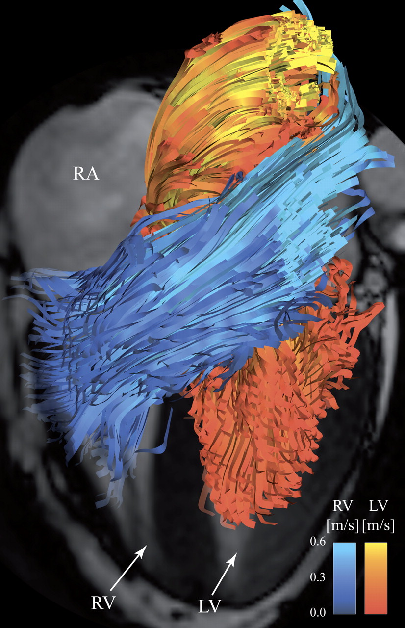
Note : RV blood flow preferentially enters the RVOT with out transiting RV body and apex.Image courtesy http://ajpheart.physiology.org/


Which is the most important part in RV ? (Among Inflow, Body, Apex, Out flow)

After reading this article it seems to me , the mechanical  function of RVOT could be most  vital. If it fails to handle the first increment  which  comes directly from  RV inflow, stasis  is likely in RV body and apex , elevating RVEDP and later promoting stasis leading to clinical events.

Clinical implication of this study

  • Differential dilatation RV chambers to pressure or volume  overload is observed .
  • We need to analyse why RV dilates in some   but   goes for hypertrophy in others when confronted with pressure overload (VPS vs PAH)
  • RV apical clot in restrictive cardiomyopathy  is a direct consequence of stasis  of blood  in RV apical zone .
  • RVOT pacing  may have a hemodynamic advantage  over RV apical pacing  . However , for anatomical reasons RV apical pacing  is  far safer than RVOT pacing where the lead  is subjected to constant life long strain due to this busy RV inflow to outflow express  high way !

Final message

Traditionally we have labeled  RV  as a  passive venous chamber .It is clearly a misnomer.It  has to handle both the venous and pumping function beat to beat with precision  without  back log .Obviously ,  RV has to think and work  more than it’s  big brother !

Reference

I wonder , if  there is  any other site other than APS . . . to  find crucial  answers in cardiac physiology  !


Right ventricle physiology blood flow  3d 4d analysisAfter thought

  • There is huge gap between physiologists  who work in research labs and the physicians at bed side .
  • I appeal all young cardiologists  to visit  APS  once in a while ,between your busy cath lab schedule and help narrow this gap.
  • Without understanding the physiology properly how are we going to intervene the pathology ?


Read Full Post »

Right ventricle is a passive venous component of the heart .It simply acts a  transit pump for blood to reach the lungs.

It  is true  , RV is dispensable in many complex congenital heart disease as we  can connect the great veins directly into the pulmonary artery  by  Fontan , Glean and it’s clones  bye passing this chamber . Still , by no means the importance of this chamber is to be underestimated.  RV dysfunction and failure  is the key to survival  many  disorders.RV shock is is cause of sudden cardiac death in acute pulmonary embolism and RV infarction .

RV is an unique muscular chamber .It is more of a triangular shape. It has  three different parts connected by three different angle .There is no true  apex  for RV , it is   connected  to Inflow and outflow in peculiar fashion .

In the  following table I have  tired to  describe  of how different parts of RV  behave in various disorders.

what is the morphology of  RV enlargement RV inflow outflow body sinus portion of RV

Read Full Post »

While many of us are preoccupied with wires and balloons ,( coronary  myopia ! )  , our radiology  colleagues are making rapid strides . Let us spend some  time  to understand  how  the myocardial segments  are inflicted the  final insult . We need to realize , there is a pattern  to  this myocardial  end game of scarring and fibrosis.

MRI is the  gold standard to assess the myocardial architecture . It has a role in both assessing the anatomy , function  , perfusion and viability .

how to differentiate ischemic dcm from idiopathic dcm myocardial scar epicardial transmural

  • LV function is assessed  by cine MRI
  • Viability  stud by  delayed enhancement MRI (DEMRI , also called as  LGE- Late Gadolinum enhancement  )
  • Myocardial scar best assessed by DEMRI*
* Why do you require DEMRI to identify scar ?
One can detect scars in plain MRI but contrasts make it better .Hence delayed enhancement in by DEMRI is used  to detect scars.
Is it ischemic  DCM or Non ischemic DCM ?  ( That is the question we commonly ask  
We rely too much on CAG anatomy for this. It can be misleading. Cine MRI with DEMRI  gives the answer straightway with high degree of accuracy  .  CAG is required in all  ,  but if it is normal , or  has insignificant lesions  , the dilemma  of ischemic DCM would continue !)
**Note ,there is one   simple algorithm proposed by the author   to  differentiate  Ischemic DCM from Idiopathic DCM  without MRI – Click here to  Link
Following  scar patterns in DEMRI help us to arrive a diagnosis.
Favors Non ischemic  DCM
  • Mid myocardial scar
  • Epicardial scars
  • Global sub-endocardial scars
  • No scar(Ironically if  no delayed  hyper-enhancement is noted it is likely to be non Ischemic DCM )
Favors ischemic DCM
  1. Regional transmural scars
  2. Localised sub-endocardial  scars
* Ischemic DCM will always involve subendocardium as ischemic wave front goes from sub-endo to epicardium.
examples for Non Ischemic DCM
  • Amyloidosis (Can be restrictive as well )
  • Chagas
  • Fabrys

Why is  scar localisation and Quantification important ?

Apart from differentiating various cardiomyopathies  it has  few clinical implication .

  • Since scar indicates irreversible damage , if extensive  it will  argue  against any re-vascularisation .
  • Scar location becomes vital if we plan CRT .It will be futile  to place a CRT lead over a scar.
  • Scars are often  form a macro re-entrant circuits for VT .Help us localize or zeroing in VT focus.
  • Scar quantification is helpful risk stratification of patients  with HOCM .and their family.
Final message
Myocardial scar location and quantification  is the new mantra in a  patient with dilated heart with cardiac failure.
It may be more important than even a coronary angiogram .MRI  will prevail over   any of the available echocardiogram modalities to assess the scar pattern.
Reference
myocardial scarring mri

Read Full Post »

Takotsubo cardiomyopathy is an  unusual response  of the left ventricle to extreme emotional stress .The catecholamine  surge  has  a profound stunning  effect of LV apex  and  a paradoxical hypercontractility of basal LV.

The exact mechanism is not clear , Following factors may contribute.

  1. Multi-vessel coronary artery spasm,
  2. Cardiac microvascular dysfunction.
  3. Abnormal myocardial fatty acid metabolism,
  4. Reperfusion injury  after an ACS *

However , the most accepted mechanism is Endogenous catecholamine-induced myocardial stunning and microinfarction

Why is LV apex alone affected  ?

The adrenergic receptor distribution is high in LV apex .They are exposed to high concentration  and gets stunned easily . Basal LV has less adrenergic innervation  , so it shows less of catecholamine toxicity , instead  it exhibits.  hyper-contractile mode. However, this rule is not absolute.

One more suggestion was apical balloons correlated with wrap around LAD.(Báñez B et all 2004)

what is the mechanism of apical ballooning syndrome 2 takotsubo cardiomyopathy

Image courtesy Circulation December 16/23, 2008 vol. 118 no. 25 2754-2762

*Some consider ACS should never be  linked to  Takotsubo.But it is not easy to differentiate.(Carrillo JACC 2009(Kosuge JACC 2010)

Reference from this site

A link to related article -Ischemic Takosubo  from this site .

https://drsvenkatesan.wordpress.com/2012/05/27/in-extreme-mental-stress-left-ventricle-becomes-a-banana/

Reference from other journals
what is the mechanism of apical ballooning syndrome takotsubo cardiomyopathy

1.Báñez B, Navarro F, Farré J et al. (2004). Tako-tsubo syndrome associated with a long course of the left anterior descending coronary artery along the apical diaphragmatic surface of the left ventricle.]”. Revista española de cardiología (in Spanish; Castilian) 57 (3): 209–16

2.Carrillo A, Fiol M, Garcia-Niebla J, Bayes de Luna A. Electrocardiographic differential diagnosis between Takotsubo syndrome and distal occlusion of LAD is not easy. J Am Coll Cardiol. Nov 2 2010;56(19

3.Dorfman TA, Iskandrian AE. Takotsubo cardiomyopathy: State-of-the-art review. J Nucl Cardiol. Jan-Feb 2009;16(1):122-34

4.Kosuge M, Ebina T, Hibi K, Morita S, Okuda J, Iwahashi N. Simple and accurate electrocardiographic criteria to differentiate takotsubo cardiomyopathy from anterior acute myocardial infarction. J Am Coll Cardiol. 2010;55(22):2514–6. doi: 10.1016/j.jacc.2009.12.059.

how to differentiate takotsubo cardiomyopathy from anterior stemi

//

Read Full Post »

The other day my fellow got a call  from surgical ward for emergency ECG opinion for a  suspected Inferior MI .It later turned out to be an acute cholecystitis.

One of the important  anatomical mis-perception  among physicians ,  is to consider  inferior, posterior  and diaphragmatic surface  of heart  as separate entities .They are all  closely linked.In fact, they  more often  mean  the same  anatomical zones !

Heart is a dynamic suspended organ within the middle mediastinum .It  can assume a vertical or horizontal position due to number of surrounding anatomical  and physiological factors. (Diaphragm, Lung , being  important ).The ratio of intra thoracic vs Intra abdominal  volume  &  pressure determine whether the posterior surface of the heart is going to face the back of chest  or simply sit and  rest on the diaphragm .We know a horizontal heart is likely to inscribe q waves  in inferior leads .

acute abdomen diaphragm inferior wall mi cholecystitis pacreatitis

Courtesy : Basic image source from digitallab3d

The  diaphragm can be termed as an  anatomical causeway , that isolates   thorax  from the  abdominal  cavity .Close encounters between the organs separated by this delicate biological  membrane is  always possible .This is especially true for electrical signals  which show little  respect for anatomical barriers .

This is the reason there are too  many abdominal conditions that mimic  inferior MI during a painful  emergency (and vice versa  when inferior  MI mimics  acute abdomen .) In  our  department , we   have witnessed  the following conditions mimicking Infero-posterior ACS.

  1. Acute ascites with polyserositis
  2. Gross obesity with APD
  3. Posterior fat pad ( Necrosis ?)
  4. Thickened pericardium
  5. Minimal posterior pericardial effusion
  6. Diaphragmatic pleurits
  7. Esophageal spasm
  8. Fundal air  trapping and ballooning after a heavy meal !
  9. Acute duodenal ulcer perforation ( With gas under diapharam causing q waves)
  10. Acute cholecystits
  11. Diphragmatic hernia
  12. Achalasia cardia
  13. Pancreatitis

Final message

Do not rush to make a diagnosis of inferior wall MI when  you encounter inferior q waves  with  or without ST /T changes , especially  when the symptoms are atypical .

Read Full Post »

The popular clinical  entity Idiopathic dilated cardiomyopathy   is often a  dust-bin diagnosis” . The fact is the word   idiopathic simply reflects  our ignorance.

For God nothing is idiopathic . . . he knows how each and every cell  would   behave  .

so , when a patient presents with progressive dilatation and  heart failure refractory to all medical  therapy he is termed as idiopathic and posted for heart transplantation. And only later , we realize the whole thing is due  a  terrible form of reversible  DCM  . That is  pheochromocytoma  induced DCM , which recurred again in the   transplanted  heart.  What a  costly  Ignorance ?

pheochromocytoma and dilated cardiomyopathy reversible dcm tachycardic

Image courtesy and source http://www.dreamstime.com.

Is sub- clinical pheo like situations rampant ?

We know  that  high levels of both epi and nor- epinephrine circulate  in cardiac failure . We presume it  to be a secondary effect .

How can  we  so sure about it ?  There  is a distinct  possibility  of   adrenal gland hyperfunction  and hyperplasia in all DCMs (Idiopathic or ischemic ! )  The dramatic beneficial effects of beta blockers in cardiac failure  will vouch for it .

So , It remains a fertile filed for the youngsters to explore . . . the hyper  adrenergic mediated reversible component of any cardiomyopathy and cardiac failure .

Final message

The default  approach  in any  patient with progressive / refractory cardiac failure   should  be  ,  to consider  whether they fit into  any form of reversible myocardial disease  .  What is idiopathic in remote clinic of   your distant  country side  may be  well recognized secondary cardiomyopathy . The irony is , even sophisticated university hospitals many times miss the true etiology as in the above case report .

                                  So, the term Idiopathic  dilated  cardiomyopathy  (iDCM )  may  aptly be named as  Ignorant  forms  of  DCM  , with an  attractive  abbreviation    . . .   iDCM

Reference

1.J Surg Educ. 2009 Mar-Apr;66(2):96-101. doi: 10.1016/j.jsurg.2008.11.004. Pheochromocytoma presenting as acute severe congestive heart failure, dilated cardiomyopathy, and severe mitral valvular regurgitation: a case report and review of the literature.

2.Kelley SR, Goel TK, Smith JM.Prog Cardiovasc Nurs. 2005 Summer;20(3):117-9. Pheochromocytoma presenting as heart failure.

3.Pheochromocytoma   masquerading as a cardiomyopathy. Garcia R, Jennings JM.  Am J Cardiol. 1972 Apr;29(4):568-71.

4.  http://www.ncbi.nlm.nih.gov/pmc/articles/PMC1894695/pdf/20070600s00025p244.pdf

pheochromocytoma and dcm dilated cardiomyopathy .catecholamine induced dcm tachycardiac cardiomyopathy

5. http://downloads.hindawi.com/crim/medicine/2011/596354.pdf

pheochromocytoma and dcm dilated cardiomyopathy .catecholamine induced dcm tachycardiac adrenal cardiomyopathy


Read Full Post »

Doppler Mitral Inflow velocity profile   is the key to  assess LV diastolic function . The ratio between  E and A has become most popular parameter .

In the absence of atrial contraction what shall we do ?

The answer is simple .  We have 2 D parameters of LV diastolic function.

LA dimension ( > 30 % basal dimension which is  usually >  4 cm  ) is a most specific marker of diastolic dysfunction in the absence of   mitral regurgitation or stenosis.

The only available  velocity E wave profile  can help .A short  E deceleration time in a short cycle  would suggest  significant diastolic dysfunction.High amplitude   E  wave  > 2  M/sec in the absence of MR  will suggest diastolic dysfunction .

Curiously  ,   it can be  assumed    an episode of   lone AF  per-se   ,  be an indicator of diastolic stress for the left atrium .

After all ,  why should a person all of a sudden develop an episode of AF .(Hypoxia, Ischemia ,  excluded )

Other parameters.

Mitral annular velocities / E propagation velocity   / E/E’  are other tissue Doppler parameters  can be used.

Pulmonary venous flow velocity is  largely not useful  (Since A reversal does not occur )

Read Full Post »

A  patient who presents with predominantly right  heart failure  is  an interesting clinical challenge . Constrictive pericarditis (CP)  remains  a popular diagnosis in this setting. However  in the bed side clinical  examination (and in cardiology Board exams )  the following  differential diagnoses are  to be  considered .( And ruled out one by one)

  1. Restrictive cardiomyopathy* especially Right  sided .In India endo myocardial fibrosis tops the list
  2. Primary Tricuspid valve disease( Tricuspid stenosis / Carcinoid etc)
  3. Chronic cor-pulmonale in terminal RV failure
  4. Silent Mitral stenosis with right heart failure
  5. Ebstein anomaly
  6. Severe forms of valvular pulmonary stenosis with RV dysfunction
  7. SVC obstruction
  8. Cirrhosis of liver
  9. Porto pulmonary hypertension

( The list is not complete , readers may contribute )

Bed side clues

  • Remember  a deep “y” descent  is  the bed side counter part of   Square root sign  recorded by  invasive RV pressure study
  • Similarly , pericardial knock is the auditory   equivalent (You hear the square root !  . . .yes  )as the ventricle thuds the rigid thickened pericardial shell in very early diastole !)
  • Pulsus paradoxus and kussmal sign can occur in both CP and RCM.
  • If a good LV apex , is  palpated it  goes against CP .
  • Please be reminded , even restrictive cardiomyopathy  will ultimately dilate their chamber pre-terminal and clinical features may be confounded with that of DCM.
  • Silent heart would suggest CP.
  • AV valve regurgitation would favor RCM
  • Features of  Pulmonary hypertension will help confirm Mitral valve disease , Cor pulmonale,
  • Deep  “y”descents  are against  any form of  Tricuspid stenosis.
  • Opening snap of mitral valve is to be distinguished from pericardial knock.( Opening snap high pitched  and occur later than   pericardial knock in diastole   , best heard in expiration )
  • Cirrhosis liver with hypo- proteinimic   fluid retention is  a traditionally close mimicker  .It  may be ruled out by the careful history taking as exertional dyspnea is an exception , if  at all , it is a very late event  in cirrhosis.
  • The issue gets further weird   as chronic constriction can lead on to chronic congestive liver and cardiac cirrhosis .
  • Severe  forms of constriction can invade the myocardium and result in features of myocardial dysfunction .It is more common than we recognise.

How to confirm ?

Following should be performed in that order

  • ECG
  • X -Ray
  • Echocardiogram
  • CT scan
  • MRI

*Cath study is no longer done (Only for academic purpose )

Final message

Even in this era of sophisticated  medical  imaging  , clinical examination  remains the key . One should  realise the importance  of  meticulous  clinical history  ,  sequential examination and interpretation .It  will   “rule out  or rule in”  majority of  cardiac disorders .

The hi tech imaging  modalities should be used only to confirm , risk stratify and  plan management . If you skip the clinical  part , one  may still arrive at a correct  diagnosis  but there is  high chances of erring in  management.

(Cardiac pearls lie in the bed side not in cath labs !   Here is  one such pearl  . Not every constriction  require surgery !

Please note about 20 % of constrictive pericarditis are  transient !)

Read Full Post »

Pulmonary  arterial hypertension (PAH ) is  an uncommon manifestation of dilated cardiomyopathy .While pulmonary venous hypertension of some degree is expected in most patients with DCM,  it is rare for these patients to go for severe arterial hypertension.

The reason for this may be the  natural history of DCM do not allow these patients to live that longer to manifest severe PAH.  Still ,  we encounter this problem  atleast in tertiary hospitals. Presence of moderate to severe PAH (> 50mm peak PAP) is a sinister sign in  DCM. They not only do badly , they also make  the transplant outcome dismal .

What causes this severe   PAH in DCM ?  The following observations are made in our institute .

Now we know , isolated  systolic dysfunction is  rarely associated with PAH  .It is the presence of  LV diastolic dysfunction (Often restrictive )  that raises the pulmonary pressures.  PAH of DCM is rarely progressive.

One important suggestion is the DCMs  which are associated with  severe  PAH may indeed represent  late stages of RCM , when the LV begin to dilate.

Associated mitral regurgitation   contributes  to PAH

Atrial fibrillation has a significant impact on elevating  pulmonary  venous and arterial  pressures in DCM.

Hypoxic PAH can occur in any medical situation  in susceptible population . DCM is no exception

For some reason  idiopathic DCM is more often result in PAH than ischemic DCM . (Is that possibel , some form of  idiopathic   PAH and DCM are etiologically  related ?)

Further , the positive inotropic agents when liberally used will worsen the diastolic  properties of LV.

Finally involvement of  right ventricle  in the cardiomyopathy  process can have an ameliorating effect on PAH.  A good RV function is essential to lift the PA systolic pressure. If RV failure is causing a low PAP , do not be happy .It simply means RV is going to  say  good bye  . . .  for the final  time !

How to manage PAH in DCM ?

There is no specific management strategy .

We do not know yet  whether Sildenafil ,  Bosentan, and Epoprostenol  have any role in this  form of  PAH. These are all basically vasodilators. It’s use in DCM is vested with a risk of  catastrophic hypotension . Of course ,  we do have a role for balanced vasodilators in cardiac failure .(As most of these patients would be already on adequate ACEI )

Presence of PAH should be considered as an independent indication for anticoagulants as in situ  pulmonary thrombus is common.

The effect of  cardiac resynchronisation therapy in reducing the PAH of DCM is not convincing.

Final message

PAH  in DCM is an unwelcome development. It makes the situation  tough .  The mechanisms are diverse  .Understanding the mechanism would help us deal  this problem better .  Conventional anti failure treatment may help  ,but  it is wiser to try  reserve drugs.

Read Full Post »

Angina occurring at night is relatively uncommon . It is  still  more rare  for angina to occur exclusively at night (With a possible exclusion of  syphilitic aortits with AR !) The underlying conditions and mechanism  of nocturnal angina  are largely unexplored. In most clinical situations nocturnal angina  is  associated with day time angina as well .

Various mechanisms are proposed

  • It is primarily due to  increased demand  (Holter monitoring has documented  brief bursts  of  HR acceleration  just before  nocturnal angina with  manifest  ST depression )
  • Increased demand  during  REM sleep .
  • Dreams  related adrenergic surge has been implicated.
  • Rarely it is due to supply side defect .
  • Coronary vaso-spasm ( Mostly  in a pre-exisiting lesion )
  • It could  simply  represent  paroxysmal nocturnal dyspnea (pnd)
  • Sleep apnea can precipitate angina  ( Ironically angina occur during   re-breathing  phase )
  • Altered hemo-rheology
  • Nocturnal gap in anti anginal medication *

* May be more  common than we realise.

Cardio vascular hemo-dynamics  at night

If we  believe , sleep is  the great relaxation , and the heart   would enjoy the   “night time”   we  are absolutely wrong . Even in sleep ,  heart has to pump the same 250 ml of blood every minute. Of course , the sleeping heart rate slows down considerably , still  it is interspersed with spikes of activity.  When the heart  rate  slows down  , diastole is prolonged , coronary blood flow  is expected to be copious  unless there is critical CAD.

                                      We  know , sleep is not a passive process  , even as the  autonomic nervous system takes complete control over the  somatic  system .The true colors of  our delicate autonomic system will come to light only during sleep.The muscle tone ,  the sympathetic drive fluctuates according  a pre-set degree . Dreams and REM sleep disturbance can have considerable impact on the sympathetic nerve terminals which ooze  catecholanines  .

Sudden awakening  from  early sleep  is vested with a risk of dangerous   spikes of adrenaline release  .This becomes especially  important in compromised coronary circulation .In fact , this is commonest  sleep -awake  sequence  in patients with nocturnal angina.

Silent ischemia at night

It is curious to note 24 hour Holter  monitoring  reveals  most episodes of ST depression at night are silent. There must be a  specific pain threshold above which a patient awakens  with angina.   The  available  studies   do not  answer this issue   and are not perfect  . We have no way to find  true   silent ischemia  during  sleep.(PET scan in thalamus ?)

Nocturnal angina  in  Aortic regurgitation

Aortic regurgitation  has special relationship with dusk  .For angina to occur AR must be severe and usually isolated .

  • Prolonged diastole at night   -Regurgitation time is prolonged .
  • Dilated LV . Increased  LV mass .Increased demand.
  • Raised LVEDP due high wall stress.
  • Diastolic coronary stealing . Venturi  effect of AR jet

Nocturnal Angina : Is it stable or unstable ?

Most  consider it   as a type of stable angina .Now ,we have reasons to suspect  it could a  marker of unstable angina as it is an  expression of rest angina .

Nocturnal angina vs nocturnal STEMI

How often an episode of nocturnal angina end up in STEMI ?

STEMI is more  common in the early hours of the day and is more related to the hemo-rheological factors  . Please  note ,  STEMI is  a supply side defect  while most episodes of nocturnal angina is due to  demand ischemia . However  it is possible   nocturnal angina episode can precipitate STEMI if  vasospasm is  the underlying mechanism  and if  it is prolonged can trigger thrombosis.

We do not know the answer as yet.

Nocturnal  Angina : Can  it  be PND equivalent ?

Paroxysmal nocturnal dyspnea (PND)  is a classic manifestation of  episodic LVF.  We  know dyspnea can be an anginal  equivalent.  What prevents angina  to  become a  dyspnea  equivalent ! ( Especially the nocturnal ones ,   since the  mechanism  of generation of PND   are very similar  to the  genesis of  angina ). It is distinctly possible  one  may  be mistaken for the  other .  Both occur when  sudden hyper-adrenergic  state  is evoked  which demands   high MVO2 .  An  ischemic heart has every reason to  respond with  angina  .

It is well known  ischemia can result in transient diastolic dysfunction and  elevate the PCWP simultaneously  and PND  would be  the sequel .  When we analysed the  nocturnal calls (  Our fellows ,  do get lots of  such calls from   general wards  at night ),  many  patients with LV dysfunction  who complained  of  classic  chest pain  had  some degree of  dyspnea  and few crackles over lung base as well  .

Nocturnal angina and obstructive sleep apnea

The incidence of nocturnal angina is more common in obese population with obstructive sleep apnea.

The reason is two-fold

1 .Hypoxia mediated

2. Inappropriate tachycardia during recovery phase

Is there any  specific management strategies  to control nocturnal  angina ?

  • General  principles apply .
  • The timing of  anti anginal medication can be adjusted . Long acting preparations taken  in  morning hours to be avoided as they do not cover night time.
  • A calcium   channel blocker   (with optional  beta blocker )  at night may be the best bet to prevent nocturnal ischemia.
  • Dinner to sleep time to be widened.
  • Heavy diet at night to be avoided.
  • Sedatives role is not clear. (Can Diazepam suppress nocturnal angina ?  If so . . .  we  can call it as anti anginal drug  . . .  is isn’t )

References

http://www.thelancet.com/journals/lancet/article/PIIS0140-6736%2884%2991693-3/abstract

http://www.ncbi.nlm.nih.gov/pubmed/8419815

http://www.nejm.org/doi/pdf/10.1056/NEJM199302043280502

  Obstructive Sleep apnea  and  Angina 1  : http://www.ncbi.nlm.nih.gov/pubmed/7715342

 Obstructive sleep apnea and Angina 2 http://content.onlinejacc.org/cgi/reprint/34/6/1744.pdf

Read Full Post »

Older Posts »