Feeds:
Posts
Comments

Posts Tagged ‘brugada syndrome’

A 5-minute session: Answers are my own. Please cross-check.

1. Is Brugada syndrome clinical or ECG diagnosis?

Always clinical. Never get confused on this.

2. Spontaneous type 1 vs Induced Type 1 (from type 2) which carries more risk?

Both are risky since they are close cousins. But, spontaneous type 1 is the dreaded devil. 

3. Is Brugada primarily a defect of myocardial depolarization or repolarisation?

Not clear. Often in both. In fact a mismatch between them. (Don’t ask how Na+ Channel defect affects repolarisation !)

4. Is Brugada VT is monomorphic, polymorphic?

Both. What determines morphology is not clear though. (All de-nova monomorphic VT will degenerate to polymorphic en route to cardiac arrest)

5. Should  Fever induced Brugada pattern be investigated further?

Better, it is not to be reported in ECG. May not be important in the majority if there is no adverse family history. (If the patient is well educated and afflicted  by Dr.Google and cardiologists can’t escape from ordering sophisticated tests)     

6. What is the overlap between ERS and Brugada?

It is all about the Idiosyncrasy of the K+ channel phenotypes ( Transmural dispersion heterogeneity )  

7. Is a benign Brugada better than a malignant ERS?

Yes, it would seem so. (Inferior or Infero -lateral ERS prone for primary VF in case they develop ischemic / ? also non-ischemic stress)

8. How important is the link between Brugada and Long QT 3 syndrome?

A rare entity, but It is double jeopardy for VT risk. The entire action potential width is vulnerable right from phase 0 to 3 or 4 A case report Sandhu A Clin Case Rep. 2017;5(8):1315-1319.

9. Is Amiodarone really contraindicated in VT?

Not really. Though Amiodarone unmasks Brugada, it can still be used during episodes of VT in patients with manifest or unmanifest Brugada. Maybe in Long QT 3 overlap, it may perpetuate the VT.

10. How important is the structural myocardial defect in Brugada?

Not important in the majority. Though localized RVOT abnormalities are noted in some..RV abaltion can be succesful in odd case.

11. What happens to the ST segment in Brugada during exercise stress?

ST-segment may normalize in some. A stress test can help to risk stratify.  Subramanian M, J Cardiovasc Electrophysiol. 2017 Jun;28(6):677-683.0

12. Which drug is probably best for Brugada as of now?

Quininde , A fairly specific blocker of Ito current. However, it needs to be used diligently. Management of Brugada Syndrome: Belhassen B, Rahkovich M, Michowitz Y, Glick A, Viskin S Circ Arrhythm Electrophysiol. 2015 Dec; 8(6):1393-402.

13. Is ICD definitive therapy?

Obviously not. But, definitely life-saving in high-risk survivors. I guess definitive therapy is possible for future generations through the science of genetic reprogramming of Na+ channels. (Of course, our planet shouldn’t succumb to man-made climatic arrhythmia, by then ) 

14. Does widespread genetic testing & screening of families help in the management and reduce anxiety?

Cracking the genomic code of cardiac ion channels is the ultimate sophistication (Blueprint of fate ?) However, there is no guarantee this information is going to ease out the family members who harbor a genocopy with or without a phenocopy. 

15. Is Brugada getting undue attention in cardiology literature compared to many other common arrhythmias?

      You can answer this …………………………………….


Further reading
Li KHC, Lee S, Yin C, et al. Brugada syndrome: A comprehensive review of pathophysiological mechanisms and risk stratification strategies [published correction appears in Int J Cardiol Heart Vasc. 2020 Dec 19;32:100699]. Int J Cardiol Heart Vasc. 2020;26:100468. Published 2020 Jan 21. doi:10.1016/j.ijcha.2020.100468


Read Full Post »

How many times you have treated cardiac arrhythmia in both emergency & non-emergency situations?

Infinite times.

How many times did you really bother to know the mechanism of a given arrhythmia before ordering medication or shocking?

Hmm,.. let me think. (Except for AVNRT/ AVRT, and few VTs, very rarely I have worried about the mechanism  !)

Why is it so? because treatment takes priority and we are able to tame the arrhythmia even without knowing the real mechanism.

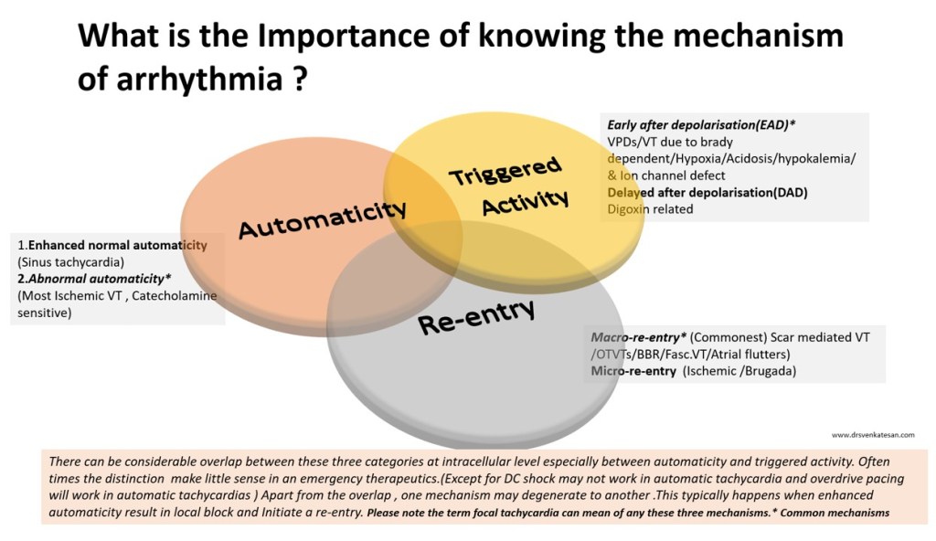
The following slide is a gross summary of the cardiac arrhythmia mechanism

Understanding cardiac arrhythmia is vitally important for a few reasons in a few settings.

  • In acute settings, we need to know automatic tachycardias will not respond to shocks. Reentry tachycardias will respond more promptly. (Of course, we may not know it till we shock ) Calcium blockers like verapamil might block triggered activity in MAT. Overdrive pacing is the answer for many automatic tachycardias and some refractory reentrant tachycardias (ATP protocols in ICD has taught us this ) 
  • In the chronic setting when you contemplate mapping, locating, and ablating arrhythmias, mechanisms are important. The task here is locating slow conduction paths and decoding the diastolic circuit around the scar  (If you plan ICD, knowledge about mechanism  becomes redundant again)

  • Finally, knowing the mechanism of arrhythmia is a fascination by itself to help understand the great subject called cardiac electrophysiology, where 100s of ion channels work nonstop drawing the action potential on a moment to moment basis sustaining our life.

A challenge

Can you localize a VT and find the mechanism in a patient who is Ischemic /hypoxic and acidotic? You can never do it. Please note, most polymorphic VTs can’t be localized. The mechanism is either automaticity, trigger activity, or even micro-reentry. You need to shock and look for the causes.(Link to How does the treatment of monomorphic VT differ from Polymorphic VT? )

Final message

Should we need to know about the mechanism of arrhythmia we treat?  Definitely yes, if you have that passion to know the truth, or else just order Amiodarone or shock and check out of CCU. (Of course, we have a very good option of calling EP consult the next day.)

 A review article on mechannism of cardiac arrhymias

Rev Esp Cardiol. 2012;65(2):174–185

Read Full Post »

Brugada syndrome is  as an  Inherited sodium Ion channel defect leading to loss of /or reduced sodium channel function.This specifically causes RV epicardial Imbalance of In-flowing(depolarising)  and out-flowing (repolarising)current , potentially triggering ventricular arrhythmia. This happens either spontaneously or during electrical stress times which include, fever, various drugs , adverse autonomic fluxes etc. So far,  we have been thinking it as primary electrical disorder with no macroscopic/ histopathologic  defects.

Newer Insights are emerging

But, how is this primary electrical disease , harbor a well demarcated  RV epicardial phenotypic substrate ? .  . . ablation of which eliminates the VT.

Zone of probable structural defect over RV epicardium (Pink zone) amplified by infusion of Ajmaline. Note the ECG showing typical ST elvation lead V1 to V3 .(Image courtesy Carlo Pappone et all  )

A recent study from Italy from the original founders  (Brugada team Ref 1 ) has confirmed RF ablation of RV epicardial tissue  is indeed feasible in many and should be considered in high risk Brugada syndrome. (Then should we suggest , ICD is no longer a choice in Brugada ?)

MRI findings in Brugada has shown some structural defects .(Ref 3,5) .It seems  Brugada is an Inherited electrical cardiomyopathy with a structural defect. (The overlap between ARVD and Brugada syndrome appear more real than we thought before ! (Ref 7 )

Final message

Still , Brugada is more of a electrical disorder,  but soon we may refer it as structural heart disease.

Reference

4.Catalano O,Antonaci S,Moro G,Magnetic resonance investigations in Brugada syndrome reveal unexpectedly high rate of structural abnormalities. Eur Heart J. 2009;30:22412248Abstract/FREE Full Text/Google Scholar
A best review comparing Brugada vs ARVC

Read Full Post »

Brugada syndrome is  probably the most fascinating discovery  in  Inherited cardiac  Ion channel dysfunction that linked the basic sciences to bedside . Its due to genetic defect in SCN gene that results in sodium channel blockade of phase  of action potential  to cause troublesome ventricular tachycardia (Phase 2 reentry ) . Now we realise, there are some phenotypic expressions to this gene defect . RV epicardial anatomical substrates are found to responsible for these Arrhythmias.

So ,does Brugada turn out to be a structural heart disease (At least histological ) ? Where is the evidence coming from ?

Now, we realise  RF ablation of epicardial aspect  of  RVOT / Adjacent anterior RV wall can eliminate Brugada pattern confirming anatomical defect  at cellular level. Our changing perception of Brugada from pure functional to anatomical cause is exciting and  intriguing as well !  ICDs were  the specific therapy so far.This discovery make it RF ablation an option.

The recent data from Brugada himself  was presented in World congress of electrophysiology / European Heart Rhythm Association (EHRA) EUROPACE-CARDIOSTIM 2016 confirming the therapeutic benefit of targeting  the epicardial structural phenotypic substrares (Ref 3 )

Final message

We have been taught  Brugada syndrome is  a primary electrical disease .It was  never considered as structural heart disease. Knowledge evolves slowly, so we shouldn’t conclude prematurely .Shall we conclude , Brugada syndrome is truly a  structural heart disease at least in some ?*. This  makes RF ablation  a new  cure for Brugada ,  making it a useful alternate modality to ICD , Of course there can be an overlap between ARVD and Brugada  syndrome. Mind you RF ablation scores over ICD on any  given day as its potential  cure , while ICD is  just a back up device and it simply wait & watch for the VT to occur.We also know  ICDs are still learning human EP data, and are  not intelligent enough to differentiate true VT from false ones with acceptable error** margin.

* Let the experts decide

**Acceptable ? What  do you mean by that ?

Reference

Questions queued

1.Is Brugada VT monomorphic , polymorphic or both ?

2.Is Amiodarone Indicated in Brugada syndrome ?

.

Read Full Post »

Have you felt like this query any time in your office ?

If “Yes” is your answer, then you are not alone .There was a unique  conference  that  took place  in 2010 to answer the same query in Rome , Italy on behalf of Italian cardiology society  , where this entity is researched more than any other place.Its worth going through this.

Reference

Read Full Post »

Ventricular tachycardia is a common cardiac arrhythmia. The significance of which can be very dangerous to relatively benign  depending upon the etiology and underlying heart disease . The ECG during VT is rarely useful to identify the etiology .Often times  ECG after reversal will  throw more light .

What are the ECG clues one should look for once VT is  reverted ?

  • Any evidence for old MI
  • Low voltage QRS/ LBBB/RBBB may indicate DCM
  • LVH -HOCM features
  • VPDs – Multiple , LBBB morphology / suggest  RVOT  VT
  • QRS slur or notching  indicating scars
  • Epsilon waves indicate  ARVD
  • RBBB pattern would  suggest  Brugada
  • Prolonged QT interval
  • Tall  T waves/ U waves /  Inverted  T -and other electrolytic abnormality.
  • Delta waves would indicate anti-dromic tachycardia.

Read Full Post »

Top 5 conditions that closely mimic and often mistaken for STEMI !

  1. Early repolarisation syndrome
  2. Left bundle branch block(LBBB)/ Left ventricular hypertrophy(LVH)
  3. Hyperkalemia
  4. Pericarditis
  5. Brugada syndrome

ERS

The repolarisation is due to  K + efflux . The  K channel porosity  is subjected to high degree of genetic  variations .If the repolarisation starts even by 10 milli- second earlier,  it would have early take off from descending  limb of R wave  and  the J point  ST segment appear elevated.

  • Common  in young  males . Especially in vago-tonic persons with relative baseline bradycardia
  • The ST elevation in ERS is often global .
  • Concavity is upwards .
  • ST elevation can be dynamic ( Further  confusing the picture ! )
  • On EST it  is expected to the  touch the baseline .
  • Benign entity in most . ( False alarm of STEMI is the major risk !)
  • There is some evidence ERS may confer a risk  of  primary VF ,  if they  experience a true STEMI  (Michel Haïssaguerre 2008  NEJM )

* STEMI in ERS :  The issue becomes too delicate ,  if  a  patient with ERS  develops  a true ACS .   ERS being a common ECG pattern in general population , it is not wise to label  every  chest pain in  ERS patient as benign . Suspicious  ones demand observation in step down units , at least !

LBBB

 “Any patient with  LBBB & chest pain . . . suspect  MI”  .

Unfortunately,  this rule is  too reverently followed by  physician community.  In fact ,  ACC/AHA guidelines  reinforced this behavior ,  as it  added a key word  in  their STEMI guidelines   “New onset”  or   “presumably new onset ”  LBBB is  an  indication for PCI/Thrombolysis    .( Physician presumption is a too delicate thread  to hang  our concepts !   )

               Every LBBB is new onset unless you have  a  documented proof otherwise  . . .   it seems to suggest !

Probably , this  is the reason many of the LBBBs are thrombolysed when they present to ER in an acute fashion . Of course , we can apply criteria of  Sgarbossa  to differentiate !  however flimsy it may appear . It  help us to exclude few benign LBBBs. Still ,  Sgarbossa will  struggle to  differentiate  an acute STEMI  in Chronic LBBB  from an  acute LBBB in  old AWMI .

Simply put . . . even old MIs  are at risk of  acute intervention if they have LBBB  and vague chest pain !

How to overcome this ?  Always rely on clinical  features  . If  STEMI is causing the LBBB ,  it  should be a large extensive one and you can not  expect the patient to be  comfortable .(Logic  would suggest necrosis of  large  parts of IVS is necessary to cause LBBB ) Chronic  LBBBs  are relatively comfortable  .

Of course , there  is one another  issue to comprehend  ie  transient ischemic LBBB .We do not know the true incidence  and long-term significance of this entity . Here , LBBB is  not due to necrosis of  the bundle but due to ischemia . (Almost impossible to differentiate it from  rate dependent LBBB  with  aberrancy  )

Role of enzymes and Echocardiogram in LBBB  and suspected STEMI .

You can always ask  for   Troponin  T / CPK MB .(They are helpful only  if 3 hours have elapsed , can we afford to wait ? ) . LBBB  due to STEMI  will  purge  a large quantum of cardiac enzymes from the infarcted zone . (So a marginal elevation is not going to help!)

Unfortunately,  LBBB  can induce wall motion defect in septum that may awkwardly simulate an ischemic wall motion. Even experts have erred in this . One clue  is,  the motion defects  can  not  extend   into anterior wall . It  is confined to septum ,the second clue  is a little delayed  post QRS  thickening of IVS (Septal beaking sign will vouch  for benign LBBB with fair degree of success  )

LVH

  • LVH can mimic a STEMI due to secondary ST/T changes . (Secondary to tall R wave )
  • LVH with incomplete LBBB  – A very common association that can further elevate ST segment in v1 to v3 .
  • Left ventricular hypertrophy  mimics old MI as poor R wave progression in V1 to  V3.
  • Contrary to our belief even Inferior  leads can  show q waves due to  inferior  septal hypertrophy.

Hyperkalemia.

With aging population and rampant  acute and chronic renal disorders it is becoming  a daily affair to get calls from medical units for ECG changes .We know  the rapidity of  efflux  potassium is responsible for ventricular re-polarisation .Phase 2, and 3 are K + exit zones. This is the same phase ST segment and T wave are inscribed.In hyperkalemia  K + accumulates inside the cell and keep  ST/T  segment  elevated .T wave also  becomes tall . It can mimic  both as hyper acute  STEMI .

Read a related article (Dialyisable current of Injury )

Pericarditis

  • ST elevation is not confined to an arterial territory
  • Can be global .(Regional ST elevation  does not exclude pericarditis)
  • ST elevation is concave upwards as in ERS

Link to Read regional pericarditis
Brugada syndrome

Brugada syndrome  is  an ECG -Clinical complex in which ST elevation in pre-cardial leads is associated with  ventricular arrhythmia. The defect lies in sodium channel . It reflects  a mis -match between RV and LV epicardial repolarisation forces .It keeps the RV epi-cardial current afloat and  the pre-cardial leads  facing the RV records ST elevation that  mimics  STEMI. It often  shows  a RBBB pattern and varying patterns of ST morphology  . The  ST segment is  also  subjected to dynamism  , due to change in autonomic tone and myocardial temperature  .(Febrile VTs)

After thoughts

Other close contenders for the top 5 slots

Myocarditis

Acute pulmonary embolism

Dissection of aorta

More

  • Acute stroke (Neurogenic ST elevation )
  • Stress cardiomyopathy (Takot Subo )
  • Acute abdominal conditions mimicking inferior STEMI.
  • Panic attacks /Anxiety states / chronic anti psychotic  medications which are known to elevate ST segments.
  • Contusion chest

(Cocaine hearts / Coronary arterial spasm / LV dyskinetic segments  and  LV aneurysms  were not nominees ! )

Read Full Post »

Brugada syndrome continues to fascinate  us for two reasons.

One , it deals with mysterious sudden  deaths of young  men and women

Two , it is one of the  fine  examples  of how  advances in molecular biology , links  physical defects in ionic channels to  sudden electrical  death (Most of them  are due to inherited defects  sodium channels  of myocyte cell membrane )

While high risk subsets of Brugada are easily managed , it is  the asymptomatic  ones  that bother us.

The following are some of the  difficult  questions ,   a  cardiologist faces when dealing with   patients , who exhibit  only Brugada pattern in ECG .

  1. Should I go for an EP study Doctor  ?
  2. Will  I  require an ICD  Doc ?
  3. Do I carry a significant risk of  dying  suddenly  ?
  4. Do  I need a genetic test for sodium channel mutation ?

Fortunately,  we can answer  all these questions with much  courage than before.

(Thanks  to the European Finger registry published in 2010  !)

“No” is the  clear  answer for all of them !

Summary from the FINGER registry. 

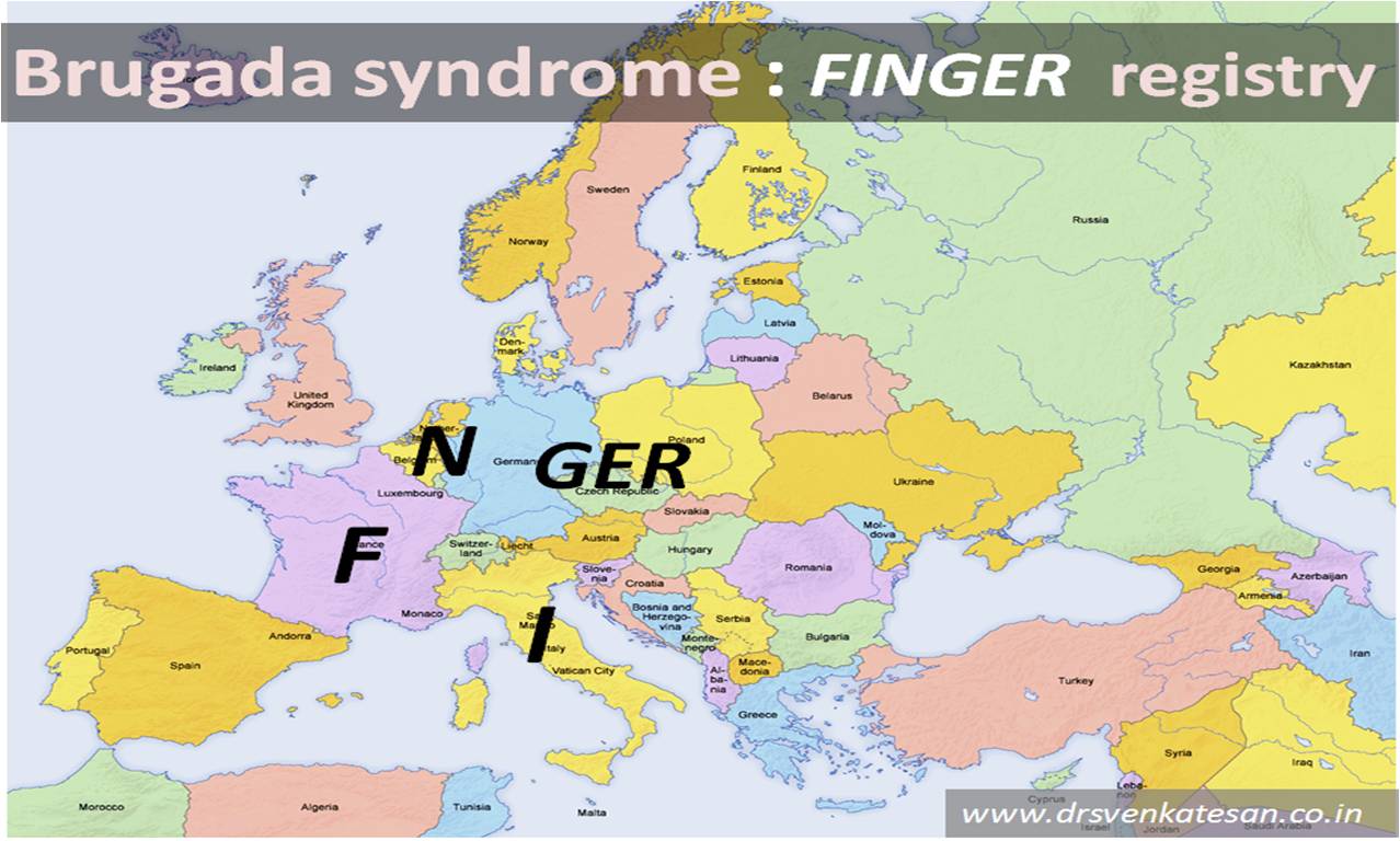
(France  , Italy, Netherlands, GERmany)

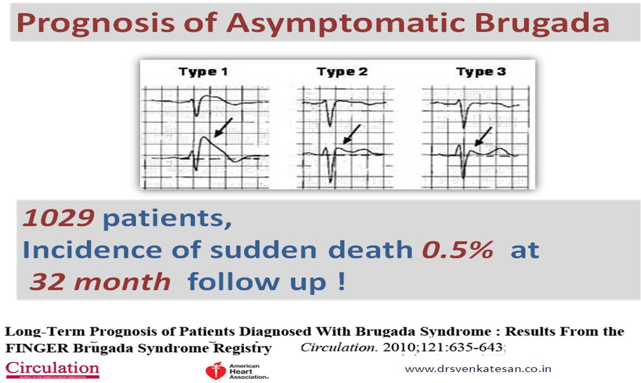
The registry included 1029 consecutive individuals

(1) Aborted SCD (6%);

(2) Syncope otherwise unexplained (30%);

(3) Asymptomatic patients (64%).

In the  follow-up of 31.9 (14 to 54.4) months . A total of  7 death occurred .

The cardiac event rates per  year was 

  • 7.7% in patients with Aborted SCD,

  • 1.9% in patients with syncope

  • 0.5% in Asymptomatic patients.

Predictors of cardiac  event

  1. Previous syncope
  2. Spontaneous type 1 ECG

Non predictors ( Surprisingly there were more non predictors ! )

  1. Gender has no predictive role
  2. Familial history of SCD,
  3. Inducibility of ventricular  tachy-arrhythmias during  EP study,
  4. Presence of an SCN5A mutation


Follow up

PRELUDE study  almost reaffirms  Finger data

(PRogrammed ELectrical stimUlation preDictive valuE)

Just publicized in JACC 2012 from the pioneer of   Brugada Silvia  Priori of   university of Pavia  Italy

Reference

http://circ.ahajournals.org/content/121/5/635.full.pdf+html

http://content.onlinejacc.org/cgi/content/abstract/59/1/37

Read Full Post »

Ever since Brugada found the unique pattern of ECG on right pre- cardial leads and its  association with  premature electrical death ,cardiac electro-physiology got a new impetus. Hundreds of articles(May be thousands !) on Brugada are  available . Many criterias  were proposed.  Brugada  and his colleagues should be credited for bringing  in such an interest in the  field of inherited ventricular arrhythmias.

On the down side ,  as we have a habit of  prematurely formulating criterias  ,  it brings  an artificial academic  barrier  Funnily , in medical  science  deviating   from a criteria (However hastily it  was  proposed  )   is a considered  big  offense Further . the hype surrounding  any new scientific  entity makes it difficult  for others  to overwrite  it .

Brugada recognized a ECG pattern with  a genetic predisposition for VT and VF  . Now , we know there are many etiologies  with a similar pattern  of ECG . What Brugada did was ,  he  exposed the tip of Iceberg called inherited ventricular arrhythmia . But the essential criteria –  Absence of structural heart disease ,  to diagnose Brugada   was  always questionable.

(Please realize , presence  or  absence of structural  heart disease depends , more  on  how advanced  our  imaging modalities are . If you can map a virtual histology of RV epicardium one may detect some  microscopic abnormality in every case of Brugada. In human biological system , God  usually bonds  structure and function too  closely  and hence  functional  abnormality rarely occur  in isolation )

Brugada is  not  a new disease ,   it is  a  recognition  of a  pattern of ECG  related  to sudden deaths . Subsequently , we  realized any dispersion in repolarisation in RV epicardial surface  , the   risk of sudden death  is increased. From the days of  Brugada  we  have  come  a long way.

What is new in Brugada syndrome  ?

(Not exactly new . . .  it is  known  for many years )

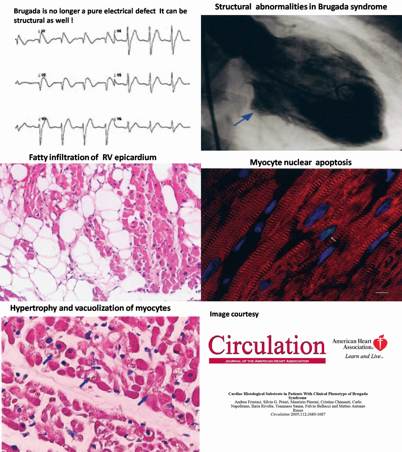
Brugada is no more an exclusive  functional disorder of  sodium channels of RV  epicardium .It can have structural defect (known & unknown ) .It may  have infective , degenerative etiology as well .

How does these structural changes appear ?

Chronic sodium channel malfunction  can result in cell membrane defects which can augment   Idio-osmole   inside  the cell and result in  apoptosis   etc .

Which comes first ,  electrical or structural abnormality ?

It is an  another  chicken- egg tale  waiting to be decoded   within the RV epicardial cells

Can wall motion defect occur in Brugada ?

Early observations done in out hospital (MMC Chennai ) has found anterior  RV free wall motion defects. Tissue Doppler studies are  being undertaken.

Final Message

The  following paper  wonderfully documents  the structural and histo-pathological  changes in RV epicardium .  This  implies ,  our belief   about this  unique electrical  disorder  is  bound to take a beating  and  we  expect a major perception makeover regarding Brugada  in the years to come .

Probably the most important paper on Brugada syndrome was published in circulation in 2005

*http://circ.ahajournals.org/cgi/content/full/112/24/3680

Read Full Post »

Older Posts »