Feeds:
Posts
Comments

Archive for the ‘Brugada syndrome’ Category

How many times you have treated cardiac arrhythmia in both emergency & non-emergency situations?

Infinite times.

How many times did you really bother to know the mechanism of a given arrhythmia before ordering medication or shocking?

Hmm,.. let me think. (Except for AVNRT/ AVRT, and few VTs, very rarely I have worried about the mechanism  !)

Why is it so? because treatment takes priority and we are able to tame the arrhythmia even without knowing the real mechanism.

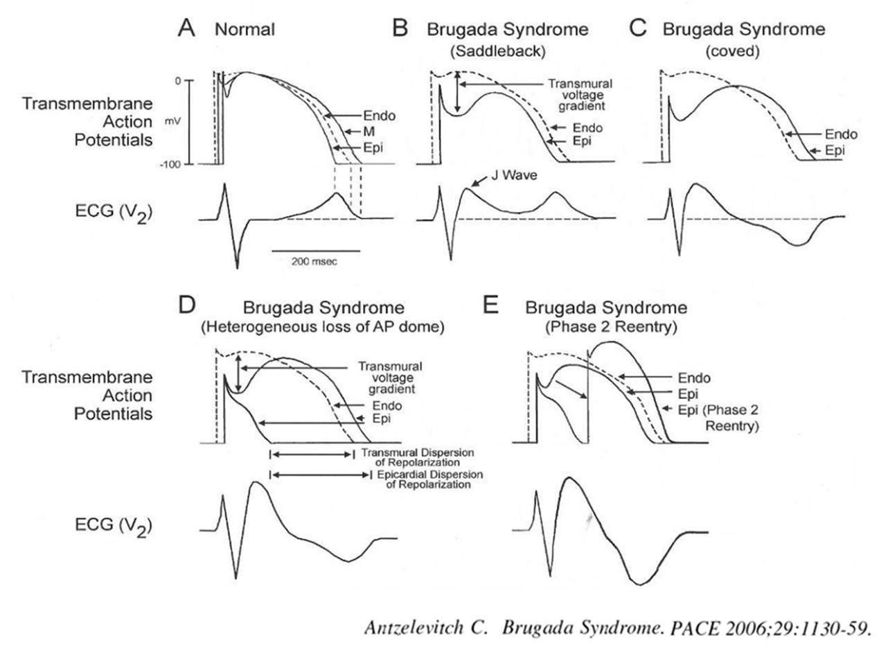
The following slide is a gross summary of the cardiac arrhythmia mechanism

Understanding cardiac arrhythmia is vitally important for a few reasons in a few settings.

  • In acute settings, we need to know automatic tachycardias will not respond to shocks. Reentry tachycardias will respond more promptly. (Of course, we may not know it till we shock ) Calcium blockers like verapamil might block triggered activity in MAT. Overdrive pacing is the answer for many automatic tachycardias and some refractory reentrant tachycardias (ATP protocols in ICD has taught us this ) 
  • In the chronic setting when you contemplate mapping, locating, and ablating arrhythmias, mechanisms are important. The task here is locating slow conduction paths and decoding the diastolic circuit around the scar  (If you plan ICD, knowledge about mechanism  becomes redundant again)

  • Finally, knowing the mechanism of arrhythmia is a fascination by itself to help understand the great subject called cardiac electrophysiology, where 100s of ion channels work nonstop drawing the action potential on a moment to moment basis sustaining our life.

A challenge

Can you localize a VT and find the mechanism in a patient who is Ischemic /hypoxic and acidotic? You can never do it. Please note, most polymorphic VTs can’t be localized. The mechanism is either automaticity, trigger activity, or even micro-reentry. You need to shock and look for the causes.(Link to How does the treatment of monomorphic VT differ from Polymorphic VT? )

Final message

Should we need to know about the mechanism of arrhythmia we treat?  Definitely yes, if you have that passion to know the truth, or else just order Amiodarone or shock and check out of CCU. (Of course, we have a very good option of calling EP consult the next day.)

 A review article on mechannism of cardiac arrhymias

Rev Esp Cardiol. 2012;65(2):174–185

Read Full Post »

Brugada syndrome is  as an  Inherited sodium Ion channel defect leading to loss of /or reduced sodium channel function.This specifically causes RV epicardial Imbalance of In-flowing(depolarising)  and out-flowing (repolarising)current , potentially triggering ventricular arrhythmia. This happens either spontaneously or during electrical stress times which include, fever, various drugs , adverse autonomic fluxes etc. So far,  we have been thinking it as primary electrical disorder with no macroscopic/ histopathologic  defects.

Newer Insights are emerging

But, how is this primary electrical disease , harbor a well demarcated  RV epicardial phenotypic substrate ? .  . . ablation of which eliminates the VT.

Zone of probable structural defect over RV epicardium (Pink zone) amplified by infusion of Ajmaline. Note the ECG showing typical ST elvation lead V1 to V3 .(Image courtesy Carlo Pappone et all  )

A recent study from Italy from the original founders  (Brugada team Ref 1 ) has confirmed RF ablation of RV epicardial tissue  is indeed feasible in many and should be considered in high risk Brugada syndrome. (Then should we suggest , ICD is no longer a choice in Brugada ?)

MRI findings in Brugada has shown some structural defects .(Ref 3,5) .It seems  Brugada is an Inherited electrical cardiomyopathy with a structural defect. (The overlap between ARVD and Brugada syndrome appear more real than we thought before ! (Ref 7 )

Final message

Still , Brugada is more of a electrical disorder,  but soon we may refer it as structural heart disease.

Reference

4.Catalano O,Antonaci S,Moro G,Magnetic resonance investigations in Brugada syndrome reveal unexpectedly high rate of structural abnormalities. Eur Heart J. 2009;30:22412248Abstract/FREE Full Text/Google Scholar
A best review comparing Brugada vs ARVC

Read Full Post »

J point is a critical point in the  ECG  when the ventricles hand over the baton in  the  electrical relay race from depolarization to repolarization .This the time the sodium  channels extinguish itself  and the potassium current begins its activity  from Phase 0 to 1 .

If the  potassium channels  activate little early and snatch the baton prematurely from sodium , we get early repolarization pattern .When this happens , the J point of ECG show a conspicuous wave  called J wave , originally  denoting  Junctional wave between QRS/ST segment  (Now  perceived as  Jitter waves ?) The other implication of premature K+ activity is , lifting up of  ST segment , making it the most common cause of non ischemic ST elevation.

* J wave in hypothermia is referred to as Osborne wave and  may not be  not related to ERS(Ref.4)

J wave and J point early repolarisation syndrome

Image source.www.cardiology.org

The Ito current is responsible for the phase  1 of action potential (AP), where a rapid outward k + ion flux take place and draws the dome of AP . The dynamics of Ito is complex .It depends  upon the density of epicardial K + channels , which are  clustered in a heterogeneous manner .There seems to be a concentration gradient   along the epicardium and endocardium , making the wave appear prominent in some. This is especially true in healthy, athletic  male population  where we have some evidence for androgen  to  play a role on how  these channels will behave.Here comes the overlap between Brugada  syndrome and ERS as well.

The subset of patients with J wave pattern were recently shown to have increased risk of primary VF due to phase 2 reentry ,  when they develop ACS. (Rather J wave pattern was more common in patients who had primary VF following STEMI(Ref 1).This resulted in a spate of worrying articles .Now we know , the  fear is  largely unfounded ,the risk is far less.

Current thinking is,  persons who have asymptomatic ERS pattern with prominent J waves should not be investigated electro-physiologically . (Please remember , every human  heart can be induced  to VF in EP lab  if appropriately  stimulated ! )

In fact , I used to tell the  young men  who  harbor  prominent J wave , as a marker of healthy heart  rather. Let us not  fear them with a remote risk  that could be as  negligible as risk of  intercontinental flight crashing into the ocean  !

References

1.Haissaguerre M, Derval N, Sacher F, et al. Sudden cardiac arrest associated with early repolarization. N Engl J Med. 2008;358:2016–2023.

2.Idiopathic Ventricular Fibrillation “Le Syndrome d’Haïssaguerre” and the Fear of J Waves , Sami Viskin, J Am Coll Cardiol. 2009;53(7):620-622. 11

Read Full Post »

Have you felt like this query any time in your office ?

If “Yes” is your answer, then you are not alone .There was a unique  conference  that  took place  in 2010 to answer the same query in Rome , Italy on behalf of Italian cardiology society  , where this entity is researched more than any other place.Its worth going through this.

Reference

Read Full Post »

Brugada syndrome is due to a genetically  impaired  sodium channel activity  ( SCN5A)  in phase o, of action potential .This results in phase 1 (Ito channel) failing to inscribe the transition between phase 0 and  1 that result in loss of  dome .This loss of dome is dominant in epicardial cells compared to endocardial cells.This result in  electrical heterogeneity and a hence a voltage gradient in repolarisation phase  that can trigger a Phase 2  reentry mediated  VT /VF.The above said defects are either dormant, manifest, self extinguishing , dynamic  subjected to autonomic tone , ambient myocardial temperature (Febrile VTs) making this a complex entity.

There are three distinct types according to surface ECG.It can be either spontaneous or induced. The arrhythmic events and prognosis and hence management differs according to the types.

mechanism of brugada syndrome three types of ecg 2All types carry  a minimal risk of SCD , variable though . Of course  syncope  has to be  much more  common. Curiously every episode of syncope is seen as naturally aborted SCD by physicians ! (No one  to be blamed for this .The definition of syncope is like that !If the patient doesn’t wake from syncope it becomes death !).

When a patient with Brugada  has a  syncope , it  doesn’t  imply  he  experienced a dreaded VT or VF.While SCD is invariably due to ventricular fibrillation , a spontaneously terminating VF  as a cause for syncope is rare in Brugada . (Ref 2 : ILRs have documented though in few)

So what exactly is the cause for syncope in Brugada ? The issue is  real  and critical in clinical decision-making. We are beginning to document variety of mechanisms. Following are the possible causes

  1. Sustained  VT or NSVT with
  2. Non sustained self terminating  VF
  3. Extreme bradycardias (Vaso vagal )
  4. AV blocks
  5. Unrelated neurogenic

Final message

It is to be strongly emphasised a significant subset of Brugada patients especially in Type 1   Brugada (spontaneous or drug induced )  the mechanism of syncope is often not related to the dreaded VT/VF. It can simply represent high vagal tone and unexplained dynamism of autonomic activity .ICD is not a default indication for all those with syncope in Brugada syndrome.Think , pause and decide when you deal with such patients. ICDs are true revolutionary devices  . . . no two thoughts about it,but it can make a hell out of heaven if used in an inappropriate situation !

Reference

Read Full Post »