Feeds:
Posts
Comments

Posts Tagged ‘tavi’

TAVI has become a popular Aortic Intervention, but the risk of coronary obstruction (Left main & RCA too) is often underestimated , it can happen during the procedure or in the long term .High degree of anticipation is essential. The exact incidence is not known as reporting the event is far less than, what it is actually. Following are the suggested precautions to prevent coronary obstruction.

Pre-Procedural Assessment

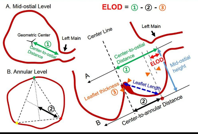
  • Low Coronary Ostial Height: Identify if the Left Main origin is (< 10–12 mm )from the aortic annular plane, that will increase  the chance of struts covering the ostia.
  • Shallow Sinuses of Valsalva : A shallow sinus( < 30 mm )leaves insufficient room for the displaced native leaflet, directing it toward the ostium.
  • Leaflet Length and Calcification: Evaluate for heavy calcification or a long native leaflet that exceeds the height of the coronary ostium, as these are easily pushed into the vessel opening.
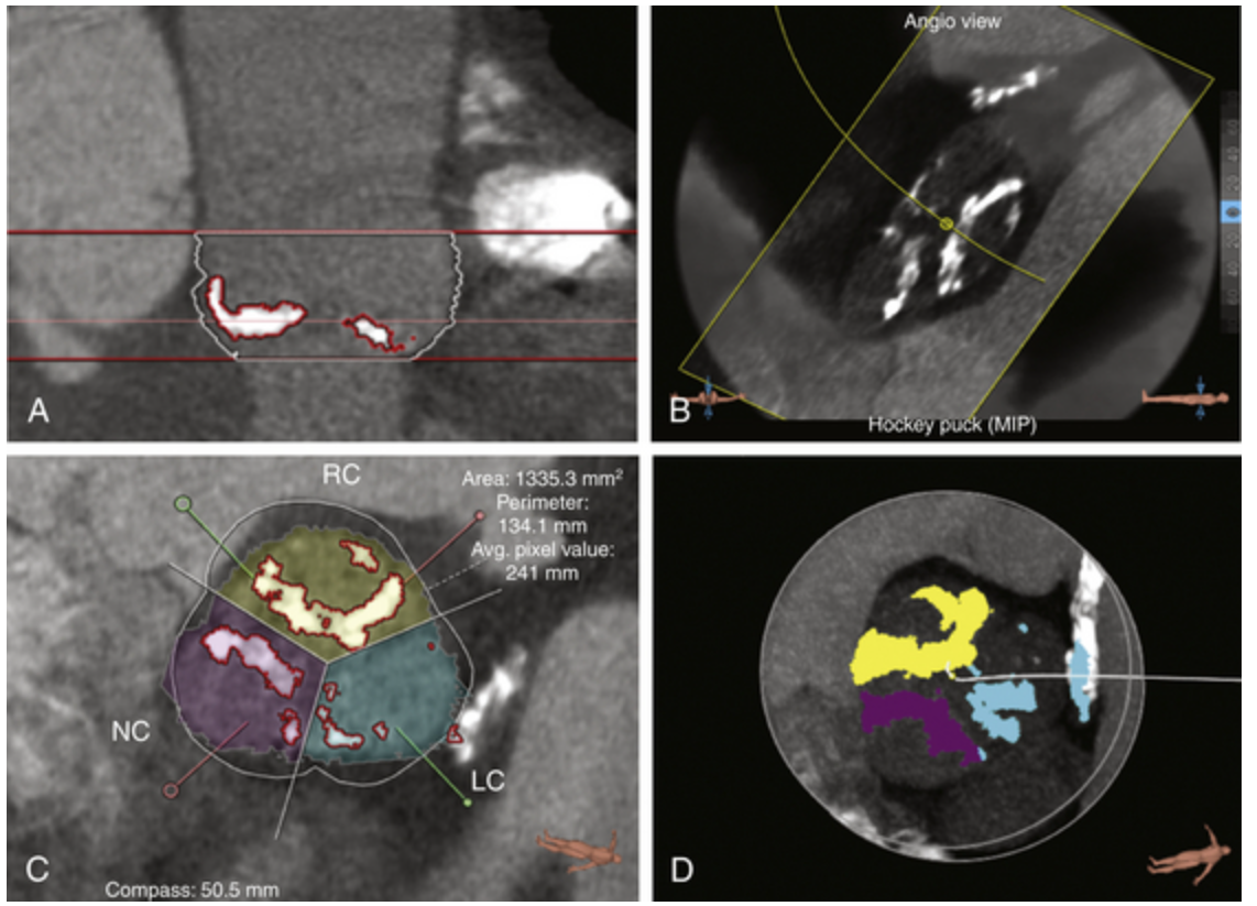
  • Virtual Valve-to-Coronary (VTC) Distance: In Valve-in-Valve (ViV) procedures, calculate the VTC distance. A computed distance less than 4 mm indicates risk of coronary obstruction.

Risk factors for potential coronary obstruction.
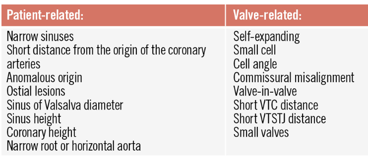
Image source Ref 2

*Leaflet length is a very critical parameter, which was ignored in the past. (It is more Important than even, the coronary height.Mind you coronary height matters little if leaflet is long enough to reach the ostium , when TAVI flushed it with wall of Aorta.)

Intra procedural precautions(In small shallow roots)

  • Chimney Stenting: Prophylactic wire in the Left Main and park an undeployed drug-eluting stent. If any segment of TAVI tends to occlude deploy the stent.
  • BASILICA/Leaflet Splitting: This is used in ultra-high risk or valve-in-valve cases, use radiofrequency energy or mechanical cutting devices to lacerate the target aortic cusp before valve deployment, forcing it to part like a curtain instead of blocking the ostium.This is not an easy procedure as it may look. Prone for its own complication.

Post-Procedural & Long-Term Management

Dual antiplatelet therapy is critical . Routine echocardiography and follow-up CT angiograms to monitor for delayed coronary obstruction or stent deformation . Documenting the valve-stent alignment is encouraged for a possible future coronary interventions.

Planning a PCI with TAVI

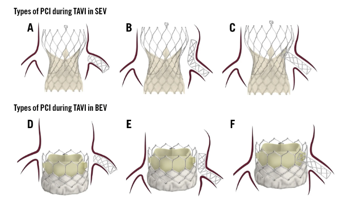
Image source : Ref 2

PCI types in different types of valves. A) PCI with minimal stent protrusion due to limited space between the valve and the STJ. B) Adequate space between the valve and the STJ, allowing for the chimney technique. C) Valve-directed stenting from the coronary ostium towards the valve frame. D) PCI with minimal protrusion, as the coronary ostium is located above the valve. E) Coronary ostium located below the valve frame, with sufficient space between the valve and the STJ to perform the chimney/snorkel technique. F) Coronary ostium located below the valve frame, with limited space between the valve and the STJ; therefore, stenting is directed from the ostium towards the valve. BEV: balloon-expandable valve; PCI: percutaneous coronary intervention; SEV: self-expanding valve; STJ: sinotubular junction; TAVI: transcatheter aortic valve implantation

Final message

TAVI is a popular Aortic Intervention, but the risk of coronary obstruction is often underestimated by cardiologists, and it can happen during the procedure or in the long term; thus, preparedness is essential.

Reference

1.Aquino_bruno.pdf TAVI and coronary interventions: indications, technical considerations, and clinical scenarios Euro Interventions Volume 22 Number 11 Jun 1, 2026

2.https://eurointervention.pcronline.com/article/tavi-and-coronary-interventions-indications-technical-considerations-and-clinical-scenarios

Read Full Post »

Learning anatomy is foundational first step in the pathway to become a great clinician. This post is written to address a potential nomenclature issue in cardiac anatomy .

What constitutes Aortic annulus.?

“Aortic annulus” by default conveys a discrete, planar, circular ring. However, from a macroscopic and histological perspective, a distinct, single-plane collagenous ring does not exist at the base of the aortic valve. Rather, aortic annulus is a complex, three-dimensional , Morpho-anatomical functional zone spanning the ventriculo-aortic junction. This brings the aortic leaflets to be arranged in a crown-like formation that crosses multiple spatial planes. The lowest points of these attachments (the nadirs) dip into the left ventricular outflow tract, while the highest points (the commissures) extend superiorly to anchor at the sinotubular junction.


Can anyone see a single plane of Aortic annulus in the above Image ? Note the leaflets , pan whole area from the virtual ring to the ST junction making the annulus a well secluded zone with from ST junction to the virtual lower ring. This aortic annular zone has clear cut boundaries, the inferior border is formed by lowermost insertion points of the aortic valve leaflets. (Which just above the virtual ring formed by sinus of valsalva)

Why the misconception ?

The misconception of a flat aortic annulus came from  extrapolation of AV valve anatomy to the semilunar valves. In the mitral and tricuspid positions, the leaflets anchor to a continuous,  planar fibrous skeleton. Consequently, their hinge lines and leaflet planes remain the same in a reatively single plane throughout the cardiac cycle.

Systolic and diastolic disconnect

This complex anatomy of aortic annulus also results in a clear disconnect between systole and diastole. During systole, the aortic leaflets open vertically like flushed doors, against the sinus walls , while  the basal plane vanish.Conversely, during diastole, the leaflets fall medially and down to meet at the center, creating a horizontal plane of coaptation.

Timing of measurement of aortic annlus

We are asked to measure the aortic annulus in peak systole. Ironically  at this point of time , every measurement becomes virtual in the aortic annular zone.  At least in diastole, the so called virtual lower end becomes real with leaflets hanging down forming a point of reference.

TAVI and Aortic annulus measuerment

The measurement of the aortic annulus has become essential due to the rise of TAVI, as CT scans accurately define the annulus compared to echocardiography, revealing the distinct height of the aortic annulus that was previously unrecognized. read below, for all the relevant measurements before doing a TAVI.

Image source : Jun-Hyok Oh IJC Heart & Vasculature Volume 37, December 2021, 100917

Newer TAVI engineering designers intentionally utilizes the entire vertical distance of the aortic root (from the leaflet nadirs to the ST junction) to anchor, seal, and position leaflets across different spatial planes to optimize physics and hemodynamics

Final message

The perception of aortic annulus as a single anatomical plane in our minds is very firm and it is due to the flaw in the way we learned cardiac anatomy. To make things right, we need to change aortic annulus as the “aortic annular zone” in anatomical literature. This change must be made at the earliest, right from the first clinical years of undergraduate education. Recognizing this is vital for understanding surgical or percutaneous repair or replacement of prosthetic valves.

Postamble (For the fellows)

When someone asks you what is aortic annular size, please ask them to confirm which dimension they are talking about. If they are surprised, teach them, there are  distinct multiple horizontal (Maximal, minimal) and vertical distance for every annulus.(Which can correlate to a certain extent to the coronary height, the TAVI guys talk about)


References

  1. Piazzese ED, de Jager SE, van Straten AH, Elenbaas TW, Soliman-Hamad MA. Surgical anatomy of the aortic valve and root—implications for valve repair. Ann Cardiothorac Surg. 2019;8(3):324-331. doi:10.21037/acs.2019.04.09
  2. Loukas M, Bilinsky S, Bilinsky E, El-Sedfy A, Anderson RH. The anatomy of the aortic root. Clin Anat. 2014;27(5):748-756. doi:10.1002/ca.22295
  3. de Kerchove L, El Khoury G. Anatomy and pathophysiology of the ventriculo-aortic junction. Ann Cardiothorac Surg. 2013;2(1):87-92. doi:10.3978/j.issn.2225-319X.2012.11.15

Read Full Post »

A. The Aortic Radiologist

B. The Principal operator of TAVI

C. The co-operator or the proctor

D. The device type, behavior & destiny

Answer : A. The radiologist, (rather the soft-warist ) tell us best landing zone in the pre procedure work up taking into the account of shape, size, leaflet morphology ,3D analysis of aortic root, and calcium distribution. He plays a vital role. Though B, is the logically correct answer, ie the expertise of the primary Operator it is not to be. Considering the complexity of the anatomy, device, and momentary longitudinal and circumferential alterations in radial forces, In reality, the answer is D.

Evidence? No one has tested so far the true intended landing zone and the final one.


One may argue , the question need to be reframed as, What are the factors that decide the landing zone in TAVI ?

Though deployment is under the control of primary operator , the valve often defies operators’ hand commands and decides to home in its own place of comfort and peace adjusting to the complex anatomy around. It never bothers about the consequences .The unpredictability of calcium crystals, the annular tensile strength, the distorted native leaflet and the blind plastering against the aortic wall all are responsible for the complications. One less talked dynamism of aortic root which finetunes micro adjustments of the valve. Para valvular leak is primarily determined by either excess (or lack )of this modulatory forces.

Following are some of the crucial factors that determine landing zone.

Anatomy of the Aortic root

 The ascending aortic curvature and angulation is a major factor of the TAVI landing site. , stretching from the aortic annulus to the proximal ascending aorta. This anatomy may vary depending on the patient and affects different types of valves (such as balloon-expandable or self-expanding). For instance, higher aortic angulation (Ex – a horizontal aorta) can influence the final depth of valve implantation, especially for self-expanding valves with longer stent frames.

The invisible radial & longitudinal forces

The resistance offered by the distorted native valve and its delicate balance with the instant radial force of in case of balloon expandable or the gradual built in force of self expandible balloon system. Apart from the radial forces. the longitudinal deformation forces in long axis as the stent expands , in the last few seconds determine the final residence of the valve.

Valve Type and Device Design

The intended implantation depth is usually planned based on the valve type—balloon-expandable valves have a short stent frame and are deployed differently than self-expanding valves with a taller frame. More precision is required in balloon expandable valves. This is because the anchoring mechanisms and the metallurgy is entirely different between the two.(SE-TAVI uses Nitinol frame, and it has wide contact area so more stable ,)

Intra-Procedural Adjustments: 

The final landing zone is also influenced by the need to avoid complications such as valve migration, aortic regurgitation, and coronary obstruction.

Final mesage

Some times, TAVI landing looks like SpaceX Dragon docking with a space station. Extreme precision is required to avoid complications. The bulk of the complications are due to inappropriate landing (too high or too low). Fortunately, aortic annulus exists in multiple transverse planes; patients can often tolerate some geographical miss. Still even a few mm error can crash the patient as well as our reputation.

Postamble

What are the chances of immediate post -procedural movement and late migration of the valve from the landing site in TAVI ?

The chances of immediate post-procedural movement and late migration of the valve from the landing site in Transcatheter Aortic Valve Implantation (TAVI) are generally low but represent serious complications when they occur. Incidence is up to 7.5%

Reference

https://www.frontiersin.org/journals/cardiovascular-medicine/articles/10.3389/fcvm.2022.928740/full

* Video source and courtesy

Read Full Post »

TAVI is becoming like PCI equivalent of aortic valve. The procedure is nothing but stenting and plastering the aortic prothesis ,with all the native diseased aortic leaflet in-situ. Pre procedure CT aortic valve mapping (rather the entire Aorta) is the key to successful outcome.

While the calcium is the essential bonding force of the valve to the aortic annulus, it can also play some serious spoilsport, along with native leaflet debri . Many times, the hardened calcium are like like sharp 3 dimensional knife hanging over there in root of aorta.

Every TAVI operator has this ligering fear . Will that speck of calcium “ice berg”, hiding 2 mm above the NCC, hit the AV node, when I deploy the valve ? Will the distorted leaflet jump few mm above and hit the coronary ostia , however high it may be. (After all , the exact final landing zone is not determined by the operator , but by the ROC curve)

Every severely calcified valve experiences a Titanic effect , fortunately most valves escape.

Realise how important the accuracy these softwares are .It is just a matter of few mm error . . Apart form calcium distribution pattern , more fundamental parameters like the annular size, shape, and optimal imaging angle are critically important. Here is brief report on various software packages available for pre procedure planning of TAVR.

Image courtesy : Thoracic Key  Assessment of aortic valve calcification.The stretch view shows dense calcification of the right coronary cusp, noncoronary cusp, and left coronary cusp. The stretch view (A), angiographic overlay (B), cross-sectional view (C), and “hockey puck” view (D) allow quantification of the calcium in the aortic valve.

The following table was curated from the respective company websites. Any further details can visit them.

SoftwareVendor/DeveloperKey Features for TAVR Pre-Planning
3mensio Structural HeartPie Medical ImagingDedicated TAVR module for automated aortic root analysis, valve sizing, and access route planning. Provides 3D visualization, calcium scoring, and virtual valve implantation. Widely used for precise measurements and procedural simulation.
OsiriX MD / HorosPixmeo (OsiriX) / Open-source (Horos)DICOM viewer with 3D multiplanar reconstruction (MPR) tools for manual valve sizing, annulus measurement, and aortic root analysis. Horos is a free alternative. Supports plugins like ProSizeAV for semi-automated sizing.
syngo.via CT Cardiac Function – Valve PilotSiemens HealthineersSemi-automated workflow for aortic valve assessment, angulation prediction, and device sizing. Includes valve pilot tools for efficient CT analysis.
HeartNavigatorPhilips HealthcareAutomated or semi-automated CT processing for TAVR, including aortic root segmentation, access route simulation, and procedural guidance. Often compared for reliability in measurements.
Valve Assist 2GE HealthcareAI-assisted tool for valve sizing, CT analysis, and planning efficiency. Focuses on automating measurements to reduce manual effort.
Mimics Enlight / Mimics PlannerMaterialiseCloud-based 3D modeling software with automated workflows for structural heart interventions, including TAVR-specific measurements, virtual valve implantation, and 3D printing support. Includes AI for segmentation.
cvi42Circle Cardiovascular ImagingAdvanced CT tools for interventional planning, including TAVR, with automation for aortic valve assessment, flow quantification, and structural heart disease management.
Intuition TAVR PlanningTeraReconComprehensive package for aortic root segmentation, centerline extraction, and pre-operative measurements. Supports advanced 3D/4D visualization for TAVR workflows.
Vitrea CT TAVR PlanningCanon Medical (Vital Images)AI-leveraged application for automated TAVR assessment, including valve sizing, access planning, and post-operative evaluation. Integrates deep learning for efficiency.

Some questions

1.Which one is most popular ?

With out doubt 3mensio is top software because of its neutrality between various TAVR valve and wide spread usage and comparisons.

2.What is the cost of these software ?

They are substantial has a monthly subscription model. 3Mensio pricing starts at approximately $500/month for 1 user, $4,000/month for 10 users.

3.Is there any Freeware for assessing Aortic root ?

Yes . OsiriX MD / Horos is a free ware, but not getting sufficient attention.

4.What is the error rate of these software ? since they are offline and often images are machine extrapolated ?

Error rate in software are well not reported. (Can’t expect the vendors to do it !) However, It must be acknowledged they are real because of the offline nature of image processing .These tools process DICOM data, in pre-trained algorithms. Errors can arise from poor CT input (e.g., motion artifacts) or extrapolation in 3D reconstruction (e.g., interpolating between slices), but studies show minimal impact with high-quality scans.

Common Error Sources: User variability, calcium blooming artifacts, or phase-specific differences in dynamic CT.

Clinical Implications: Errors in sizing can lead to complications like paravalvular leak (if undersized) or embolism (if oversized), but validation shows risks are low (e.g., <2 mm differences rarely affect outcomes). Multi-reader or expert double check is encouraged to improve accuracy.

Read Full Post »

We know TAVI is in the striking distance , to literally take over most aortic valve interventions. From a humble beginning from very high surgical risk with prohibitive comorbidity, now it has almost touched the totally asymptomatic, relatively morbid-free patients. Thanks to the hardware, expertise, and motivation from multiple forces.

While the numbers increase, still the debate between SAVR and TAVR is riddled with speculation, skepticism, and absolute confidence. (Reason: TAVI is a passively fixed valve in a blind procedure at a self-selected annular plane, with no option to remove the crushed native leaflet debris and the resultant complications. Lastly, TAVI’s lifespan* is currently less than half of a mechanical valve. *Expected to improve with polymer valves)

The latest trial to join the litereture is EARLY TAVR in October 2024

Here is a brief, personal comment about the paper for non-academic consumption. Look carefully at the 15th second of the video. Pause it, look at the number over there on the bar of unplanned hospitalisation.

It is a staggering 41.7% in clinical surveillance group, twice more than TAVI group, pathologically tilting the conclusion of the study.

Video source and courtesy https://youtu.be/3wwQEEG4aWg

By the way, what is that unplanned hospital admission? Who is planning that admission in the asymptomatic control group? If 41% of people in the clinical surveillance group needed hospital admission, what does it mean? Does that mean clinical surveillance was so poor that they were rushed to the hospital despite being asymptomatic and stable in the surveillance period?

Why should totally asymptomatic patients get admitted in the control arm, in such huge numbers? You can presume what could be the reason. My guess is too sinister.

Another issue plaguing the RCTs for decades, is continuing even in 2025. That is putting together death, stroke, and unplanned hospital admission as a combined endpoint in the same basket. This is the familiar old cheat story i.e., used to intentionally torture the truth.

Final message

Any student with basic sense of statisitcs can interpret the result of this landmark trial from NEJM correctly. The question we need to ask is, what are the triggers for those unplanned hospital admissions?

Further, it is good for NEJM (and the medical community) not to accept any papers, if the studys’ endpoints are not appropriate or defined with the intention to manipulate, which happens in many sponsored trials.

Read Full Post »

This image comes with courtesy of the Journal of SCAI Jai Parekh, Vikram Sharma, Jared Robl,et al Journal of the Society for Cardiovascular Angiography & Interventions 3 (2024) 101310

What is your diagnosis ?

I thought, it was pacemaker extrusion. It was indeed a close answer, still terribly wrong. It is an intentional exterior placement of a permanent pacemaker generator mimicking an extrusion due to pocket infection. Here is a patient, where a permanent pacemaker was kept temporarily for a few weeks or a month in high-risk reversible complete heart block situations. This typically occurs after an inferior posterior myocardial infarction, drug-induced CHB.

Currently, with the arrrival of TAVR, CHB has beceome a glamorous complication and is getting wider attention. This happens due to the anatomical uncertainties where the inferior landing zone of TAVI is pre-destained and is beyond our control. This is more true in the self expanding Core valve platform . When the lower edge treaspass the non-coronary cusp- membranous septal junction, it hits perfectly the compact post-penetrating bundle of His, confering a high risk of CHB.

Still, the good thing is some of them recover as the pressure edema regress .Putting a PPM in all such patients was considered mandatory or even a vanity in the past. Now we realise it is an additional metallic luggage in an already strained heart, Temporary-PPM the oxymoronic innovation is perfect option in this setting.

Final message

A typical external temporary pacemaker can be kept for up to 2 weeks maximum. (We have kept it for a month or so) It’s done via the jugular, subclavian, or even femoral. If the underlying condition demands more time for recovery of CHB, many do a regular permanent pacemaker.

Now , we have this unique option of using PPM as TPM. This is not a new concept though. It was used few decades ago .Has come back in more centers .Thanks to TAVI and its specific complications.

Reference

1.Rodés-Cabau J. Ellenbogen K.A. Krahn A.D. et al. Management of conduction disturbances associated with transcatheter aortic valve replacement: JACC Scientific Expert Panel. J Am Coll Cardiol. 2019; 74: 1086-1106.

2. Leong D, Sovari AA, Ehdaie A, Chakravarty T, Liu Q, Jilaihawi H, Makkar R, Wang X, Cingolani E, Shehata M. Permanent-temporary pacemakers in the management of patients with conduction abnormalities after transcatheter aortic valve replacement. J Interv Card Electrophysiol. 2018 Jun;52(1):111-116. doi: 10.1007/s10840-018-0345-z. Epub 2018 Mar 12. PMID: 29532275.

Read Full Post »

Welcome to the  future of  valvular heart disease . This is just the beginning.Expect more dramatic break through  . (Already mitral valve prototype is in advanced stages of development .)

Currently we have two approved percutaneous aortic valves for use in isolated Aortic stenosis. The Edwards valve is popular in  USA  ( 2011 ) and Medtronic  is used extensively in  Europe (From 2007)

Though both valves appear suitable .There are major differences in the concept , design , and technique of implantation .

tavi edward sapiens vs medtronic core valve

Reference

Major  issues to be addressed. Late onset Para valvular leak :
Please remember, these valves are not sutured around the aortic annulus ,  which our surgeons do it meticulously . The force that keep the valve  within the  aortic root is nothing but the disease process itself . The stiffened, elastic aortic root .(Does it appear  foolish to expect the diseased  aorta to hold the valve in situ ? but that is the reality  ! )
If the aortic root  dilates  for  some reason  which is very likely in  atherosclerotic  process    the very foundation of valve is shaken and para valvular leak is certain.

Read Full Post »

Cardiologists are  closing in ,  trying to capture the final frontiers. The  trans-cutaneous Aortic valve Implantation now has  a two year follow up. (NEJM March 2012  Issue) . The results are encouraging .

While two companies are fighting for the supremacy in TAVI ,   the real  threat is for the cardiac surgeons. Currently Edward  Sapiens  has an edge over Medtronic core valve as it  has a provision to redeploy or fine-tune the  final geo- position.

Reference

PARTNER 1

PARTNER 2

Medtronic core valve

Open access  article  by Martin Leon

http://www.rmmj.org.il/userimages/22/1/PublishFiles/25Article.pdf

Read Full Post »

There was a time  , even  cardiac catheterisation was contraindicated if the aortic valve  is  significantly calcified. LV angiogram was judiciously  avoided in all such patients . Why ? A significant increase in disabling strokes were witnessed .Those were the time  a sense of  fear (common sense ?)   prevailed . Every one was following this dictum with sanctity .

Now in 2010 .TAVI has  arrived with great fanfare . We not only cross the calcific valve , we literally play  a violent contact sport   in the aortic root  for over two hours with all sorts of pushes  and passes  on  a  fragile valve.And  we are happy to  claim that  stroke rate is comparable to aortic valve surgery and TAVI is not-inferior to AVR in high risk surgeries .

How is this possible ? As the times  changed ?  Is it true , our stroke  fears are just imaginations  or have we lost our  faculty of  reasoning and  sense ? (Will it be logical to  fund a research  if someone claims a  surgical  technique  to replace  aortic valve in  a beating heart without aortic cross clamping !)

Data shows  even if  distal protection devices are  used the stroke rates  can reach to  objectionable levels .It remained  a mystery ,  at least to me how no body was  questioning this ? I was happy to find this editorial in NEJM which  just stopped  short  of   banishing  this modality in its current form.

http://www.nejm.org/doi/full/10.1056/NEJMe1103978

What price it asks ?  and leaves the readers to guess  the answer ? NEJM wants to be too decent and polite , but in science politeness is generally not required  ,  as long as  your  observations are  correct !

For all those enthusiastic  interventional cardiologists  here is  a positive message .

Nothing comes easy in science.Great  inventions do have problems  initially .  Without  major hurdles  there can be no progress ! It is  because of   you  modern cardiology is making giant strides . Remember  the early days of angioplasty , early days of pacemaker  .  But  please realise  the most important issue  is ,  whatever  we   innovate or discover it  should be shown   superior to the  best  existing modality in all aspects(Technique,  procedural  complications, long term  outcome ,costs, side effects etc  ) .It is awful  to note   new drugs or devices  are  rarely compared with  the best treatment that is currently available .

A  new  treatment that simply  complements  or proves  non-inferiority  can never be considered an invention. How can we   portray radio frequency  renal denervation (  a complex  lab procedure ) for controlling blood pressure   as a great innovation for man kind  while we  have   so many drugs and  modalities  available  at a fraction of the cost  with  little  consequence .

Reference

http://www.escardio.org/congresses/esc-2009/news/Pages/Transcatheter-Aortic-Valve-Implantation.aspx

Read Full Post »