Feeds:
Posts
Comments

Posts Tagged ‘proximal lad’

Proximal LAD lesions require  specific and early Intervention.Hence we need to know what exactly  we mean by proximal LAD disease.Unfortunately , it means different things to different cardiologists .There is no dispute regarding the  origin of  LAD since it begins with bifurcation point  .The problem comes with  this question !

Up to what distance LAD can be termed as proximal ?

  1. Bifurcation  to   “First   diagonal” of  any size
  2. Bifurcation  to   “First Major diagonal”
  3. Bifurcation to     “First septal”  of any size
  4. Bifurcation to    “First  major septal”
  5. Bifurcation  to   “Any major  first branch ” (Either septal or diagonal )

Answer : I think  4 is the correct answer . But many believe  5 can be correct as well !

Why  there is  confusion in the  definition of proximal LAD ?

This is because the first branch of LAD itself is not a  constant one  . It can either be a septal  or  uncommonly  a diagonal.

It should be noted , the septal and  the diagonal  branches  neither respect   seniority  nor follow a  hierarchy .The first diagonal may be diminutive while the   second or third diagonal may be major one  and vice versa .Further  ,  there can be a trade of  in length and caliber of   septal and diagonal branches  .This  phenomenon is also  common between  diagonals  and   OMs  . All these confound the picture .

Cardiologists even though they are  primarily physicians they are  pro-anatomy  like surgeons when it comes to coronary interventions .

                                  In the strict sense ,  we  need to differentiate a  lesion  from being   physiologically proximal  or anatomically proximal  !

Is there a proximal LAD equivalent ?

There are three  situations  this can occur .

  • Some times a lesion  by  definition may not fit in  as proximal  LAD  but physiologically  few major diagonals  will arise after the lesion.
  • Other situation is , LAD lesion may be  mid or distal but  a major first  diagonal may be diseased  , making it  equivalent  to proximal LAD in terms of physiology.
  • A mid LAD  with a large OM lesion which is running in the D1  territory

Final message

It is ironical  millions of cardiology interventions happen  for proximal  LAD lesions  every year without  even  proper understanding of what we mean by  it ! Youngsters are argued to ponder  over this issue whenever  they indulge in  such cases for revascularisation!

Reference

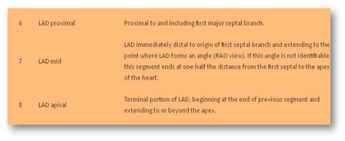
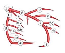
Text books differ in their definition about proximal LAD. Currently , the  SYNTAX  scoring system  has defined the coronary segments in a practical way.

http://www.syntaxscore.com/index.php?option=com_content&view=category&layout=blog&id=1&Itemid=32

Definition from SYNTAX
Read Full Post »

Very often in clinical  practice  cardiologists are asked to R/O significant coronary artery disease in asymptomatic persons .This population includes  people with multiple risk factors like diabetes, HT dyslipidemia  and non specific ST/T changes in ECG.

Many of us have lost the confidence of   ruling out CAD   in these population without looking at their  coronary angiogram.

Is it a right way of practicing cardiology ?

What we need to realise is,  we are asked  to rule out any critical lesions that are going to make a impact on these  other wise comfortable patients.  Nothing wrong if you miss a 30% lesion in PDA or OMs or diagonals !

Can we do this without doing coronary angiogram ?

Yes ,  we can .

Step by step  Ask these questions

  1. Ask the patient , if  he /she   can climb three  flight of  stairs  without any difficulty or
  2. Walk briskly for  20 minutes (5km/hr)

If yes , give  a   certificate   that he  has no critical  left main or proximal LAD  disease.

If you do not believe in his words , put him on a tread mill ,  if he crosses   stage  3   Bruce in TMT ( 9 mts)

give the above certificate  “with a frame”  now .

For still suspicious  physicians ,  We have  one more  investigation called  echocardiography !

Echo : The forgotten tool  for screening left main lesion.

Modern day echo machines have a  3mm resolution power (Many have 2mm ) .While ,  we are expected to look for 3mm vegetation to R/O Infective endocarditis , rarely is  a  cardiologist ,  tuned to  look for the left main ostium  in routine echocardiography  which averages 4-5mm is size. (Left main by echo link to another article)

In short axis  view just tilt at the level of pulmonary valves  (Atrio- pulmonary sulcus) one can visualise the left main ostium and the proximal left main emerging from the 4 o clock position. If you are lucky you can see the entire left main.

If nothing satisfies the physician (Or the patient)  ,Refer him for sliced CT scan , catheter coronary angiogram , or a  nuclear Imaging .Be ready for the attendant anxiety, interpretation errors, corporate  pressures , urge to  balloon ,  kick backs etc etc

By the way , how can  one  be happy by ruling out only left main disease ?  Is it not other lesions possible ?

Experience (Not science) has taught us  no  critical coronary obstruction is  possible ,  if  a patient walks for  9 minutes  in treadmill (10METS).

Even if it is there (A remote chance)  there is little documented benefit of any revascularisation procedure.

Counter point ?

Is it not a “crazy idea  to rely on patients history in ruling out  CAD   in these era , where   angiograms relayed  live  into   cardiologists  ipad  ?

Science has no value if it is not applied  for the patients welfare. Meticulous clinical  examination (And application of mind)  is the foundation stone on which  any medical investigation and therapy  should be based  upon. Most of the inappropriate coronary revascularisation are due to  neglect   of  this vital  component of clinical examination.

(I wonder ,  is it  really possible  these ” acts of omission”   be  deliberate some times  ! )

Final message

Clinical interrogation  may  miss an insignificant  CAD  ,  but it can never miss a critical CAD* .


Do not do coronary angiogram routinely to R/O  CAD.

It is not the way cardiology is to be practiced !

If only we apply  those  simple,  time tested concepts in every day practice we not only  save millions of  Rupees ,   but also thousands of futile   diagnostic tests and associated untoward effects can be avoided.

* Senstivity of  ruling out any CAD is about 70% , but it’s capcity to R/O critical CAD approaches 100%.

Reference:

Please refer your own Brain.

Read Full Post »

T waves attract less  attention in STEMI ,except for the  fact   tall T waves  implies   hyper acute phase of  STEMI.

What is the duration of hyper acute phase ?

  1. Few seconds
  2. Few minutes
  3. An hour
  4. Few hours
  5. Any of the above

Answer

No one exactly knows  .It can  be highly variable .  So , 5  could be  the correct answer .  

 * Most importantly  hyper acute phase  need not occur in all patients with STEMI as suggested in experimental models.

Some  observations in T wave behavior in STEMI

Mechanism of hyper acute  T waves

It is the pottsium channel dynamics.Transient intracellular hyperkalemia  is thought to be responsible.

T wave as marker of  reperfusion

Inverted T wave in precordial leads are a good marker of IRA patency  especially in LAD

Slowly evolving STEMI

This is relatively  new concept . STEMI with a prolonged hyper acute phase  ,  ie ,  T waves ” dilly dallying”  for hours or even few days have been recognised. (This was  refered  to pre-infarction angina in the past )

This sort of T wave behavior makes it difficult to diagnose STEMI.Enzymes will help , still  thrombolytic guidelines  demand us to wait till ST elevation to occur. This is  unfortunate .But as physicians we are  justified to thrombolyse tall T waves with a clinical ACS .The other simple solution is to shift the patient to cath lab to find what exactly is happening in the LAD ! 

Now , what is new about  T waves in STEMI ?

It is  the localizing value  in LAD infarct

A tall persistent  hyper acute T wave  helps us to localise a LAD lesion .This paper from Netherlands ,  clearly  confirms this observation. The study was done from a primary PCI cohort,   a perfect setting to assess the  T wave behavior  in the early minutes /hours of  STEMI .

Other mysteries about T waves in STEMI

Does hyperacute T waves  occur in infero-posterior STEMI ?

I would believe it is very rare .Our CCU has not seen any tall T waves in inferior lead. Further analysis of the  data from the  above study could answer this question .

How often a  hyperacute T waves transform into NSTEMI ?

This again is not clear.Most of the hyper acute T will evolve as STEMI .But  , nothing prevents it to evolve as NSTEMI a well . After all , a hyper acute T   MI can  spontaneously lyse in a lucky few , ( Who has that critical  mass of natural  circulating TPA )  .If  these natural lytic forces are only partially successful , it may evolve into de nova NSTEMI.

Bi-phasic T waves in ACS.

A benign looking T waves with terminal negativity in precordial leads  can some times be a deadly marker of critical LAD disease.This has been notorious to cause deaths in young men which often correlates with the widow maker lesion in LAD.

What is a slowly evolving STEMI ?

Prolonged tall T wave phase  possibly   indicate , the myocardium is relatively resistant to hypoxic damage .

The most bizarre aspect in our understanding about ACS pathophysiology  is the concept of  time window , based on which , all our  ACS therapeutics revolve !

Does all myocardial   cells  have a same ischemic shelf  life ?  Can some patients  be  blessed with  resistant myocardial cells   when confronted with hypoxia or ischemia ?

                                 It is well-known  , in some hearts ,  the  muscles go for necrosis within  30 minutes of  ischemia,  while some hearts can not be infarcted even after 24 hours of occlusion .So , slowly evolving STEMI is a feature of  myocardial ischemic resistance .This is not  a new phenomenon as we have extensively studied about the concept   ischemic preconditioning .

We wonder there is something more to it . . .  the quantum of preconditioning  can be inherited .Further  , we are grossly ignorant about  the molecular secrets of  non ischemic metabolic  preconditioning  .

Final message

                         T waves attract less  attention in STEMI . Cardiologists are often tuned to look only the ST segment , after all ,  ACS  itself is classified based on  the behavior of this segment.(STEMI/NSTEMI) . We need to recognise ,there is a significant subset of ACS   affecting exclusively T waves.  Shall we call T elevation  MI ? ( TEMI )

Do not ignore T waves in STEMI. It has more hidden electrophysiological  treasures that  is waiting to be explored .

Read Full Post »

Can we advice CABG for single vessel disease  ?

Yes, CABG  may be indicated  in

  • Critical , proximal , complex  LAD disease   with or without  ostium involvement.
  • Many of the bifurcation lesions with large and significant  side branch
  • Small caliber LAD with diffuse disease .

When these occur  in diabetic  subjects , the  indication for CABG is more certain .

* Present generation cardiologists  would feel  every  lesion  is  stentable and should not be referred to the surgeon .But it should be emphasized here,   technical feasibility alone  ,  does not  imply  PCI is superior and ideal in all coronary interventions.

Can we do a CABG  in  single vessel disease  with  normal  LAD ?

CABG is  very rarely  indicated   for isolated RCA or LCX disease. It should be consciously avoided in this patient population.

This is because the at risk myocardium  supplied by these vessels are far less than that of LAD. PCI  is  preferred    in these vessels .(Ofcourse , after considering medical management  ) .

CABG is  ,  too traumatic a  surgery , to  offer  in this  low  risk  coronary  lesions.

Exceptions

CABG  can still be done in following situations  for non LAD single vessel disease.

  • Left dominant circulation  with  complex lesions in LCX /OMs.
  • It is common to see diffuse , long segment  and severe disease of RCA with normal LAD /LCX system .PCI is not feasible in this subset.
  • Failed PCI
  • Recurrent instent restenosis.
  • Bail out CABG after a acute complication during PCI

One should remember ,  inability to do a PCI  does not  mean ,  the patient  should   land in surgeon’s table .We should recall , from our memory medical management is an effective and established form of treatment in single vessel disease ( Mainly for non LAD , and some cases of LAD also !)


Read Full Post »

pci-ptca-ebm-stent

Scientifically ,  the  indication for coronary revascularisation   should be  based on following

  1. Patient’s  symptom ( more specifically angina , dyspnea is less important !)
  2. Prov0kable  ischemia  ( A significantly positive stress test )
  3. Signifcant LV dysfunction with  documented  viable myocardium &  residual ischemia
  4. A revascularisation eligible coronary anatomy * TVD/Left main/Proximal LAD etc ( *Either 1, 2 or 3 should be  present  in addition )
  5. All emergency PCI during STEMI /High risk NSTEMI

Practically ,

A CAD  patient  may fulfill  “Any of the above 5 or  “None of the above 5” ,  but ,  if   a coronary obstruction  was  revealed  by coronary angiogram  and if he  fulfils The 6th criteria , he becomes  eligible for  revascualrisation

6th criteria

If the patient has  enough monetary   resources (by self  ) or by  an  insurance company  to take care of PCI /CABG *

*The sixth  criteria overrides all other criteria in many of the cath labs .Of course , there are few genuine ones still  fighting hard , to keep the commerce out ,  from contaminating cardiology !

Read Full Post »

Why PCI  in   left main CAD is considered  an inferior modality than CABG ?

CABG is superior to PCI for the  simple reason it provides complete revascularisation virtually in all  patients with LMCAD , while PCI is possible only in a fraction of patients with LMCAD.

If  we take 100 patients  with left main  disease may be ten (At best !)   would be  suitable for PCI ! In other words PCI is contraindicated in vast majority of LMCAD  by technical criteria alone , while there can never be a contraindication for CABG in patients with LMCAD.(Except  when , comorbidity precludes surgery )

Why  PCI in  LMCAD difficult ?

It is  dependent on  technicalities

CABG does not tackle a lesion,  it simply avoids it  and by passes it ” No great brains required”

while PCI takes on the plaque frontally ,  in the dangerous  terrain of  left main artery  itself !

so,  much caution,  planing ,  logistics are required . Further ,  if there is a complication there is a potential

for catastrophe  as the only  supply line is cut off . This is the reason , cardiologists were worried to try this on

unprotected left main. (Protected LMCAD refers to left main disease following CABG  wherein atleast   LAD or LCX is  grafted )

Points to ponder in LMCAD

  • PCI is suited for isolated discrete LM disease.In realty  this is seen in less  than 5-8 % CAD.
  • LMCAD is very often associated  with  critical and multivessel distal CAD . So these patients will be candidates for CABG.
  • Left main ostium or LAD ostial  involvement makes PCI a tougher exercise
  • Calcification is more common in LMCAD that  again makes PCI difficult.

The following article in Feb 2009 is a major blow for proponents of  PCI for left main

http://circ.ahajournals.org/cgi/content/extract/119/7/1013

left-main

http://content.onlinejacc.org/cgi/content/abstract/51/5/538?ijkey=84c977d189e84327c3abbd4c1228de17dd99048a&keytype2=tf_ipsecsha

Final message

  • Conquering left main disease is an interventionist’s  ultimate dream.
  • But, before that they have  to tackle the bifurcation lesions .This is of vital importance, because 2/3 rd of left main  patients have  some form of bifurcation lesions. Current techniques , hardware  and outcomes are far below the idealistic solutions in bifurcation lesions.
  • Till that time ,  CABG would  remain the only choice for all , but for  a small fraction of isolated  left main disease where PCI may be possible.

Read Full Post »

Coronary artery disease has a strong   predilection  to involve proximal segments.

But in a significant population it affects only the distal vascular bed ! What is the extent of this problem ?

This paper was presented in  the annual sessions of Cardiological society of India , Mumbai 2005

Down load presentation

distal-cad-csi-2005

Read Full Post »