Feeds:
Posts
Comments

Posts Tagged ‘left main disease’

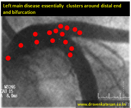
Conquering  left main disease is considered as crowning glory for the Interventional cardiologists. For over three decades , CABG has remained the undisputed modality which is being challenged  today. Fortunately, the Incidence of true isolated  left main disease is  low .(If Medina bifurcation subset is excluded)


left main

With growing expertise , advanced hardware and Imaging ( like a 360 degree OCT fly through view ) one can virtually sit inside the left main and complete a PCI .

Still , coronary care is much . . . much  . . . more than a technology in transit !

Most importantly, these complex PCIs require rigorous maintenance protocol  with meticulous platelet knockout drugs , patient compliance and the genetic fate of drug efficacy . (Clopidogrel has since entered the final laps of inefficiency while Ticagrelor has some more time I guess !)

What is the current thinking  about  unprotected left main PCI ? Let us know it from real life experts !

For those answered , yes to  the above question please leave this page , as the following question might  trouble you much !

While competent surgeons are waiting to tackle left main by surgical means ,there are many centers which are Inclined towards  PCI though we lack long-term outcome (At least 10 years like CABG )

Why do you think this is happening ? Are you ready for another crooked poll ?!  

What exactly is left main disease ?

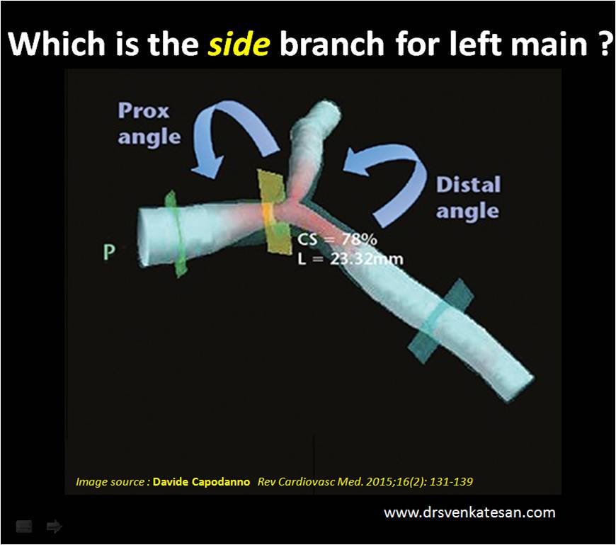
Some of  us also suffer from a knowledge gap and tend to think  Bifurcation lesions  and left main disease are two distinct entities .The fact of the matter is , significant subset of bifurcation lesions are Indeed either left main equivalents or true left mains ( Medina 1,1,1 would constitute > 50 % all  bifurc lesions )  If you include Invisible left main lesions in Medina ( 0,1,1 or 0,0,1 ) detected by IVUS/OCT  it might reach easily cross 90% (Scientific guess !)  Does that mean we have to think CABG even for all complex bifurcation lesions ? and reserve left main disease for isolated discrete mid shaft or ostial left main ?

Final message 

My observation (Sincere to my limited conscience !) at least in this part of the world is : Left main Interventions are  “perceived as pride” and its more related to “show of expertise” and is little to do with patient outcome.Unfortunately , cardiologists should not be blamed for it in isolation as the studies they follow are conflicted.

Forget SYNTAX/PRECOMBAT trials, the two famous studies EXCEL (Favor PCI) and NOBLE were published in 2016 made our life tough .One suggested PCI is acceptable /on par with CABG, while the  other one put CABG superior , ensuring clarity  replaced with confusion ! When we have a dispute , logic would suggest we should fall back on the status quo ie “CABG is superior” unless proved convincingly. Many sections of cardiology society failed to appreciate this.

Post PCI thoughts

*It may not be that hard to do a complex PCI . But, it’s never easier to understand current cardiology literature that is supposed to raise our intellect , which has a direct relevance to patient welfare. Note, many crucial , high stake studies  tend to play academic deceit games  with  linguistic and statistical hyperboles like Non Inferior , likely superiority , Never inferior , near equipoise , regression of hazards, virtual follow-up in  real vs trial world etc , etc !

I can only hope for a better scientific world !

Reference 

  1. Which is the best option for left main disease PCI or CABG ?  Journal of Individual wisdom and evidence based conscience : Volume 1 Chapter 1- Coronary Intellect : Pages 0 to ∞ Jan 2018.

Read Full Post »

Bifurcation  angioplasty is a  newly conquered(Or not yet !)  target  for Interventional cardiologists.We have come a long way  in planning  interventions  for left main  with state of the art  hardware, expertise and  image assistance .However , every  classification , approach, strategy  for BFL talks about tackling the main and  side branches meticulously.

Still . . . one question  is not answered clearly is  . . .

A mini MCQ.

Answer: Open for contribution.

My inference

*It all depends upon the Indication and Individual arterial ischemic burden. In ACS, if  LAD territory is infarcted and beyond 24 hours.LAD becomes a  side kick to the vital LCX which supplies  the remaining life sustaining myocardium which includes the critical basal segments.

Final message 

Since , the risks involved in the interventions of  left main and its bifurcation is inherently linked  with , what exactly we mean (and do ! ) to the side branch .Its mandatory we spare few intellectual moments before our hands invade the coronary battle zone.

Read Full Post »

Medina classification  is the most popular angiographic classification  of bifurcation lesions based on the presence or absence lesions at the three levels  of branching  (0,0,0 ) to (1,1,1). The popularity of this scheme is essentially due to its simplicity.

It can further be subdivided according to angle and size .Though there are three angles possible it is the angle of LM with LCX that matters most.

T shaped  left main. Angle of LM-LCX is around 90 Degrees

Y shaped left main. Angle of LM- LCX is > 120 Degrees

Three types of Y according to size of branch vessel size.

Y1 Large left main divided two equal LAD, LCX.

Y2 Left main and one of its branches are equal

Y3 All three are equal diameter.

Here is a series of  lectures on left main (Probably the best I guess  !)  from Dr.Boris Varshisky ,Hadassah University hospital  Jeruselam.He critically discusses about the   nuances of left main disease from pathology, technical and therapeutic considerations.

Spend some time on these videos , you should be able to learn about

  • Distribution of left main disease
  • The complexities in defining the true shapes of of left main ostia .(Ostial sharing between LCX and LAD ?)
  • Lesion based strategy
  • Carinal shift vs plaque shift
  • Stent sizing in Y 3 left main

and much , much  more !

Read Full Post »

Distribution of Left main disease.

  1. Ostial
  2. Ostio-proximal (Within 1 cm of  origin )
  3. Shaft -Discrete  mid left main
  4. Shaft -Diffuse
  5. Isolated distal shaft( 1.0.0)
  6. Bifurcation ( Medina 1.1.0 -LAD)*
  7. Bifurcation (Median 1.1.0-LCX)
  8. Bifurcation ( Median 1.1.1)*
  9. Trifurcation ( With ramus )

* These three locations account for nearly 75% of all left main lesions.

left main disease coronary angiogram

We know atherosclerosis is  a branch point disease .Normal left main measures 1 mm to 20mm.The shorter the left main lesser is the the incidence of LMD. Short left main can not engage the atherosclerosis much (No left main = No left main disease ) However ,very short left mains  may increase ostial lesions .

  1. The commonest left main lesion is distal left main with one of the branch involvement (1.1.0.LAD is more common )
  2. Least common entity is discrete mid shaft lesion.

Simple strategy.

First dictum : All complex looking LMDs should be referred to a good  surgeon.

Final dictum : Remember medical management for left main disease is still an accepted strategy in stable , non flow limiting situations .

Interventional  Cardiologists  feel they have the exclusive rights   to indulge between these two  spectrum of LMD .May be true! But extreme caution is required as we are playing  our game in the most critical  coronary high way .

Some suggestions and thoughts.

  • 50 % diameter stenosis is significant. But significance does not mean we should tackle the lesion by aggression.
  • Symptomatic flow limiting lesion only to be intervened . (Flow limiting means both angiographic and a stress test .FFR <.8 is also an index for flow limiting .Symptom means Angina on exertion )
  • IVUS, OCT, FFR,NIR ,SYNTAX  are not path breaking tools .They essentially  add  more glamor  to left main disease than anything .
  • Most bifurcation LMDs are  managed by single stent with stent jailing the major side branch (Yes side branch can be LCX !)
  • However ,two stent strategies is not banished .It can be vastly  superior in some selected cases .(Especially with huge plaque load at carina )But needs expertise .
  • In very small vessels two stent strategies are risky .

Reference (2012 update)

left main disease  coroanry angiogram management  Fajadet

PRECOMBAT left main disease south korea everolimus
Link to related articles in this site

Read Full Post »

Very often in clinical  practice  cardiologists are asked to R/O significant coronary artery disease in asymptomatic persons .This population includes  people with multiple risk factors like diabetes, HT dyslipidemia  and non specific ST/T changes in ECG.

Many of us have lost the confidence of   ruling out CAD   in these population without looking at their  coronary angiogram.

Is it a right way of practicing cardiology ?

What we need to realise is,  we are asked  to rule out any critical lesions that are going to make a impact on these  other wise comfortable patients.  Nothing wrong if you miss a 30% lesion in PDA or OMs or diagonals !

Can we do this without doing coronary angiogram ?

Yes ,  we can .

Step by step  Ask these questions

  1. Ask the patient , if  he /she   can climb three  flight of  stairs  without any difficulty or
  2. Walk briskly for  20 minutes (5km/hr)

If yes , give  a   certificate   that he  has no critical  left main or proximal LAD  disease.

If you do not believe in his words , put him on a tread mill ,  if he crosses   stage  3   Bruce in TMT ( 9 mts)

give the above certificate  “with a frame”  now .

For still suspicious  physicians ,  We have  one more  investigation called  echocardiography !

Echo : The forgotten tool  for screening left main lesion.

Modern day echo machines have a  3mm resolution power (Many have 2mm ) .While ,  we are expected to look for 3mm vegetation to R/O Infective endocarditis , rarely is  a  cardiologist ,  tuned to  look for the left main ostium  in routine echocardiography  which averages 4-5mm is size. (Left main by echo link to another article)

In short axis  view just tilt at the level of pulmonary valves  (Atrio- pulmonary sulcus) one can visualise the left main ostium and the proximal left main emerging from the 4 o clock position. If you are lucky you can see the entire left main.

If nothing satisfies the physician (Or the patient)  ,Refer him for sliced CT scan , catheter coronary angiogram , or a  nuclear Imaging .Be ready for the attendant anxiety, interpretation errors, corporate  pressures , urge to  balloon ,  kick backs etc etc

By the way , how can  one  be happy by ruling out only left main disease ?  Is it not other lesions possible ?

Experience (Not science) has taught us  no  critical coronary obstruction is  possible ,  if  a patient walks for  9 minutes  in treadmill (10METS).

Even if it is there (A remote chance)  there is little documented benefit of any revascularisation procedure.

Counter point ?

Is it not a “crazy idea  to rely on patients history in ruling out  CAD   in these era , where   angiograms relayed  live  into   cardiologists  ipad  ?

Science has no value if it is not applied  for the patients welfare. Meticulous clinical  examination (And application of mind)  is the foundation stone on which  any medical investigation and therapy  should be based  upon. Most of the inappropriate coronary revascularisation are due to  neglect   of  this vital  component of clinical examination.

(I wonder ,  is it  really possible  these ” acts of omission”   be  deliberate some times  ! )

Final message

Clinical interrogation  may  miss an insignificant  CAD  ,  but it can never miss a critical CAD* .


Do not do coronary angiogram routinely to R/O  CAD.

It is not the way cardiology is to be practiced !

If only we apply  those  simple,  time tested concepts in every day practice we not only  save millions of  Rupees ,   but also thousands of futile   diagnostic tests and associated untoward effects can be avoided.

* Senstivity of  ruling out any CAD is about 70% , but it’s capcity to R/O critical CAD approaches 100%.

Reference:

Please refer your own Brain.

Read Full Post »

Imaging  coroanry artery is  generally  in the   domain of interventional cardiologists. MDCT has helped us to change that.

The  humble echocardiography can   identify the origin* of   coronary arteries   in  most   persons. The resolution power of modern day echocardiography is  2mm and the left main  ostium is >3.5mm in 99%  of population . If some body says one can’t  visualise the coronary artery by echo ,   it can only reflect their ignorance or lack of patience to get an optimal image. Of course technological limitations are there.

*  To be emphasised again , only the origin can be identified.

Can we identify ostial leftmain or proximal  left main disease  by echocardiography ?

It should be possible in  few .

Can we place  a doppler sample volume  within  the left main and measure coronary flow velocity ?

When obsterticians are able to  assess the  uterine artery flow  in a bulky uterus ,  it should be possible to do the same in  a coronary artery . Motion artifacts is the issue in the heart.  Micro sample voulme (<1mm) are expected in the future  that will make a non invasive coronary flow assesment a distinct possibility.

Read Full Post »

Exercise stress testing(EST)  is one of the common investigation modality in the evaluation of CAD.he indication for EST  generally fall into two broad categories.

  • Diagnostic in patients suspected to have CAD
  • Prognostic evaluation in patients with established CAD .9Many times after a coronary angiogram)

Currently there is a major shift in our thinking,  patients with  classical angina  may undergo coronary angiogram  directly .This is understandable as the stress test  has little to   improve  diagnostic  sensitivity and specificity in patents with clinically obvious CAD.

So , it is now becoming clear , the diagnostic  value  is  increasingly  restricted in the evaluation of  o atypical chest pain .

What is a strongly positive response ?

  • Gross ST segment depression > 2-3mm
  • Occurring in stage one
  • Fall in blood pressure
  • Prolonged angina into recovery

What is the angiographic  correlates of strongly positive EST?

  • Critical left main disease
  • Near total proximal LAD /LCX
  • A severely compromised bifurcation lesion

Morphological correlation

  • These patients  often have eccentric lesions with irregular margins.
  • unstable  lesions
  • Lack collaterals

What is the effect of vigorous  excercise on a critical flow limiting lesion ?

The shear stress over the plaque  increases  with  exercise  and  the  transcoronary gradient can reach a theoretical 60-90mmhg .One can imagine the what this stress can do to the  unstable lipid core .This is the reason unstable angina is an absolute contraindication  to EST.

What does a strongly positive EST imply for the patient ?

  • It indicates he needs urgent CAG and  most likely an immediate revascularisation.
  • Often , these patients have prolonged angina , and mandates admission in a coronary care unit.
  • there has been many incidence of ACS in these  patients  within 24hours of EST.
  • Lives have been lost  on their  way back    ,   as  these patients are sent home , as EST is a  OP procedure .

Final message

  1. It need to be realised a strongly positive response to EST  could  be a  clinical equivalent of  unstable angina .
  2. The common response  from a   physician or cardiologist    after witnessing  a  gross ST depression to EST  would be   “Had  I known this  I would have sent him straight into cathlab instead of EST ”
  3. If only , we give little ear to our patient’s  history we can pick the high risk clue in 9 out of 10 cases !
  4. It can be argued ,  a strongly   positive  EST  by itself  is  “A  clinical diagnostic  failure”  ,   ie  failure  of the physician  to recognise  the likely hood of strongly  positive EST ie a left main disease.
  5. These patients  should never be sent home immediately  after the EST .This is fraught with a risk SCD
  6. Most of them will require observation in step down unit for 24 hours  and if feasible they should be posted for coronary angiogram in the earliest available slot.

Read Full Post »

The term  left main disease (LMD)  invariably  creates  a  near  panic  reaction in   many of the   contemporary cardiologists . It may be acceptable   in  a broader sense, but it need to be realised , there  is a significant group of patients  with  isolated  non critical  LMD . Many times , these patients can be managed effectively with  intensive medical management.

However , the  following rules may be applied in the management of non critical isolated LMD

  • In patients  who present with unstable angina  ,there is nothing called non critical LMD .Any degree of lesion (Even a 20%)  is significant .
  • Lesions with irregular margins, hanging eccentric  plaques are always critical irrespective of obstruction.
  • LMD involving LAD /LCX ostium need to be tackled as an  emergency .

What are the safe left main disease ?

  • Isolated tapering  left  main artery .
  • LMD  with <  50% lesion.
  • A left main patient who is pain-free on a tread mill > 10 METS
  • Left main with stable angina responding well to medical therapy
  • New onset left main disease in a patient with functional LIMA to LAD /LCX *

*This is sometimes called protected  LMD.  Protects what ? Protects the LAD ,   in case of  complication occurring during LM stenting . If the  function of  LIMA graft is good enough to  protect LAD , why should we attempt to open  the diseased LM in the first place ?  It is an unanswered question !

Why is it riskier  to  stent an  insignificant  LMD or stable  LMD ?

A  left main artery ,  engulfed with a  50%   stable plaque is less riskier to develop an  ACS than an  artificially  normalised  left main lumen with a stent. This is especially true for the  drug eluting stents which need life long  dual antiplatelet therapy as the drug which is supposed  to  prevent  the  restenosis ,  interferes  with  the normal endothelialisation over the stent .

In effect,   PCI   especially  with a DES for a hemodynamically insignificant lesion is fraught with a risk of converting a stable  lesion into  potentially vulnerable lesion !

Final message

A discerning  reader may ask , is it possible at all ? . . .to  have a   patient  with LMD  & enjoying  good exercise capacity ?

Yes , it  may be  rare , but not “non existent” .  Remember ,  one of   the common cause  for  rarity in medicine is ” non recognition of a  fact” or   otherwise  called ” Ignorance”

It is an irony , LMD is considered  by many as a  homogenous  entity ,  even as we  acknowledge  there is a  huge spectrum of lesions among left main disease . There is a distinct (although small !  )  subset of LMD * where medical treatment could be ideal and PCI  may even carry greater hazard.

*The most important caveat in assessing a LMD  lies  in the   50% criteria. Calipers  we use ( often visual )are never going to estimate the lesion correctly considering the importance of  Glagovian  phenomenon . As of now ,  we have no simple means to  measure the vulnerability of a left main plaque .Thermography, OCR/Raman spectroscopy/ RF intravascular ultrasound would probable redefine the indications for intervention in LMD.

Legal issue in LMD

Can we  defend in the  court of law, if a patient loses  his life,  who was adviced  medical management for LMD ?

Any thing can be defended in this funny world of  judiciary . A person who kills in broad day light,  hundreds of  innocent lives can argue  he has never seen a gun ! and he may even,  be  acquitted  for want of evidence  !

How can we   prove  with evidence , the  death in question  occurred  “only because ” he was  adviced medical management ?

No court on the earth can prove it !

So , an occasional life lost due to an unintentional  judgment  error can  easily be argued in favor of the noble profession . Scientific  guidelines are only recommendations .If a person with a  significant LMD  due to  a smooth stable plaque , who has  little  symptoms , carry on with his daily activities comfortably  , his  cardiologist has every right to advice him  medical management.  The doctor , can not be penalised , provided  , he has explained  to the patient ,  that  he is deviating from the official guideline only  for the benefit of   the patient’s  health and  he   has  fully understood the issue.

Read further , for  more controversy !

Land mark  randomised control trials (RCTs) are generally  done in specialised centres with high degree of expertise . They rarely represent the real world patients  seen in  the remote towns (or even  cities ) of the  developing countries  .We can not equate a PCI  done in an  angiographic core laboratory , say in Cleveland or Mayo clinic  ,  with that of  cath labs  ,  that  works  with par time staff and non dedicated cardiologists . So , in these situations  intensive medical therapy (which do not have a geographical variation in efficacy! ) would score over complex procedures .

Read Full Post »

Left main disease is the  most dangerous subset of CAD population .The danger is attributable more on the perceived fear  of  sudden occlusion .How often this occurs in stable , left main plaques is not known.

There is a significant group of patients with isolated ,  asymptomatic ,  non flow limiting , leftmain  disease with stable , smooth plaques. The ideal management for this group of CAD is not clear.

left-main-d

Advising a  CABG /PCI  is  an easy and very practical  decision ! That’s what the current guideline also suggest

But is there scientific evidence  to do that ? Many times practical approach  could be    synonymous with  an  unscientific approach.

  • PCI has a potential to  convert  a stable plaque into  a vulnerable one (Metalled plaque is not inert )
  • CABG will reduce the flow across , the  already narrowed left main and  there is  a likely hood of rapid  progression of native left main disease

So what is left  ?

If it is a stable plaque ,  and does not limit the flow both at rest on exercise * medical management will be optimal.

* Excercise stress test must be done

Read this article from the circulation ,  that suggests  a role for medical management for left main disease

http://circ.ahajournals.org/cgi/content/full/118/4/422

left-main-circualtion1

Read Full Post »