Feeds:
Posts
Comments

Posts Tagged ‘left main artery imaging’

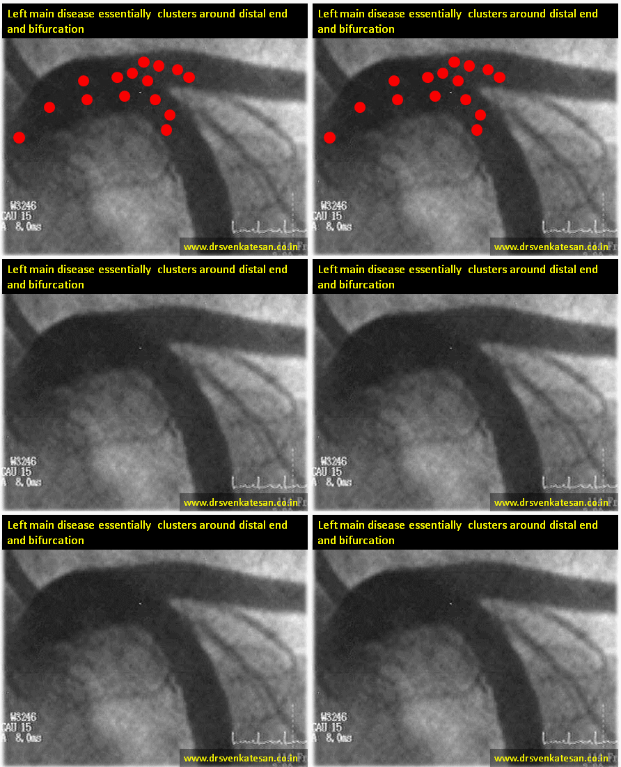
Distribution of Left main disease.

  1. Ostial
  2. Ostio-proximal (Within 1 cm of  origin )
  3. Shaft -Discrete  mid left main
  4. Shaft -Diffuse
  5. Isolated distal shaft( 1.0.0)
  6. Bifurcation ( Medina 1.1.0 -LAD)*
  7. Bifurcation (Median 1.1.0-LCX)
  8. Bifurcation ( Median 1.1.1)*
  9. Trifurcation ( With ramus )

* These three locations account for nearly 75% of all left main lesions.

left main disease coronary angiogram

We know atherosclerosis is  a branch point disease .Normal left main measures 1 mm to 20mm.The shorter the left main lesser is the the incidence of LMD. Short left main can not engage the atherosclerosis much (No left main = No left main disease ) However ,very short left mains  may increase ostial lesions .

  1. The commonest left main lesion is distal left main with one of the branch involvement (1.1.0.LAD is more common )
  2. Least common entity is discrete mid shaft lesion.

Simple strategy.

First dictum : All complex looking LMDs should be referred to a good  surgeon.

Final dictum : Remember medical management for left main disease is still an accepted strategy in stable , non flow limiting situations .

Interventional  Cardiologists  feel they have the exclusive rights   to indulge between these two  spectrum of LMD .May be true! But extreme caution is required as we are playing  our game in the most critical  coronary high way .

Some suggestions and thoughts.

  • 50 % diameter stenosis is significant. But significance does not mean we should tackle the lesion by aggression.
  • Symptomatic flow limiting lesion only to be intervened . (Flow limiting means both angiographic and a stress test .FFR <.8 is also an index for flow limiting .Symptom means Angina on exertion )
  • IVUS, OCT, FFR,NIR ,SYNTAX  are not path breaking tools .They essentially  add  more glamor  to left main disease than anything .
  • Most bifurcation LMDs are  managed by single stent with stent jailing the major side branch (Yes side branch can be LCX !)
  • However ,two stent strategies is not banished .It can be vastly  superior in some selected cases .(Especially with huge plaque load at carina )But needs expertise .
  • In very small vessels two stent strategies are risky .

Reference (2012 update)
left main disease  coroanry angiogram management  Fajadet

PRECOMBAT left main disease south korea everolimus
Link to related articles in this site

Read Full Post »

Left main coronary artery is  considered as the sanctum sanatorium  for  the cardiologists .

One would wish  to rule out  disease of left main  in any given  patient with CAD.

Though there are strong clinical predictors of  LMD, this  segment of the coronary artery  tends to  throw   surprises.

A  strongly positive stress test,  ST elevation in AVR  , fall in blood pressure with exertion  are good markers of left main disease.

Still,  in the era of  optical coherence tomography (OCT )  and IVUS  , we do  have a simple tool that can image the left main coronary artery fairly accurately .

We know the  resolution power of  routine trans thoracic echo  is 3mm and above  . (It can detect vegetation of that size easily !)

So , it can easily accomplish  the task of  imaging the  left main ostium .(which is a minimum of  4-5mm diameter )

How to image left main by echo ?

  • Parasteranal long axis  or short axis  the ideal view. Short axis would also  help.
  • Normal left main is easily diagnosed  by two parallel  lines . ( See above picture )
  • Plaques are  diagnosed when this line is  distorted  and filled by haziness.
  • Significant ostio proximal  lesion must never be missed by TTE .However distal left main can not be assessed in most .
  • Doppler assessment may not be possible in all as pulse doppler sample volume can not be placed in left main.
  • Trans esophageal echo would increase the yield.

Final message

Processing power of echo machines  and  their image quality has improved  vastly over the years. The existing literature about left main imaging  by echo are based on old generation machines. The data are as obsolete as those  machines . This has to be kept in mind.

I wonder why most cardiologist are averse ( rather feel guilty ) to report  the  status of  left  main  artery  by  echo cardiography .

Every patient with  a  positive TMT must undergo a  focused echocardiogram  of  left main . You will be rewarded with a  good glimpse of the sacred segment  of coronary artery 9 out of 10 times  !

So , can we shoot the Left main  at the bed side  ?

Yes definitely  . . . if only we wish to !

* A correction

The left coronary visualised in this parasternal Long axis view is in fact exceptional. The ostium and shaft often better seen in short axis in around 3-4 O clock position.

Read Full Post »