Feeds:
Posts
Comments

Posts Tagged ‘asd rims’

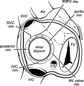
The inferior vena cava (IVC) rim is the most critical rim for successful transcatheter ASD device closure due to its role in preventing device embolization and prolapse. Absence or deficiency of the IVC rim is often a contraindication, as it leads to higher rates of procedural failure compared to other rims like the aortic or SVC.

Anatomical Reasons

The IVC rim forms the posteroinferior border of the secundum ASD, measured in the bicaval TEE view as the distance from the defect’s inferior margin to the IVC ostium. Without ≥5 mm of adequate IVC rim, the device’s right atrial disc lacks stable septal anchorage inferiorly, allowing slippage toward the compliant, funnel-shaped IVC-RA junction. This contrasts with superior rims (SVC), where the SVC-RA junction provides firm foundation.

A dislodged device Video source : APCIS 2016 Jae Young Choi (Yonsei University, Korea) 

Physical and hemodynamic forces at IVC:RA junction

High-volume IVC blood flow (2-3 L/min, 60-70% of venous return) directly strikes the inferior septum and device, exerting continuous shear force that can dislodge a poorly anchored disc, causing left disc prolapse into the RA or embolization. SVC flow (1 L/min) is less voluminous and angled favorably, reducing its destabilizing effect.

If there is good rims on all other sides (270 degreees) can we proceed with ASD device ?

Some cardiologst believe so. Some institutions have reported ASD device closure in deficnet IVC rims as well . This is not respecting the evidence (Ref 4,5) But, the fact seems to be different. The IVC’s perpendicular, high-momentum jet amplifies dislodgement. Even good  superior/anterior rims fail to counter this inferior “pull,” as the device tends to tilt  around the waist, under the  dyanmic atrial pressures and flow. Hence IVC deficiency  predisposes to instability despite the availablity of 270° of other rims.

One caution : A common mistake done is eustachian valves is taken as false IVC rim and the device is implanted and later facing issuers. So, a meticulous TEE imaging is neccessary .

Reference

  1. Amin Z. Considerations for ASD closure. Cardiac Interventions Today. 2014;7(2):1–8.
    Link: https://citoday.com/articles/2014-mar-apr/considerations-for-asd-closure
  2. Amedro P, Bayburt S, Assaidi A, Kreitmann B, Habib G, Fouilloux V, Fraisse A. Should transcatheter closure of atrial septal defects with inferior-posterior deficient rim still be attempted? J Thorac Dis. 2019 Mar;11(3):708-716. doi: 10.21037/jtd.2019.02.89. PMID: 31019758; PMCID: PMC6462671.
  3. Song J. Comprehensive understanding of atrial septal defects by imaging studies for successful transcatheter closure. Korean J Pediatr. 2014 Jul;57(7):297-303. doi: 10.3345/kjp.2014.57.7.297. Epub 2014 Jul 23. PMID: 25114689; PMCID: PMC4127391.
  4. Remadevi KS, Francis E, Kumar RK. Catheter closure of atrial septal defects with deficient inferior vena cava rim under transesophageal echo guidance. Catheter Cardiovasc Interv. 2009 Jan 1;73(1):90-6. doi: 10.1002/ccd.21756. PMID: 19089959.

Postamble

Can we create a neo IVC rim by some means?, (Ex by agumenting the lower end of the IVC with stent , that can protrude clost ASD and act as a rim . We have strated closing the complex sinus venosus ASD, so , this might also be possible)

Read Full Post »

Bicaval view is an Important TEE view to visualize, the LA, IAS, and right atrium. I used to have some trouble getting oriented to this view. Hence this post. It is obtained in the 90-120 degree view at the mid esophageal position. Imagine the patient is lying on his left side and the probe comes from above down between the spine and heart to the LA from within the esophagus. This is the best view to see IAS in the profile.(Subcostal TTE can also do it) Note how the LA hugs the right atrium which is actually an ill-defined (In TEE I mean) common meeting point of both IVC and SVC. Also important is the relationship of RUPV with SVC & the horizontally running RPA sitting right over the top of LA.

The relationship between RUPV and SVC is crucial in device closure of large ASD, especially in sinus venous defect.

Clinical Importance of this view

  • Very useful in ASD rim morphology especially in the posterosuperior rim.
  • Delineates clearly the defect boundaries in SVC ASD.

Sinus venosus defect: Image source not known. Thanks to the creator.

  • This view doesn’t miss even the smallest PFO (With Contrast )
  • Can be used to guide IAS puncture in structural heart Interventions.
  • IVC /SVC mass extension into RA well visualized.

RA myxoma attached to septum: Image source -Michael Essandoh from Research gate

Final message

Getting oriented to TEE  planes and images is so useful in structural heart interventions, like TAVRs, mitral clip, LAA occluder, tandem heart, valve in valves, etc. It is indeed a tough exercise and requires re-learning of cardiac anatomy with fluoroscopic overlay*.I wish, I go back and sit with first-year medical school students and start all over again.

*Current hybrid cath labs do provide Echo/Fluro co-registration, still it demands core 3D anatomical Imagination.

Read Full Post »


asd-closure-device-www-drsvenkatesan-com

The following  factors are critical determinants of the success of ASD device closure

  1. Location of the defect ( Only ostium secundum )
  2. Size of the defect (<35mm .Never forget  simple truth , larger the defect shorter would be the  rim )
  3. Shape  of the defect (Please note ,none are strictly circular but most devices are ! )
  4. Eccentricity of the defect (RA aspect of ASD need not match LA aspect)
  5. Length of the rim ( 5mm said to be adequate)
  6. Thickness of the rim ( Least respected parameter .Thin filament like rims are notorious  in sagging the device into RA)
  7. Pre and Per- operative TEE (As Vital as the procedure)
  8. Technical expertise . (This includes extreme patience  of the primary operator .Most sub optimal results and complications are related to this.
  9. Good team ( Not every  interventional  cardiologist should  attempt this !)
  10. Courage to abandon the procedure
  11. Device brand (Probably less important )

Read Full Post »

This article   provides  you every thing  you  want to learn  about ASD device clsoure .The anatomy , the art of doing TEE in cath lab etc.Do not ever shy away from lesser known journals .It is simply amazing  to find  hidden treasures .Thanks to  Mr Tim BernersLee  invenor of the Internet !


mexican cardiology journal


Sample this arricle

 http://www.elsevier.pt/en/pdf/90140903/S300/


Read Full Post »

ASD device closure as a modality is constantly improving  . . . but  the consensus is  , it is  yet to catch up with  of  good old surgical  outcome . The key to success is not only in the device but hugely dependent on the technique and pre-procedure evaluation  .In fact , the pre procedure TEE imaging technique  is as important as the procedure itself.

There are lots of discussion about this particular issue. TEE is mandatory we know  but now we realise it is  still better to have a  Real time 3dimensional  (RT3D ) TEE . Rim  size  and ASD  morphology estimation is  the primary aim.

There are  at least 6 named rims for ASD. For a circular  orifice  it  may not be logical to have a fixed number of  6  rims . Ideally the entire circumference must have a rim .( This happens in  central defects )In many,  the complex anatomy of IAS does not allow this. So we are compelled to fix the number of rims to six.

  1. Aortic (Superoanterior),
  2. Mitral (AV valve/ Inferoaterior)
  3. SVC  (Superoposterior),
  4. IVC  (Inferoposterior),
  5. Posterior ( Atrial free wall ).
  6. Coronary sinus rim

One can realise how important these rims are , as  they are the   foundation tissues on which the device is going to be seated for the rest of the patients life.

When do you call a rim is adequate sized ?

5mm is  considered suffice. But it varies depending upon the device and expertise.

Can we deploy an ASD device  in patients   with deficient rims?

Logically the answer is expected  is   “No” but  , many have liberalized the criteria now , after realizing   one may  not have 5mm rim in all six sites in a given patient. If you follow this criteria strictly   you can’t do more than few devices a year !

What is the resolution power of TEE can it miss a 3mm rim  ?

TEE has a good resolution it should pickup any thing equal to 2mm or more.

Which is most important rim and which is the least important rim ?


What are the potential complications that can arise if ASD device is deployed with a critically low rim ?

Having discussed  that every rim is equally vital  ,  we  need to answer this sort of questions  often .  I am waiting to get the  practical tips for the above issue from  my experienced colleagues .  I shall post it soon .

It is sometimes assumed Aortic rim may not be that important .Here is a   good discussion  for  ASD closure with deficient aortic rim from Saudi Arabia  . http://www.rmsolutions.net/rmfiles/SHA21/028002.pdf

Meanwhile let us learn . . .

How to perform the “all important” pre- procedure TEE ?

The following article which also  includes video clippings will be immensely useful for all those enthusiastic cardiologists.Thanks to JACC  for making this link free .

Three cheers to AMRITA team from India

http://imaging.onlinejacc.org/cgi/content-nw/full/2/10/1238/

A stylish article on the topic

Read Full Post »