Feeds:
Posts
Comments

Archive for the ‘Atrial fibrillation’ Category

Answer  is question is wrong : RAA clot do occur in AF and severe right heart failure.It is less often recognised , since echo views are difficult and clinical events are silent.

RAA right atrial appendage clot tee echocardiographyBrief account of RAA clot formation

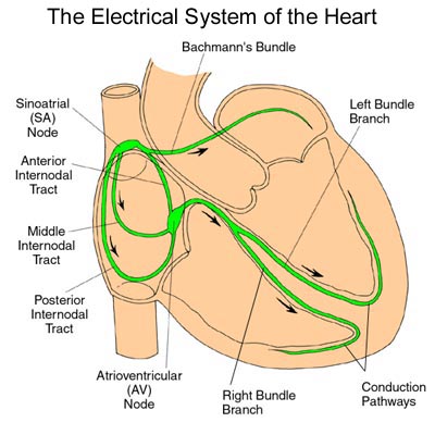
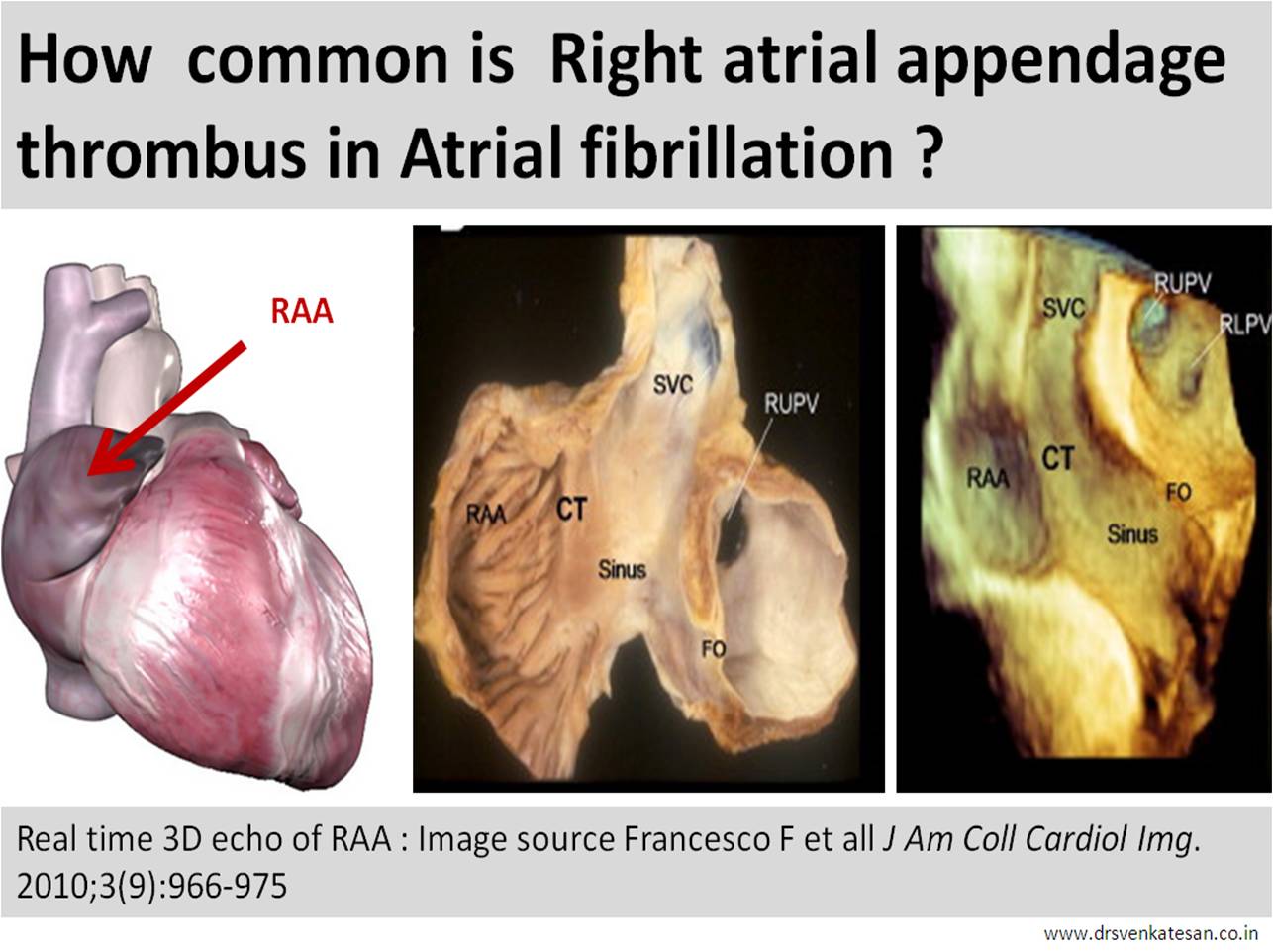
  • RAA is broad flat ,thin ,  chamber comparable to elephant’s ear.The ostium is not that distinct as the body as it  blends  with crista  terminalis .
  • Rough pectinate muscles  should make it prone for thrombus.Further , RAA has more sluggish flow than LAA  increasing the propensity for thrombus.However , the flat nature of the chamber , absence of tortuous tracts , constant  SVC flow which is abutting the  RAA can counteract this.
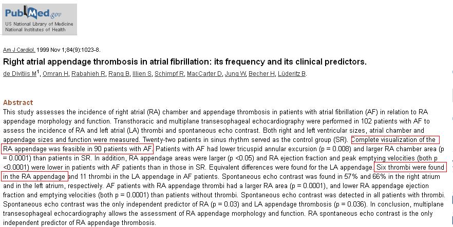
  • RAA clots are  less recognised as echo views are difficult .TEE is often required.
  • Overall RAA clot is 50% less common than LAA.
  • RAA clot should be specifically looked  for  in chronic AF and any severe right heart failure. (Unlike MR jet TR jet has less efficiency in flushing the  Right atrium )
  • Finally,clinical events from RAA clot are less conspicuous as the emboli reaches the pulmonary  bed silently.Unlike its colleague on the left side it  neither triggers TIA nor a stroke !

Reference

right atrial appendage clot raa clot in af atrial fibrillation

1. Buğan B, Baysan O, Demirkol S, Güngör M, Yokuşoğlu M. Right atrial appendage thrombus in a heart failure patient with sinus rhythm. Gulhane Med J. 2011; 53(3): 214-215.


2.Subramaniam B, Riley MF, Panzica PJ, Manning WJ. Transesophageal echocardiographic assessment of right atrial appendage anatomy and function: comparison with the left atrial appendage and implications for local thrombus formation. J Am Soc Echocardiogr.; 2006; 19(4):429-33.

3.Sahin T, Ural D, Kilic T, Bildirici U, Kozdag G, Agacdiken A, Ural E. Right atrial appendage function in different etiologies of permanent atrial fibrillation: a transesophageal echocardiography and tissue Doppler imaging study. Echocardiography;2010; 27(4):384-93

4 .Ozer O, Sari I, Davutoglu V. Right atrial appendage: forgotten part of the heart in atrial fibrillation. Clin Appl Thromb Hemost; 2010; 16(2): 218-20


Read Full Post »

electrical CONDUCTION OF HEART

Image source and courtesy http://www.heart-consult.com

Answer

I am afraid the 4th response is closer to truth .Readers may share their thoughts. If there are three distinct pathways   spreading widely connecting the two spacious chambers and   converging again with  precision at the compact  AV node , it  is a  marvel .

Further , If these pathways are real ,  we must  experience different types of  inter nodal re-entrant  tachycardias.Of-course ,we do come across few macro re-entrant tachycardia in the form of atypical atrial flutters  They need a close  watch .Tracking these arrhythmia may throw light on existence of these pathways.

However, the presence of nodal approaches  with preferential inputs to AV node from different parts of atria would indeed  suggest existence of such pathways !

Further study

What does  sophisticated carto and other electro anatomic mapping say about these inter nodal pathways ?

Reference
An excellent article from imperial college London
Atrial anatomy and inter nodal pathway thorel bachman wenkeback

Heterogeneous three-dimensional anatomical and electrophysiological model of human atria . Seemann G, Höper C, Sachse FB, et al. Institute of Biomedical Engineering, University Karlsruhe (TH), Kaiserstrasse 12, 76128 Karlsruhe, Germany. Transact A Math Phys Eng Sci 2006 Jun 15; 364(1843) :1465-81.

Read Full Post »

No procedure is  impossible in medicine    . . . but it should be useful for the patient !
Where will you ablate ? What is the purpose ?
The much  hyped*  ablation of pulmonary vein  is never going to help in rheumatic heart since  arrhythmia focus occurs at  random . How can you  locate a   focus of AF over a  huge , scarred  left atrium  ? ( which looks like a lunar surface  sprinkled with a rocky terrain ! )
Gross specimen
                                      Note the huge , scarred LA .It would
                                      be a wild guess to locate the true focus
                                      of AF . (Image source : http://www.e-heart.org)
So , the other option was disconnecting atria electrically . In the past  surgeons advocated  linear  or multiple incisions as in Maze and  Corridor procedure  after mitral commissurotomy .This  helped to a certain extent , still effective , organised atrial contraction was not restored in many.
Now , some electrophysiologists tried to do the same with catheters without much benefit.* Please realise , pulmonary vein ablation even in lone and ischemic AF is struggling  with a concept collision !
Can Right atrial focus trigger and  sustain  AF in RHD ?
Autopsy studies reveal tricuspid valve scarring in 33%  of all RHD .Since RHD is a diffuse process , RA lesions can  be very well be the focus .  It is not an easy task to identify the real culprit focus. So ,concentrating  LA  for ablation may end up in futility.
There are only few studies available on RF ablation in RHD  .This one from Istanbul ,Turkey  and it  does not favor it as recurrence rate is still significant .
rf ablation in rheumatic atrial fibrillation
Final message
Approach  to AF in RHD  : Opening up the mitral valve (or replacing it )  and controlling ventricular rate  with beta /calcium blocker along with  adequate  oral  anti-coagulation substantially reduce the risk of embolic events .One may never need to contemplate restoring sinus rhythm  in rheumatic atrial fibrillation.

//

Read Full Post »

« Newer Posts