Myocardial development is a complex process that begins in the precardiac mesoderm, and is regulated by number of genes.

After formation of tubular heart , the initial increase in ventricular mass is achieved by development of trabeculations. Trabecular compaction coincides with genesis of coronary circulation, and results in formation of ventricular chambers.The hallmark of sponge-like myocardium is delayed and poor compactive forces.
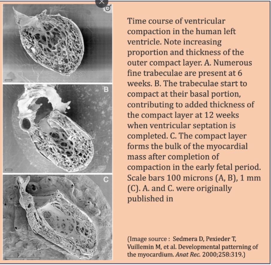
Time line of ventricular compaction
A deranged compaction process need not be macroscopic. It can be very localized, regional, or global. The timing and the quantum of compaction is important , that injects the contractile power to the ventricle . It is increasingly observed in echocardiography that a small percentage of the population has more echo-free zones in LV myocardium than others. They could represent potential weak spots, at times of mechanical stress.
A curious case of normal Echo
Would like to share few images from a routine echocardiography in a healthy young female.
The apparent absence of mid myocardial shadow was not entirely a surprise though .At the same time, it raises some curious thoughts as to why certain myocardial areas are not well visualized by the ultrasound.
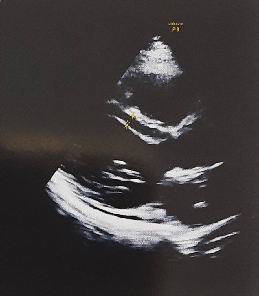

How echo is able to pick up only endocardium and epicardium , making entire myocardium look like an empty shell .
Still, LV is contracting vigorously, implying muscle mass is just not visible to ultrasound eye.
Clinical implication
These echo free dark zones in IVS or LV is so common, one can safely ignore, but its worth recalling few entities, that can be related to this. Intramyocardial hematoma, dissection and ultimately rupture (A case report) when they happen to develop an ACS -STEMI. We know , free wall and IVS rupture and mechanical complication occur signiifcant population of STEMI. Though, we can easily blame it on fate, may be these are the ones , who harbor such silent echo free slits due to defects in compacting genes making the myocardium soft , spongy that gives way wihtout a fight at times of tissue necrosis.
Can non-compaction occur without LV contractile dysfunction ?
Unlikely , is the likely answer from most of us. But , routine TTE might miss subsclinical LV dysfunction . We know , now degree of LV fucntion is directly related to the imaging modal;ity we depende to define it.
Can a consistency or sponginess of a myocardium be detected by echo?“`
As of now, it is not possible. This might become a reality when the science of tactile haptics enters the ultrasound domain.
Final message
“The act of observing changes the observed”
Non compacted LV is casually used term nowadays. The title of this post was made Intentionally provocative to stress a point, that what appears as non- compaction can be observed in normal persons.. However, request the fellows to look little deeper into the myocardial architecture, especially when you witness large echo-free zones.
Mind you, this is different from the well-defined condition of classical non-compaction with excessive deep trabeculations. Don’t know how to name this. This is different. Maybe “Isolated Intra-mural partial non-compaction” is an ideal term.
Counterpoint
When I discussed this with an expert colleague, he said, “No, you’re imagining a non-existing entity.” The ultrasonic interface with myocardium and the interstitial echoes that define the echo texture is so variable.Let us see.time will tell .(See this video and case report )
Postamble
This post is meant to be looked up purely from an academic perspective. Reporting such entities, unless you are absolutely sure, should be avoided, as it ends up increasing the anxiety of our beloved patients.
Reference
2.Kirby ML. Cardiac Development. New York: Oxford University Press; 2007. [Google Scholar] [Ref list]


