Feeds:
Posts
Comments

Archive for November, 2023

Hypertension is a prevalent condition in the general population, as is mitral regurgitation (MR). For most of us, HT traditionally conveys a “singular meaning” that is, high pressure within blood vessels. We often forget that the origin of blood pressure begins right inside the heart, i.e, the left ventricle, which is guarded by two valves – the aortic and mitral. (Though we are aware, LVH is the classical response to HT),

Obviously, there will be signiifcant consequences to the structural integrity of these valves when LV pressure is raised beyond the tolerable limit of endocardial layers that line these valves. Mind you, the bottom of the mitral valve is whipped from beneath 100 tousand times times a day & 2.5 billion times in life time at an average pressure of 140 mmHg, and during exertion, it can reach up to 200 mmHg .Apart from hemodynamic damage, It should be noted MR in HT can be consequence of altered geometery duE to LVH, (Concentric Initially & eccentric in late stages)

The MR-HT link : Not to be missed

It is basic bed-side teaching that any isometric activity will push the blood from LVOT towards the Mitral valve, if it is leaking. Hence, it may not be a great discovery to show that HT will aggravate MR. Now, what is the new message you are trying to tell? There is big data (Really massive one, available from 5 million HT patients Rahimi K, et al 2017) that confirms with authority that HT patients are at risk of developing both primary degenerative and secondary MR at a later age.

If HT accentuates MR , why vasodilators didn’t show much beneift in MR ?

It is true routine vasodilators didn’t do much in regression of MR, but the subgroup analysis of those patients who have intrinsic HT or tendency of hypertensive response during exertion did benefit from vasodilator. So, it is mandatory that anti-HT drug titration is an essential strategy to arrest progression MR or prevent new onset MR.( Mojadidi M, JACC  2014)

Since we can’t selectively identify who will benefit from vasodilator therapy, it is always worth a trial of ACEI or even Amlodipine in patients with significant MR.(Note guidelines prohibit vasodilator therapy in MR unless it is Ischemic or non Ischemic DCM with functional or secondary) One clinical clue is, if a HT patient shows undue fatigue, one must suspect he or she is prone to develop MR on exertion as forward cardiac output is interfered with and fatigue results. (Special efforts must be taken to ensure a competent MV in HT patients) .

*Special effort means just a simple echocardiogram. In India, it can be taken for as little as $20 in any cardiac clinics or labs scattered across major cities. (Curiously, in Western countries, it costs $1000 and has a worrying waiting time as well, so it really becomes special effort!)

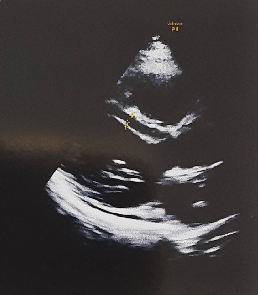
Assessing MR in echocardiogram: A well known tip

All of us know how tricky is, to assess and grade MR accurately. As discussed above it can as labile as systemic BP. Try to document the Heart rate and BP at the time of assesment.

Does Aoritc valve gets damaged with hypertension ?

Logically, high blood pressure is expected to damage the aortic valve more than the mitral valve. Does that happen? When the high-pressure ventricle contracts, the aortic valve is not resisting the flow like the mitral valve. It opens respectfully, so guess which valve is likely to get injured more. (However, it must be noted that the diastolic pressure exerts pressure on the far side of the aortic valve and can trigger aortic regurgitation and degeneration on the far side of the aortic valve. While high systolic jets can dilate or dissect the aorta. ) So, let me clarify this post doesn’t convey a meaning that Aortic valve is sort of protected against HT related Injury.

Final message

It is unacademic to delink mitral regurgitation (MR) from systemic hypertension, both in etiological and therapeutic aspects. Hence, it is wiser to include MR within the “Complication basket” of hypertensive heart disease. Recognising it and tackling in a timely manner will reap definite symptomatic benefit.This simple concept should be emphasized to our students.

Reference

1.Rahimi K, Mohseni H, Otto CM, Conrad N, Tran J, Nazarzadeh M, Woodward M, Dwyer T, MacMahon S. Elevated blood pressure and risk of mitral regurgitation: A longitudinal cohort study of 5.5 million United Kingdom adults. PLoS Med. 2017 Oct 17;14(10):e1002404.

Read Full Post »

A brief learning session with cardiology fellow about a possible new concept in Left heart failure.

What is the commmest cause for acute pulmonary edema?

Left sided heart failure”

“Can you be more specific , Left sided means ?

“I meant LV failure , typically DCM of any cause or Severe un-controlled HT”

“Ok .good. Does Left Heart failure include mitral valve dysfunction also ?

“Yes sir, very much. Classical mitral stenosis and Isolated MR can cause pulmonary edema. In fact, acute AR Iis also part of left heart disease”

So far, so good, now coming to the complex part of left heart

Can LA fail in isolation independent of LV , ie I mean with normal Left ventricle ?

I am not sure. Can we call new onset atrial fibrillation as a primary atrial failure that can result in pulmonary edema?

Excellent. You are absolutely right. But I am talking about mechanical atrial failure, not electrical. Are you aware that most of the time AF is a well tolerated arrhythmia , it can even be silent in many cases. This is because the pumping function of the atria contributes only 20-25% to LV filling. This can easily be compensated by augmented LV suction force , provided the baseline LV function is normal.

Have you heard about ACM. Atial cardiomyopathy?

“No sir”,

“You will hear more about it soon” (Ref 1) Scientists, especially Echo guys are closing in on this concept. We know, the atria has three functional components, namely conduit, reservoir, and pumping. Curiously, we have realized that the pumping function of LA may not be that critical from indirect observations from some land mark studies . (Rate control vs rhythm control studies in AF are a powerful proof on the atrial pumping function .(AFFIRM/RACE etc ) I don’t know, whether I am right in saying the above statement.

It is tempting to conclude , only if all the three functional components of atria gets affected , then only primary atrial dysfunction can be diagnosed. The concept is more complex than we realise. In diastole , pulmonary vein, LA , LV all work as single functional unit. Only in systole, we see them as different things.

Atrial DCM

Like LV systolic function, which is coupled with RV in parallel , LA function is closely knitted to LV in series during diastole. The key to suspect or diagnose this entity is to demonstrate dissociation of LVEDP with LA mean pressure & PCWP. This is not an easy job in bedside. Isolated Increase in LA volume without any reason , is one clue. LA ejection fraction is possible marker. (Kanagala P, . Int J Cardiovasc Imaging. 2020)

Final message

We are in the early days of understanding primary atrial mechanical failure, Atrial cardiomyopathy (ACM) or atrial DCM is being proposed as separate entity. It is very likely, some subsets of HFpEF might turn out to be primary atrial disease, depending on the level of investigation we do.

Reference

1.Li M, Ning Y, Tse G, Saguner AM, Wei M, Day JD, Luo G, Li G. Atrial cardiomyopathy: from cell to bedside. ESC Heart Fail. 2022 Dec;9(6):3768-3784. doi: 10.1002/ehf2.14089.

2.Patel, R.B., Lam, C.S.P., Svedlund, S. et al. Disproportionate left atrial myopathy in heart failure with preserved ejection fraction among participants of the PROMIS-HFpEF study. Sci Rep 11, 4885 (2021). https://doi.org/10.1038/s41598-021-84133-9

Read Full Post »

Myocardial development is a complex process that begins in the precardiac mesoderm, and is regulated by number of genes.

Duelen R, Sampaolesi M. Stem Cell Technology in Cardiac Regeneration: A Pluripotent Stem Cell Promise. EBioMedicine. 2017 Feb;16:30-40.

After formation of tubular heart , the initial increase in ventricular mass is achieved by development of trabeculations. Trabecular compaction coincides with genesis of coronary circulation, and results in formation of ventricular chambers.The hallmark of sponge-like myocardium is delayed and poor compactive forces.

Time line of ventricular compaction

A deranged compaction process need not be macroscopic. It can be very localized, regional, or global. The timing and the quantum of compaction is important , that injects the contractile power to the ventricle . It is increasingly observed in echocardiography that a small percentage of the population has more echo-free zones in LV myocardium than others. They could represent potential weak spots, at times of mechanical stress.

A curious case of normal Echo

Would like to share few images from a routine echocardiography in a healthy young female.

The apparent absence of mid myocardial shadow was not entirely a surprise though .At the same time, it raises some curious thoughts as to why certain myocardial areas are not well visualized by the ultrasound.

Look at the IVS, why should mid myocardium is echo free (measured 5mm) and appear de-laminated.
Only endocardium and epicardium are echogenic .

How echo is able to pick up only endocardium and epicardium , making entire myocardium look like an empty shell .

Still, LV is contracting vigorously, implying muscle mass is just not visible to ultrasound eye.

Clinical implication

These echo free dark zones in IVS or LV is so common, one can safely ignore, but its worth recalling few entities, that can be related to this. Intramyocardial hematoma, dissection and ultimately rupture (A case report) when they happen to develop an ACS -STEMI. We know , free wall and IVS rupture and mechanical complication occur signiifcant population of STEMI. Though, we can easily blame it on fate, may be these are the ones , who harbor such silent echo free slits due to defects in compacting genes making the myocardium soft , spongy that gives way wihtout a fight at times of tissue necrosis.

Can non-compaction occur without LV contractile dysfunction ?

Unlikely , is the likely answer from most of us. But , routine TTE might miss subsclinical LV dysfunction . We know , now degree of LV fucntion is directly related to the imaging modal;ity we depende to define it.

Can a consistency or sponginess of a myocardium be detected by echo?“`

As of now, it is not possible. This might become a reality when the science of tactile haptics enters the ultrasound domain.

Final message

“The act of observing changes the observed”

Non compacted LV is casually used term nowadays. The title of this post was made Intentionally provocative to stress a point, that what appears as non- compaction can be observed in normal persons.. However, request the fellows to look little deeper into the myocardial architecture, especially when you witness large echo-free zones.

Mind you, this is different from the well-defined condition of classical non-compaction with excessive deep trabeculations. Don’t know how to name this. This is different. Maybe “Isolated Intra-mural partial non-compaction” is an ideal term.

Counterpoint

When I discussed this with an expert colleague, he said, “No, you’re imagining a non-existing entity.” The ultrasonic interface with myocardium and the interstitial echoes that define the echo texture is so variable.Let us see.time will tell .(See this video and case report )

Postamble

This post is meant to be looked up purely from an academic perspective. Reporting such entities, unless you are absolutely sure, should be avoided, as it ends up increasing the anxiety of our beloved patients.

Reference

1.Sedmera D, McQuinn T. Embryogenesis of the heart muscle. Heart Fail Clin. 2008 Jul;4(3):235-45. doi: 10.1016/j.hfc.2008.02.007. PMID: 18598977; PMCID: PMC2715960.

2.Kirby ML. Cardiac Development. New York: Oxford University Press; 2007. [Google Scholar] [Ref list]

Read Full Post »

« Newer Posts